Talar neck fractures and complications AO Trauma Advanced


")


 • Fixation at")


- Slides: 29
Talar neck fractures and complications AO Trauma Advanced Principles Course
Learning objectives • Classify talar neck fractures and identify associated injuries • Describe the blood supply to the talus and its implications in treatment options and prognosis • Select a surgical approach appropriate to the fracture pattern • Evaluate fixation options • Identify complications (avascular necrosis, neurovascular injuries, malunion, infection)
Anatomy • 60− 70% articular cartilage • No muscular attachments • Complex articulations • Neck deviation
Blood supply • Extraosseus blood supply via three vessels • Peroneal • Anterior tibial • Posterior tibial • Major blood supply to body is the artery of the tarsal canal • Deltoid and sinus tarsi vessels provide significant minor vascularity
Blood supply
Mechanism • Shear force (not forced dorsiflexion)
Fractures of the talus • Talar neck • Talar body • Talar head • Lateral process • Posteromedial • Extruded talus
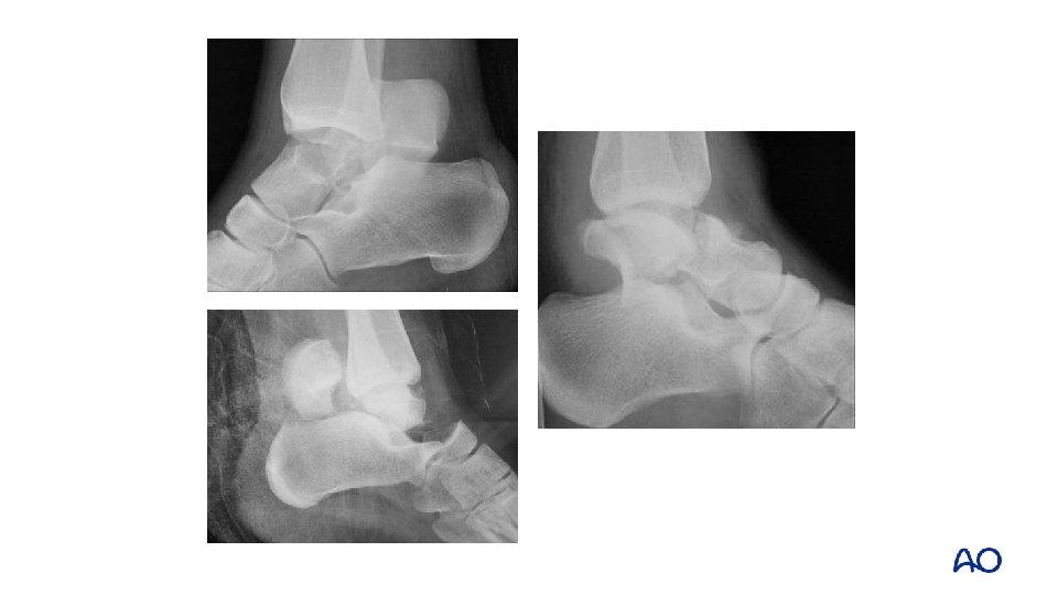
Talar neck fractures: Hawkins, AO/OTA Classification 81. 2 Type I/81. 2. A Type II/81. 2. B* Type III/81. 2. C * Type IV/81. 2. D* *Qualification: a. Simple, b. Multifragmentary
Talar emergencies? • Dislocated closed fractures • Open fractures • Closed fractures with soft tissue at risk • Neurovascular compromise • All talar neck fractures?
Talar emergencies? • Of 109 experts surveyed, 89 responded as follows: • 60% said waiting more than 8 hours is acceptable • 46% said waiting more than 24 hours is acceptable • Clinical studies show neither improved outcomes nor reduced rates of avascular necrosis after emergent definite fixation of all talar neck fractures
Goals of open reduction • Restoration of anatomy • Alignment of talar neck • Maximization of revascularization • Allow early ROM
Approaches • Anteromedial • Anterolateral • Posterolateral • +/- malleolar osteotomy • +/- fibular osteotomy
Medial approach
Lateral approach
Anteromedial and anterolateral incisions
Minimal soft-tissue stripping Medial Lateral
Surgical tactic • Consider dual exposures • Intraoperative distractor • Clean subtalar joint • Leave deltoid ligament intact • Span comminution (dorsomedial) • Avoid varus
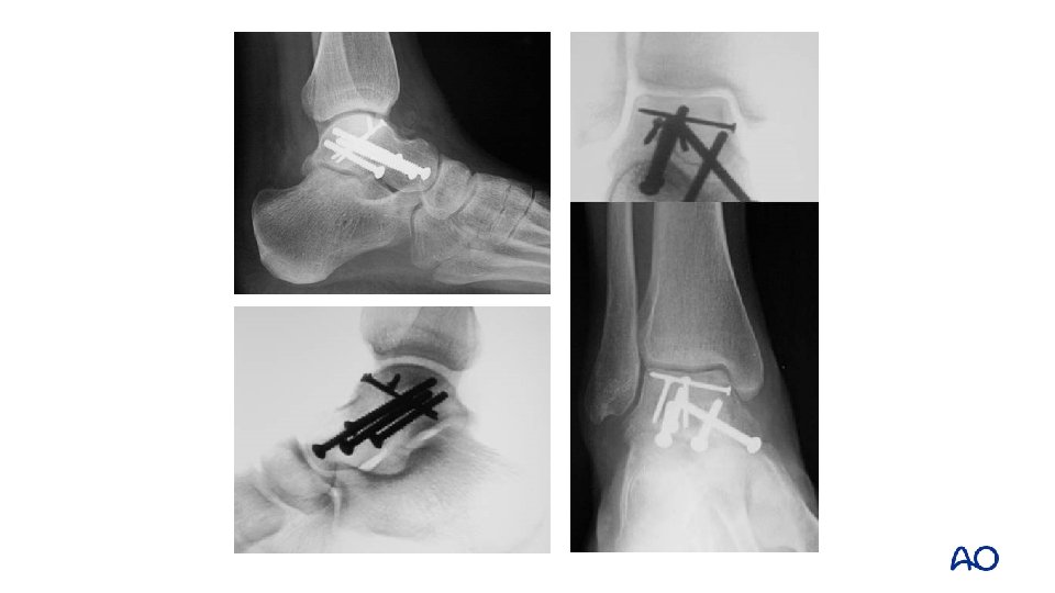
Surgical tactic • Reduce the tension side first (lateral and plantar) • Fixation at nonarticular talus • Countersink screws at talar head
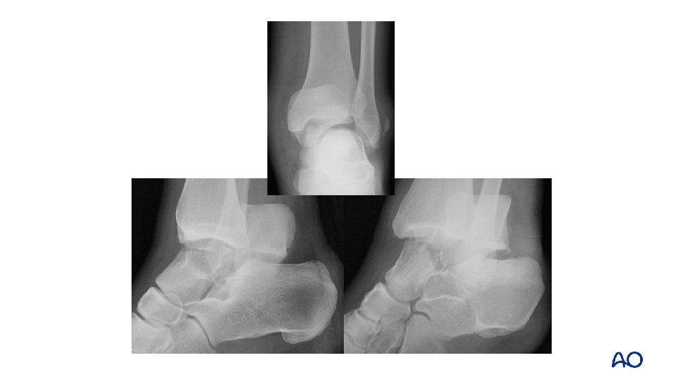
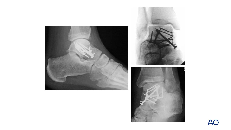
Plate fixation of talar neck fractures • Good results in 23 patients
18 -year-old woman, motor vehicle accident
Osteonecrosis 49% with radiographic evidence 63% with collapse 37% without collapse • 31% get avascular necrosis (AVN) with collapse • Hawkins II: 39% (9/23) • Hawkins III: 64% (9/14) • P = 0. 18
Functional outcomes In a study following 70 displaced talar neck fractures over 10 years: • 26 (37%) required secondary surgical procedures • Secondary procedures increased with time • Good function if no complications (? ) • Worse functional result with malalignment, subtalar arthritis • AVN low (9%)
Open reduction and stable fixation In a study following 26 isolated, displaced talar neck and body fractures for a minimum of 4 years • Union in 88%; all closed and healed (time independent) • Subtalar arthritis universally observed • AVN in 50% overall, 86% in open fractures • Conclusion: • Delay in treatment does not affect outcome • Arthritis more common than AVN • Open fractures problematic
Postoperative treatment—talar neck fractures • Active ankle and subtalar range of motion when wounds sealed • Nonweight bearing for 12 weeks • Plain x-ray evaluations • Weight bearing despite questionable vascularity
Take-home message • Accurate reduction is critical • Dual approaches • Avoid varus • Osteonecrosis does not equate to a poor result • Arthritis is common