
t s r i f n i V







- Slides: 31
t s r i f n i V D d n g a n i R n T e e r NB, c s er t s e trim E. Keshavarz
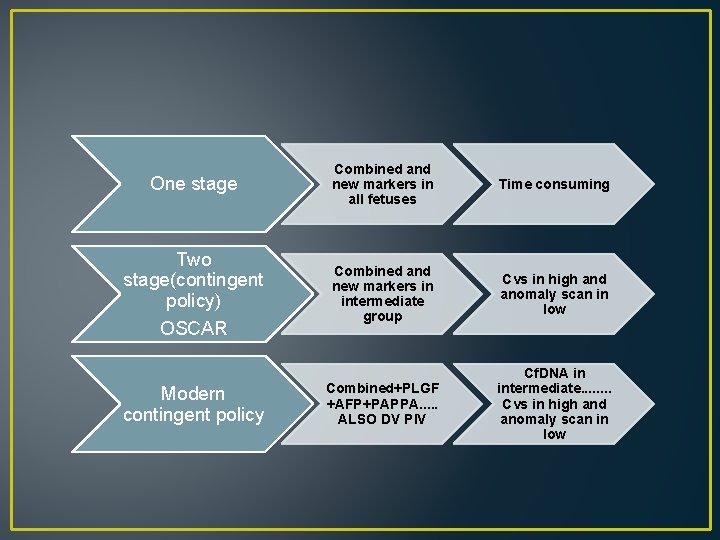
>=1/50 CVS(70 -90 day) Intermediate <1/50, >1/1000 Detailed US for looking the new US markers Low <=1/1000 US at W 20 High
One stage Combined and new markers in all fetuses Time consuming Two stage(contingent policy) OSCAR Combined and new markers in intermediate group Cvs in high and anomaly scan in low Combined+PLGF +AFP+PAPPA. . . ALSO DV PIV Cf. DNA in intermediate. . . . Cvs in high and anomaly scan in low Modern contingent policy
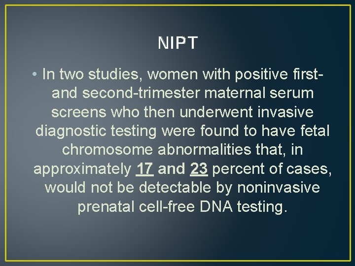
NIPT • In two studies, women with positive firstand second-trimester maternal serum screens who then underwent invasive diagnostic testing were found to have fetal chromosome abnormalities that, in approximately 17 and 23 percent of cases, would not be detectable by noninvasive prenatal cell-free DNA testing.
New US markers • 1 -NB • 2 -Facial angle • 2 -DV PI • 3 -Ductus venusus flow • 4 -Trichospid flow • 3 -TR
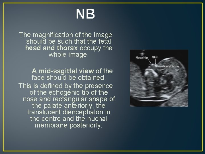
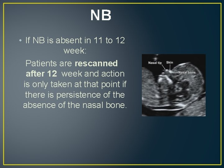
NB The magnification of the image should be such that the fetal head and thorax occupy the whole image. A mid-sagittal view of the face should be obtained. This is defined by the presence of the echogenic tip of the nose and rectangular shape of the palate anteriorly, the translucent diencephalon in the centre and the nuchal membrane posteriorly.
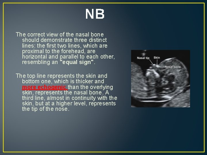
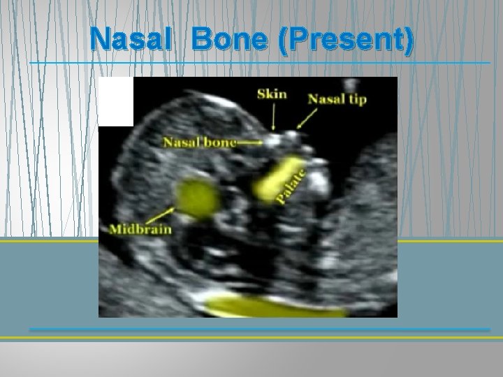
NB The correct view of the nasal bone should demonstrate three distinct lines: the first two lines, which are proximal to the forehead, are horizontal and parallel to each other, resembling an "equal sign". The top line represents the skin and bottom one, which is thicker and more echogenic than the overlying skin, represents the nasal bone. A third line, almost in continuity with the skin, but at a higher level, represents the tip of the nose.
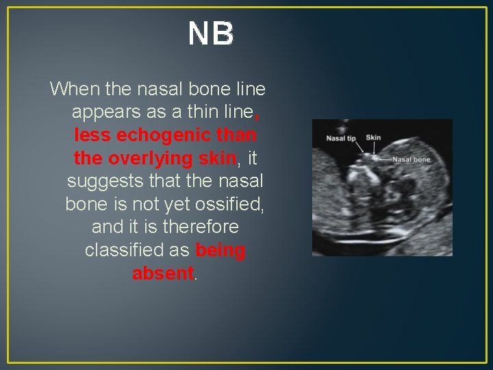
NB When the nasal bone line appears as a thin line, less echogenic than the overlying skin, it suggests that the nasal bone is not yet ossified, and it is therefore classified as being absent.
Nasal Bone (Present)
Nasal Bone (Absent)
NB • If NB is absent in 11 to 12 week: Patients are rescanned after 12 week and action is only taken at that point if there is persistence of the absence of the nasal bone.
• Firstly calculates a risk based on maternal age, fetal NT and maternal serum free β-h. CG and PAPP-A. • If the risk is more than 1 in 50 and the nasal bone is normal the risk does not change. • If the risk is 1 in 50 to 1 in 1, 000 and the nasal bone is normal the risk is usually reduced. If the nasal bone is absent the risk is always increased.
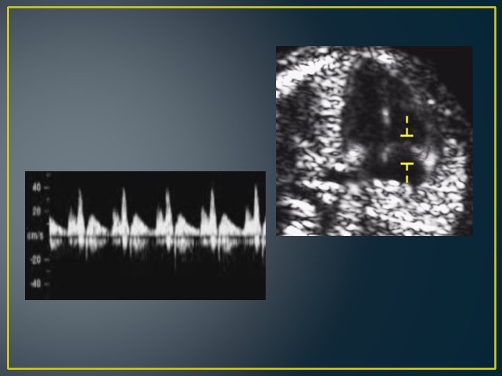
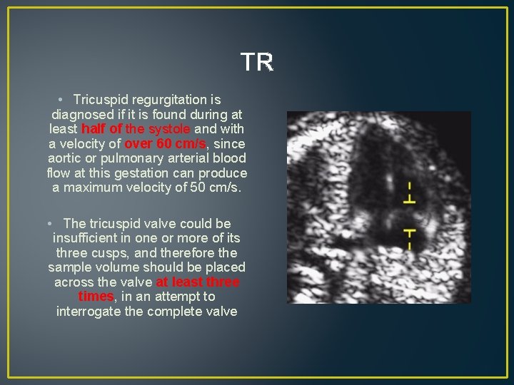
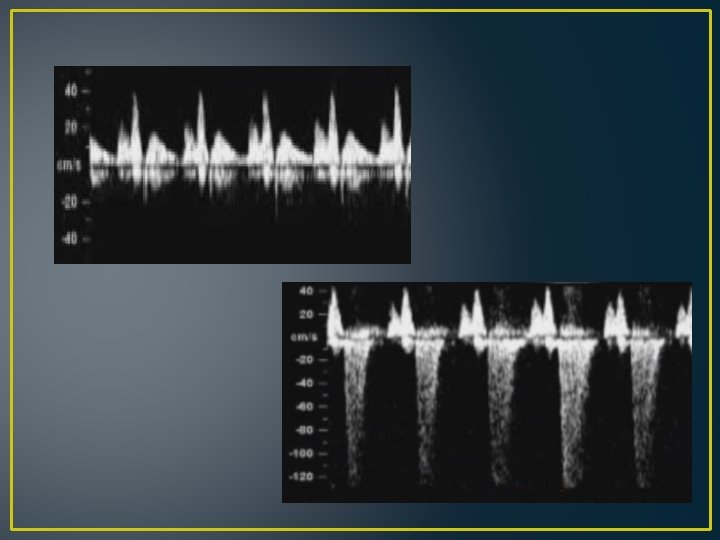
TR • The magnification of the image should be such that the fetal thorax occupies most of the image. • An apical four-chamber view of the fetal heart should be obtained. • A pulsed-wave Doppler sample volume of 2. 0 to 3. 0 mm should be positioned across the tricuspid valve so that the angle to the direction of flow is less than 30 degrees from the direction of the inter-ventricular septum.
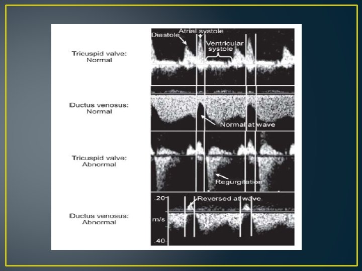
TR • Tricuspid regurgitation is diagnosed if it is found during at least half of the systole and with a velocity of over 60 cm/s, since aortic or pulmonary arterial blood flow at this gestation can produce a maximum velocity of 50 cm/s. • The tricuspid valve could be insufficient in one or more of its three cusps, and therefore the sample volume should be placed across the valve at least three times, in an attempt to interrogate the complete valve
• The incidence of tricuspid regurgitation is related to NT and CRL as well as aneuploidy, being more common when the NT is high and the CRL is low. • If the risk is more than 1 in 50 and tricuspid flow is normal the risk does not change. • If the risk is 1 in 50 to 1 in 1, 000 and the tricuspid flow is normal the risk is usually reduced. If there is tricuspid regurgitation the risk is always increased.
• In addition, there is an increased risk for cardiac defects and therefore such patients should have a follow up specialist fetal cardiac scan.
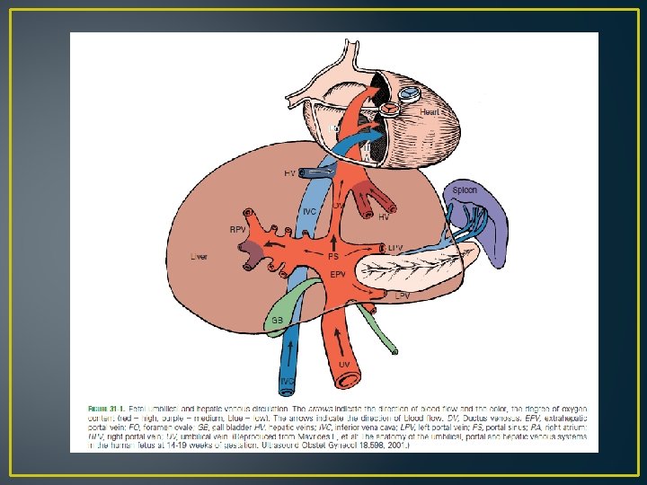
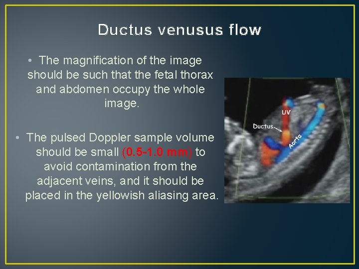
Ductus venusus flow • The magnification of the image should be such that the fetal thorax and abdomen occupy the whole image. • The pulsed Doppler sample volume should be small (0. 5 -1. 0 mm) to avoid contamination from the adjacent veins, and it should be placed in the yellowish aliasing area.
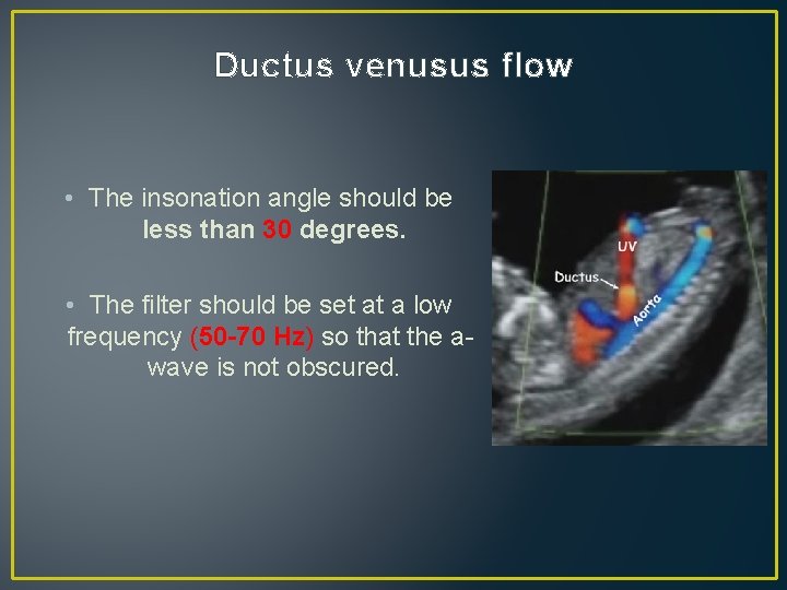
Ductus venusus flow • The insonation angle should be less than 30 degrees. • The filter should be set at a low frequency (50 -70 Hz) so that the awave is not obscured.
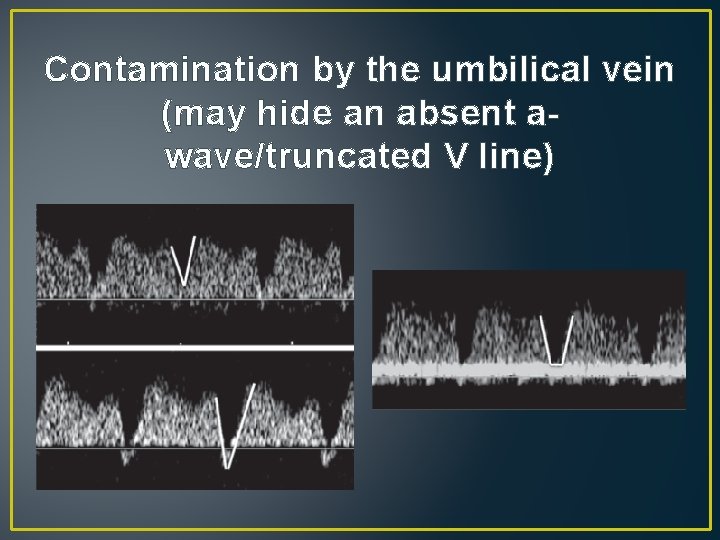
Contamination by the umbilical vein (may hide an absent awave/truncated V line)
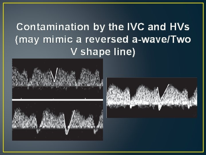
Contamination by the IVC and HVs (may mimic a reversed a-wave/Two V shape line)
• If the risk is more than 1 in 50 and ductus venosus flow is normal the risk does not change. • If the risk is 1 in 50 to 1 in 1, 000 and the ductus venosus flow is normal the risk is usually reduced. If there is reversed a-wave the risk is always increased
• In addition, there is an increased risk for cardiac defects and therefore such patients should have a follow up specialist fetal cardiac scan.
• The incidence of reversed ductus venosus a-wave is related to NT and CRL as well as aneuploidy, being more common when the NT is high , PAPP-A is low and the CRL is low. • 1 -Atrial contraction wave is greater than in later gestation. • 2 -Cardiac afterload is greater in the first trimester because of higher placental resistance. • 3 -Only a small impairment of the cardiac diastolic function might be enough for cardiac dysfunction to become evident as a reversed a-wave
• Abnormal ductus venosus flow in the first trimester may be a predictor of twin-to-twin transfusion syndrome (TTTS). • Previous studies reported that another early manifestation of TTTS is increased NT in at least 1 of the fetuses or a large discordance in NT between the 2 fetuses. • Multiple regression analysis demonstrated that in the prediction of TTTS there was no significant improvement by the addition of NT discordance to the finding of reversed a-wave.
Hey DI…VI…. I will patiently hunting you Thank you