SYSTEMIC SCLEROSIS DR EMAD ABOKHABAR LECTURER OF INTERNAL

SYSTEMIC SCLEROSIS DR EMAD ABOKHABAR LECTURER OF INTERNAL MEDICINE AND NEPHRLOGY SOHAG UNIVERSITY

Introduction n n Chronic systemic disorder of unknown etiology. Characterised by thickening of the skin (scleroderma) and distincitive involvement of multiple internal organs most notably lungs , GIT , heart , kidneys. Skin induration limited to fingers( sclerodactyly) No skin induration (SSc sine scleroderma) RAYNAUD’S phenomenon is predominant feature seen.

Definition SSc is a multisystemic , autoimmune disease affecting small arteries, microvessels and fibroblasts resulting in vascular obliteration, collagen accumulation and scarring (fibrosis) of skin and internal organs. Serologic specificity of the disease is the presence of ANA , directed against cellular nuclear enzymes, like DNA topoisomerase -1 (anti –Topo 1 ) and RNA polymerase, as well as centromeric proteins (anticentromere Ab)
 criteria for the classification of systemic")
Diagnostic criteria The AMERICAN COLLEGE OF RHEUMATOLOGY (ACR) criteria for the classification of systemic sclerosis. One major criteria, two or more minor criteria for diagnosis. MAJOR criterion : PROXIMAL scleroderma –characterized by SYMMETRICAL thickening, tightening and induration of the skin of the fingers and the skin proximal to the MCP /MTP joints. these changes may affect the entire extremity, face, neck, trunk. MINOR 1. SCLERODACTYLY – thickening, induration, tightening of the skin limited only to fingers. 2. DIGITAL PITTING SCARS/LOSS OF SUBSTANCE FROM THE FINGER PAD –due to ischemia. 3. BIBASILAR PULMONARY FIBROSIS- b/l reticular pattern of linear or lineonodular densities in basilar portions of the lung on CXR. diffuse mottling/honey comb lung not attributable to primary pulmonary disease

Pathology Following pathogenic mechanisms always present: Endothelial cell injury Fibroblast activation Cellular and humoral immunologic derangement. Activation of the immune system is an outstanding disease feature. –autoantibodies, perivascular lymphocytes (cd 4 T ) Chronic forms of GVHD shares features of SSc.

Clinical description The condition may be divided into different subtypes DIFFUSE CUTANEOUS SSc (Dc. SSc) LIMITED CUTANEOUS SSc (Lc. SSc ) SYSTEMIC SCLEROSIS SINE SCLERODERMA Dc. SSc – abrupt in onset, RP is common, thickening of trunk , acral skin edema –earliest features, PF, renal crises. Lc. SSc – skin induration limited to hands, face, feet—CREST. SSSc ---visceral disease without skin involvement.

SUBSETS OF SYSTEMIC SCLEROSIS

Clinical features RAYNAUD’S PHENOMENON: RF ü Episodic vasoconstriction in the fingers and toes. ü Tip of the nose , earlobes can also be affected. ü Triggers – exposure to cold, temperature, stress, vibration. ü frequency , severity in winter. ü Typical attack : PALLOR CYANOSIS ERYTHEMA. Vasoconstriction ischemia reperfusion. • women > men ü PRIMARY –exaggerated physiological response to cold.

Raynaud’s phenomenon § Primary – no underlying causes § Positive family h/o § Absence of digital necrosis § No ulceration , gangrene § Negative ANA test § Secondary ---- > 30 yrs § More severe § Assosciated with ischemia, infarction of digits.

SKIN features • Clinically evident skin thickening is the HALLMARK of SSc—distinguishes it from others. . • Symmetrical , B/L • Skin is firm , coarse and thickened. • Darkly pigmented extremities and trunk. • Diffuse tanning in the absence of exposure to sun – very early feature. • Vitiligo like hypopigmentation –in dark skinned individuals. • Pigment loss spares perifollicluar areas – salt and pepper appearance of skin –scalp , upper back , chest. • Loss of transverse creases on dorsum of fingers • Fixed flexion deformity of fingers

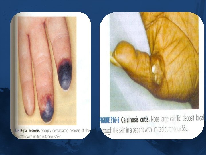
§ FACE – mouth fish appearance – § Skin is firmly bound to subcutaneous fat – thinning and atrophy. ü MACULAR telangiectasia –localised scleroderma ü ATROPHY of skin – slow healing ulceration on extensor surface of PIP joints. ü DIGTIAL pits – healed ischemic ulcers. ü Resorption of terminal phalanges – acro osteolysis. ü Calcium deposits in skin , soft tissue. . CREST syndrome – ü Finger pads, palms, extensor surfaces. ü Firm , non tender subcutaneous lumps– ulcerate through skin –chalky white matter.

Salt and pepper appearance of skin

Skin features Calcinosis telangiectsias

Pulmonary features • Leading cause of death. • ILD, PAH • Less frequently –aspiration pneumonitis , pulmonary hemorrhage , obliterative bronchiolitis , pleural involvement , restrictive ventilatory defect due to chest wall fibrosis , spontaneous pneumothorax. • broncho alveolar carcinoma • Asymptomatic until advanced.

Pulmonary Arterial Hypertension • Mean Pulmonary arterial pressure > 25 mm Hg at rest. • 12 -25% have PAH • In assosciation with ILD/solitary. • Usually downhill course –RHF –death. • Asymptomatic, exertional dyspnea. • PASP >40 mm Hg at rest in 2 d echo. • Right heart catheterisation accurate. • Increased BNP. • Prognosis degree of pulmonary artery pressure elevation.

GASTROINTESTINAL • • 90% Pts. Asymptomatic /weight loss Abnormal motility of intestines. Prominent atrophy , fibrosis of smooth muscle, intact mucosa, obliterative small vessel vasculopathy – all over GIT. • Oral aperture • Periodontal disease • GERD – early • LES pressure • Increased emptying time. • Gastroparesis – early satiety • Distention, abd pain • Increased reflux

GIT • GASTRIC VASCULAR ECTASIAs – seen in the antrum. • Subepithelial lesions • Diffuse small vessel vsculopathy • Watermelon appearance • Recurrent GI bleed occult • Fat , protein malabsorption, vitamin b 12 • Wide mouth sacculations in the colon occur – perforation. • Pneumatosis cystoides intestinalis • Primary biliary cirrhosis may coexist. • May present with acute abdominal pain

RENAL • HTN, chronic non progressive proteinuria. • SCLERODERMA renal CRISIS –dreadful complication of SSc. • 20 -25% pts • < 4 yrs of onset of diseases • It is due to OBLITERATIVE VASCULOPATHY of renal cortical arteries. • RBF JGA hyperplasia renin secretion RAAS renal vasoconstriction malignant HTN. • Localised scleroderma –infrequent development.

RENAL • Presentation –abrupt onset of malignant HTN, severe headache, blurred vision, chest pain. • 10% normal BP – normotensive renal crises. • Urinalysis -- proteinuria, microscopic hematuria, fragmented RBCs • Rapidly progressive oliguric renal failure follows • Creatnine > 3 mg/dl at presentation –poor prognosis. —permanent hemodialysis – high mortality, • Prompt use of ACEI to make BP under

Cardiac § § § § Myocardial , pericardial , conduction abnormalities. Can occur secondary to renal , pulmonary involvement. LVDD –due to HTN , myocardial fibrosis. Asymptomatic until HF , arryhthmias occurs. Myocarditis in assosciation with inflammatroy polymyositis. Conduction defects due to fibrosis of conduction system. Pericardial effusion.

Musculoskeletal CARPAL TUNNEL SYNDROME. Early disease –generalised arthralgia, stiffness. Joint mobility affected in diffuse disease. HANDS –PIP joints, wrists. Tendon friction rubs are present. —extensive fibrosis, adhesion of tendon sheaths. Muscle weakness due to disuse, malnutrition. Chronic noninflammatroy myopathy is usually seen. Bone resorption –a freq late complication –terminal phalanges –loss of distal tufts –ACRO OSTEOLYSIS. Mandible affected—biting difficulty.

Other disease manifestations § Dry eyes, dry mouth –SICCA complex. § Biopsy of the minor salivaryglands –fibrosis rather than lymphocytic infitration. (sjogren’s syndrome) § Hypothyroidism –fibrosis of gland. § CNS is generally spared. Sensory trigeminal neuropathy due to fibrosis /vasculopathy can occur. § Pregnancy -adverse outcomes § Erectile dysfunction in the male.

Laboratory features • Anemia – frequent • Most common is mild normocytic /microcytic anemia – chronic inflammation • Serum iron is low or normal, ferritin is elevated. • Iron deficiency anemia –occ ult GI bleed (WATERMELON STOMACH), chronic esophagitis. • Macrocytic anemia – vitmainb 12, folate, bacterial overgrowth, drugs. • Thrombocytopenia , leukopenia –DRUG toxicity. • ESR is normal • Increased ESR –MYOSITIS. / MALIGNANCY

ANTINUCLEAR ANTIBODIES ü All patients of SSc ü Highly specific are anti topoisomerase –I (Scl 70) and anti centromere Ab ü Topoisomerase I ab 31% of dc. SSc , 13% of lc. SSc ü Anticentromere ab in 38% of lc. SSc, 2% of dc. SSc. ü Antibody titers correlate with disease severity.

Differential diagnosis ü ü ü Based on vascular changes: 1. PRIMARY raynaud’s phenomenon 2. physical trauma –jack hammer 3. chemical exposure 4. drugs toxins – toxic oil syndrome, ergotamine, Bblockers , carbidopa, 5 HT. 5. SLE, DM/PM, RA. , cryoglobulinemia. Based on skin changes 1. localized scleroderma 2. scleroderma like skin changes 3. metabolic –genetic disorders –scleredema/scleromyxedema Based on visceral involvement 1. idiopathic pulmonary HTN 2. idiopathic pulmonary fibrosis 3. sarcoidosis 4. amyloidosis

Treatment ü No therapy has been shown significantly to alter the natural h/o SSc till date. ü Multiple interventions are avialable in alleviating the symptoms –slowing progression of organ damage. ü Combination of drugs are used. ü Physician –patient relationship to be mainatined.

Disease modifying treatments • Immunosuppressive agents –modest or no benefit in Rx of SSc. • Glucocorticoids - stiffness and aching in pts with early stage disease. • Do not influence the progression of skin/internal organ involvement. • ↑risk of scleroderma renal crises at high doses. • Avoid if possible. • Low dose , brief periods. • Cyclophosphamide –daily oral/IV in SSc related ILD. • Reduces the progression of ILD , stabilises, improvement in skin induration. • Early stage SSc/extensive pulmonary involvement – candidate

Disease modifying treatments • Methotrexate – modest therapeutic benefit • Potential profibrotic effects --? Use in fibrotic phase • Mycophenolate mofetil – improvement in skin induration • Immunomodulation – cyclosporine, azathioprine, extraxorporeal photopheresis, thalidomide, rapamycin. • Immune ablation with high dose chemotherapy followed by autologous peripheral stem cell reconstitution • Stem cell transplantation –experimental.

Rx of GI complications n n n Elevate head end of the bed. Eat frequent small meals PPIs , H 2 B at higher doses are effective. Laser photocoagulation –recurrent bleeding from watermelon stomach Metronidazole, erythromycin, tetracycline –bacterial overgrowth. Parenteral nutrition in case of severe disease.

Rx of PAH § Symptomatic – oral endothelin receptor antagonist § Diuretics § Oral anticoagulants § Digoxin § 0 2 –nasal cannula § Sildenafil – short term benefit § Lung transplantation

Rx of Renal Crises • • Medical emergency Avoid NSAIDs, glucocorticoids Rx –ACEI , short term dialysis. Kidney transplantation.

Skin care o o o o 5 mg prednisone D penicillamine Cyclophosphamide Regular skin massage Telangiectasia –laser pulse dyed laser Finger tip ulcerations –occlusive dressings Infected ulcers –topical Abs Surgical debridement.

Poor prognosis § § § § Male gender young age of onset African american race Extensive skin thickening Truncal involvement Visceral organ involvement Topoisomerase I ab Increased ESR , anemia , proteinuria on initial presentation –high mortality…

Take home message ü SSc is a chronic systemic disorder. ü RULE out other causes. ü Anti topoisomerase –dc. SSc, anti centromere ab Lc. SSc ü After diagnosis risk stratify the patient. ü Regular follow up and monitoring. ü Patient education is needed. ü Treatment is individualised. ü Renal crises is an emergency. ü ACEI are to be started in a pt with HTN. ü No treatment for calcinosis cutis

- Slides: 36