SYSTEMIC OPHTHALMOLOGY OCULAR MANIFESTATIONS OF DIABETES MELLITUS AND

SYSTEMIC OPHTHALMOLOGY -OCULAR MANIFESTATIONS OF DIABETES MELLITUS AND HYPERTENSION DR. ADNAN DEPARTMENT OF OPHTHALMOLOGY CHRI

ORBITORHINOMUCORMYCOSIS ORBITAL CELLULITIS

LIDS AND THE ADNEXA • Hordeolum externum

• Hordeolum internum

• BLEPHARITIS

CONJUNCTIVA • Xerosis of the conjunctiva.

CORNEA • The cornea shows a decreased nerve fiber density and nerve conductivity causing decreased corneal sensation.

• bacterial and fungal keratitis • neurotrophic non healing corneal ulcers

DIABETES AND GLAUCOMA • Many studies suggest a direct correlation between diabetes and Primary open angle glaucoma (POAG).

IRIS • Rubeosis iridis.

PUPIL • Diabetics generally have a smaller pupil and show latency to dilatation with instillation of mydriatics during examination.

LENS • Transient fluctuating myopia • cortical cataract and the posterior sub capsular cataract.

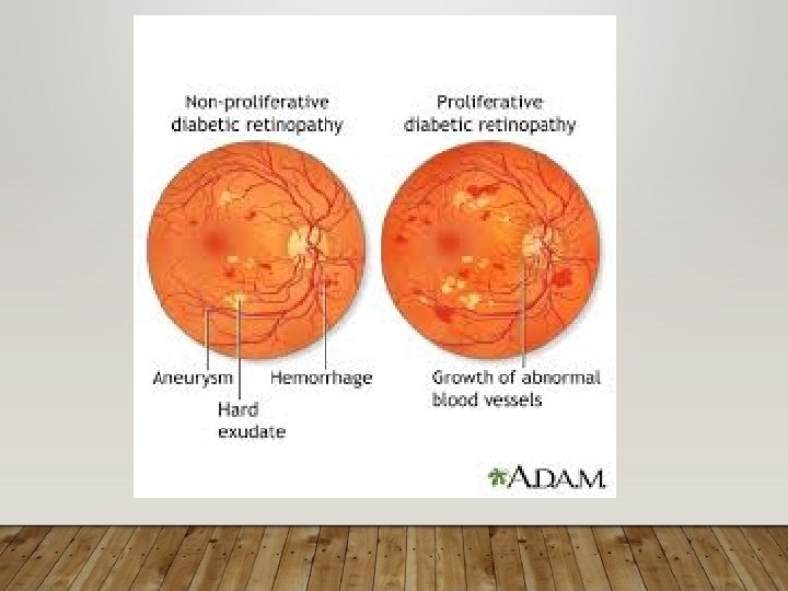
VITREOUS HAEMORRHAGE • Vitreous haemorrhage is commonly associated with proliferative diabetic retinopathy.

ASTEROID HYALOSIS CALCIUM SOAPS WITH LIPOIDS 5. 4% AMONG DIABETICS
 is probably up to 40%.")
DIABETIC RETINOPATHY • The prevalence of Diabetic retinopathy (DR) is probably up to 40%. It is more common in type 1 DM than in type 2 DM and sight threatening disease is seen in up to 10%

FOLLOW UP

CENTRAL RETINAL VEIN OCCLUSION
. • Cranial")
NEURO-OPHTHALMIC MANIFESTATIONS • Non arteritic type of anterior ischemic optic neuropathy (NAION). • Cranial nerve palsies (Oculomotor and Abducens) and ophthalmoplegia.

TRANSIENT REFRACTIVE CHANGES • Changes in the state of lens hydration & alteration of the refractive index of the lens due to osmotic changes. • Hypermetropia: in hypoglycemia • Myopia: hyperglycemia. NO GLASSES SHOULD BE PRESCRIBED IN UNCONTROLLED DM

HYPERTENSION

SCH

• Hypertensive retinopathy • Hypertensive optic neuropathy • Hypertensive choroidopathy

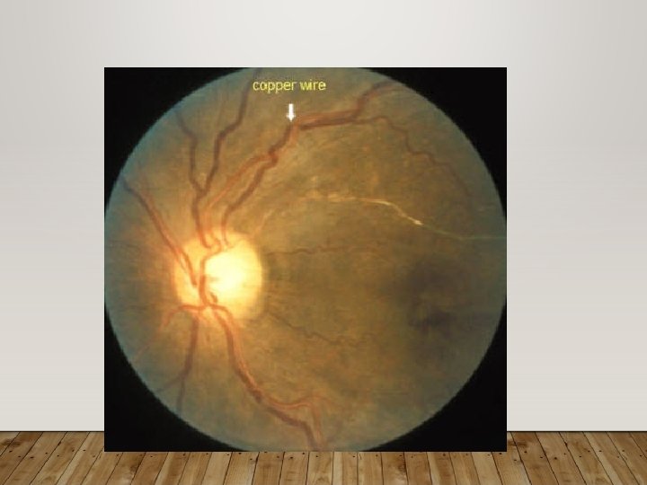
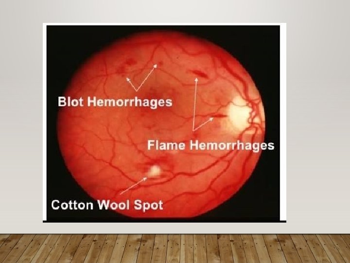
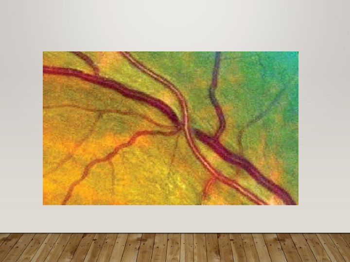
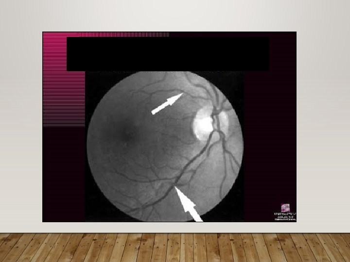
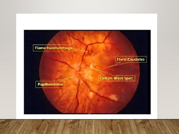
CHANGES IN HYPERTENSIVE RETINOPATHY • Focal arteriolar narrowing and arterial venous nicking – sclerosis • Flame hemorrhages • blot hemorrhages • Microaneurysm • hard exudates • Cotton wool spots • Optic disc swelling
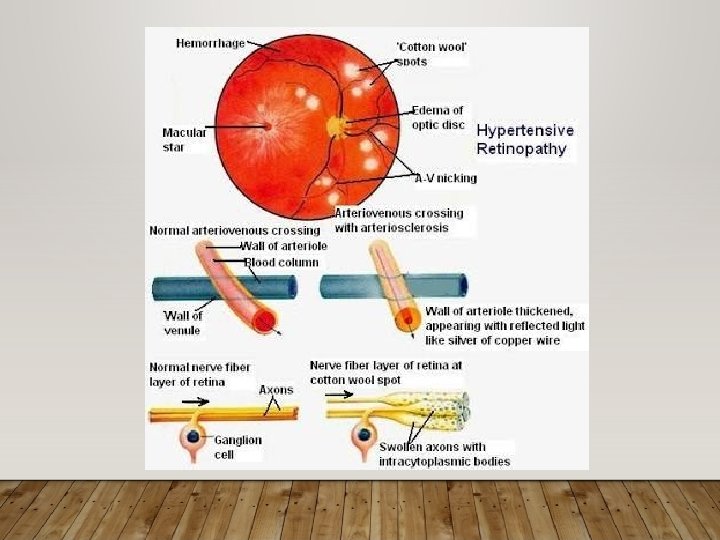

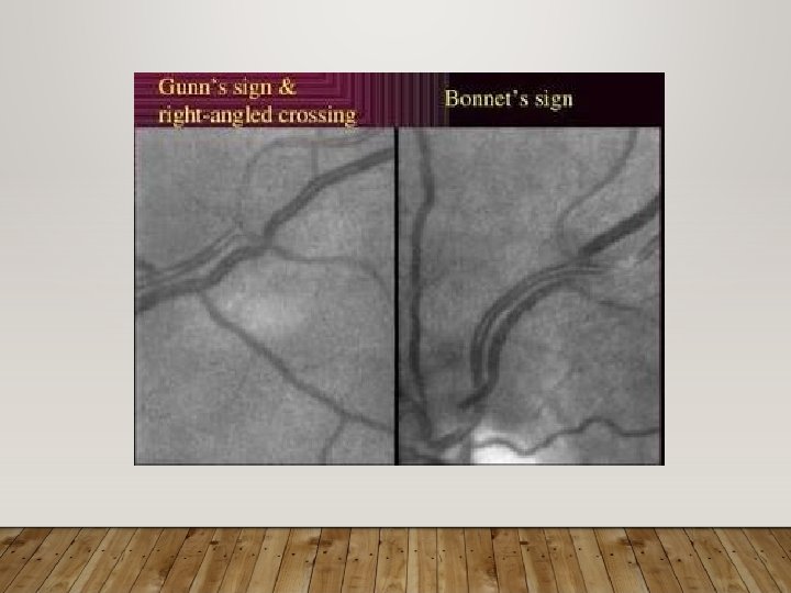
• AV crossing changes: • Venous deflection – salus sign • Venous nipping • Distal banking – gunn sign – bonnet sign
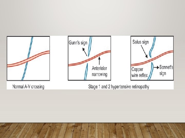

CLASSIFICATION • Keith Wagner and Barker Grade 1 - Mild to moderate narrowing Grade 2 - a. There is moderate to marked narrowing of retinal arterioles. b. Copper wire reflex c. Typical arteriovenous crossing changes Grade 3 – a. retinal arteriolar narrowing and focal constriction. b. Silver wire appearance c. Retinal oedema d. Cotton wool or soft exudates e. Superficial flame-shaped haemorrhages

• Grade 4 : grade 3 and macular star with papilledema

Modified Scheie Classification of "Hypertensive Retinopathy": • Grade 0 - No changes • Grade 1 - Barely detectable arterial narrowing • Grade 2 - Obvious arterial narrowing with focal irregularities • Grade 3 - Grade 2 plus retinal hemorrhages and/orexudates • Grade 4 - Grade 3 plus disc swelling

2021/6/5 HYPERTENSIVE CHOROIDOPATHY • Typically occurs in young patients • episode of acute hypertension associated with preeclampsia, pheochromocytoma, or renal hypertension • Elschnig spots - non perfused choriocapillaries • Siegrist streaks - hyperpigmented streaks

2021/6/5

2021/6/5

MANAGEMENT • Prompt control of BP • Grade IV retinopathy/ Papilloedema: matter of urgent referral

CONCLUSION • The physician should get ophthalmological opinion from the date of diagnosis of diabetes and hypertension and every year for early detection and prevention of its complications.
- Slides: 43