Systemic Lupus Erythematosus INTRODUCTION Systemic Lupus erythematosus SLE

 is a syndrome of unknown aetiology most")


. Bertsias G K et al. Ann Rheum")


 •")


 appears to be mediated by anti-platelet antibodies or/and anti-phospholipid")
, its cause is probably a combination")

 acute lupus pneumonitis: fever, dyspnea, cough with scanty sputum, hemoptysis,")


")


 is characterized by recurrent arterial and /or")
 Cutaneous necrosis of the legs. Fiehn C et al. Ann Rheum Dis 2001;")
. Notice the contrast between the involved left")


 cell it has been superseded by the")

 pattern of linear bright green staining around the")


 • Anti-Native DNA Antibody (Anti-n. DNA) •")
 • Most useful in SLE • Sensitive but not specific for")


 antigen • Specific for SLE • Present")


 Clinical feature seizure , psychosis , organ brain syndrome")


 Plasma exchange/ IVIG NSAIDS")
 are used preventively to reduce the")
")
 complications (Ca, vit D, bisphonates) • Aggressive BP and")

 high-grade fever, toxaemia, severe mucocutaneous manifestations, marked photosensitivity, moderate")
 organ/life-threatening features such as : focal/diffuse proliferative glomerulonephritis with")
 antiphospholipid syndrome (recurrent DVT, CVAs, recurrent foetal")
, which is then maintained with")

, 3. Steroids-injections,")
 lupus nephritis. 39 Systemic lupus erythematosus patients with proliferative")


, fever, malaise, and malar rash(2)")


 CRITERIA FOR DIAGNOSIS OF SLE • • Serositis (Pleurisy,")


, azathioprine")

- Slides: 145
Systemic Lupus Erythematosus
INTRODUCTION Systemic Lupus erythematosus ( SLE ) is a syndrome of unknown aetiology most commonly affecting young women. Virtually any organ of the body may be involved. Typically the course of the disease is a series of remissions and exacerbations. With good management, the ten years survival may be over 90%.
Etiology and Pathogenesis of SLE
1. Genetic factor • • Many studies have described familial aggregation of SLE. 5 -13% of lupus have at least one first or second degree relative with lupus It was found a 24 -58% concordance in monozygotic twins. 2 -5% concordance in dizygotic twins or siblings. The risk of a child developing lupus born from a mother (or father) with lupus is calculated to be 3 -4% at worst.
• What are the reasons of Genetic susceptibility? 1. It seems likely that most of the genes predisposing to SLE are normal. 2. An individual inherits an unlucky combination of normal genetic polymorphisms, each of which permit a little immune overreponse, or presentation of high quantities of target antigens in certain tissues. The combination of which is just enough to permit SLE to evolve after some environmental stimulus. 3. C 2, C 4, C 1 q deficiencies, DR 2, DR 3, 1 q 41 -42 region, Fc-r RIIA, IL 10 and Bcl polymorphisms.
2. Environmental factors 1. 2. UV light, especially UVB, flares SLE in most patients. It is unclear whether exposure to UV light can initiate the lupus, but onset after a sunburn is not unusual. There is good evidence that exposure of skin to UV light alters the location and chemistry of DNA as well as the availability of Ro and RNP antigens. Drug-induced lupus. Drugs ( hydralazine, procainamide, beta-blokers, isoniazid, penicillamine) can induce lupus. Drug-induce lupus may resemble SLE both clinically and serologically. Usually the disease is mild, and renal and neurological complications are rare. Generally, lupus that is caused by a drug exposure goes away once the drug is stopped.
3. Allergy. Does it induce lupus flare? No direct evidence. 4. Infection. There has been continuing interest in the possibility that infectious agents might initiate or flare SLE. Mechanism might include molecular mimicry between external Ag and a self-Ag, epitope spreading, nonspecific activation of T or B cells. There has been recent interest in EB, CMV and other virus.
3. Sex hormones • Female : Male=9: 1 • The sex difference is most prominent during the female reproductive years. • In mice, castrating females and /or providing androgens or antiestrogens protects from disease, whereas castrating males and providing estrogens accelerates and worsens SLE.
• The metabolish of sex hormone is abnormal in some lupus patients. Men and women with lupus metabolized testosterone more rapidly than normal, and estrogenic metabolites of estradial persist longer in women. • Neuroendocrine system. Hyperprolactinemia, abnormalities in hypothalamic and/or pituitary function.
4. Abnormal immune system • Sustained presence of autoantigens: increased apoptosis , impaired clearance of apoptosis • Hyperactivity in B and T lymphocyte. • Increased expression of surface molecules participating in cell activation in both B- and T-cell. • Overproduction of IL-6 and IL-10 • Defective regulatory mechanism.
Autoimmine Diseases Autoimmune diseases result from a break down of self tolerance
Autoantibodies to DNA, RNA, and a host of other cell nucleus antigens. Circulating immune complexes are frequently observed and these may deposit in the kidney, skin, brain, lung, and other tissues. It causes inflammation and tissue damage by a number of mechanism, notably fixation and activation of the complement system.
Overview of the pathogenesis of SLE Infection UV light Self Ag External Ag Skin cell Genetic susceptibility APC T cell IC APC Defective IC clearance B cell Ab Target
SLE Tissue damage occurs by: • 1. The formation of immune complexes (type III hypersensitivity) • 2. Antibody mediated injury to blood cells (type II hypersensitivity) The mere presence of the autoantibodies seen in these disorders can not be the sole cause of these disorders as the antigens for all these antibodies should normally be sequestered inside cells and not exposed the antibodies in the extracellular environment.
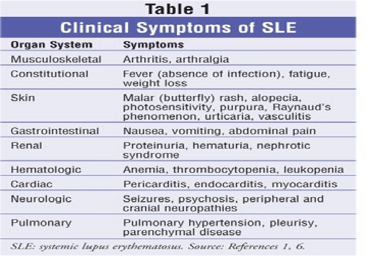
Clinical manifestations of SLE
In systemic lupus erythematosus all pathways lead to endogenous nucleic acids-mediated production of interferon (IFNα). Bertsias G K et al. Ann Rheum Dis 2010; 69: 1603 -1611 © 2010 by BMJ Publishing Group Ltd and European League Against Rheumatism
Clinical manifestations of SLE
The clinical spectrum of SLE is very broad It make SLE both fascinating but potentially difficult to diagnose and manage.
Disease course of systemic lupus erythematosus (SLE). Bertsias G K et al. Ann Rheum Dis 2010; 69: 1603 -1611 © 2010 by BMJ Publishing Group Ltd and European League Against Rheumatism
General symptoms • The most common symptoms listed as initial complaints are fatigue, fever, and weight loss. Fever: fever secondary to active disease was recorded from 50% to 86%. No fever curve or pattern is characteristic. It can be difficult, but very important to distinguish the fever of SLE from that caused by complicating infections.
• Fatigue is common in patients with SLE, especially during periods of disease activity. It is also often the only symptom that remains after treatment of acute flares. Low grade fever, anemia, or any source of inflammation can result in fatigue.
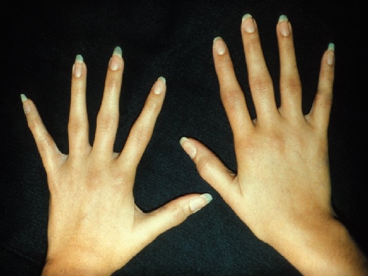
• Raynaud’s phenomenon is commonly found in lupus. It lack specificity. (a triphasic reaction of distal digits to cold or emotion, in which the skin colour changes from white to blue to red)
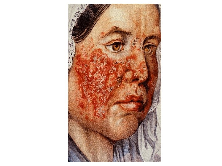
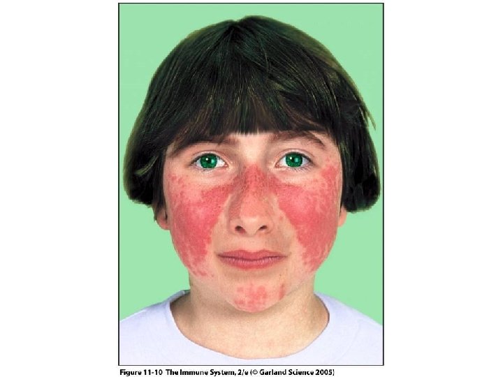
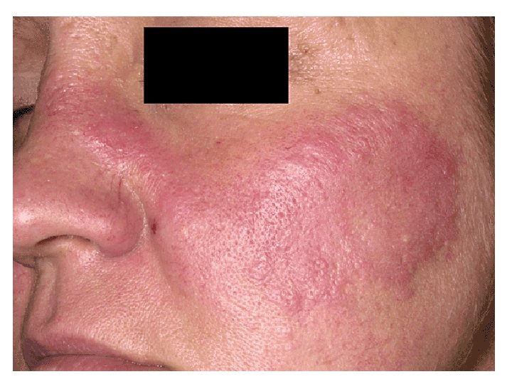
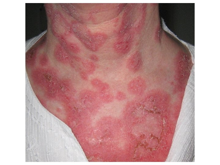
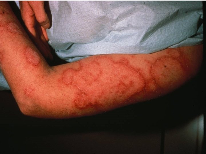
Dermatological involvement • • • Up to 85% of SLE Butterfly rash Maculopapular eruption Discoid lupus Relapsing nodular non-suppurative panniculitis Vasculitic skin lesin Livedo reticularis Purpuric lesions Alopecia Oral ulcer
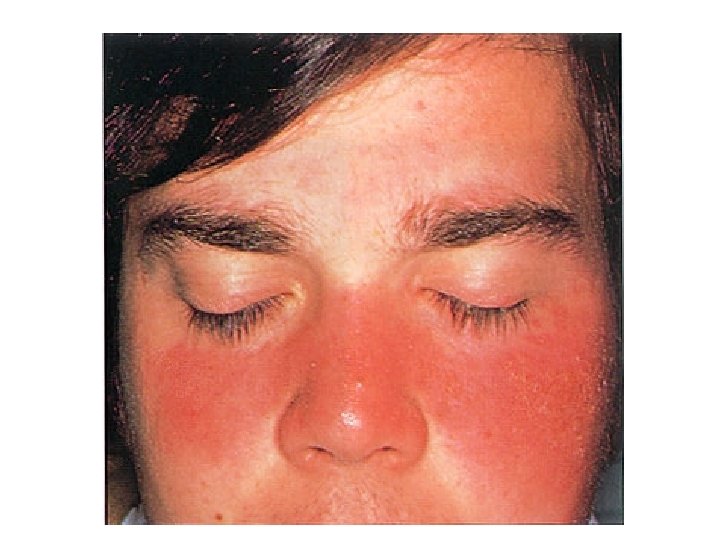
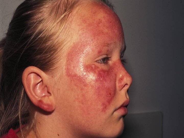
Photosensitivity sun poisoning rash
Skin clinical and pathology changes: a typical lupus skin rash seen in July 2003 (A) and in September 2003 (B). Gensburger D et al. Ann Rheum Dis 2005; 64: 153 -155 © 2005 by BMJ Publishing Group Ltd and European League Against Rheumatism
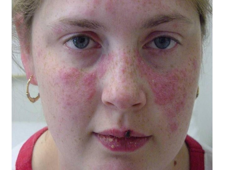
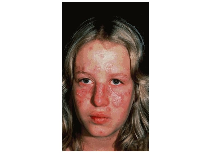
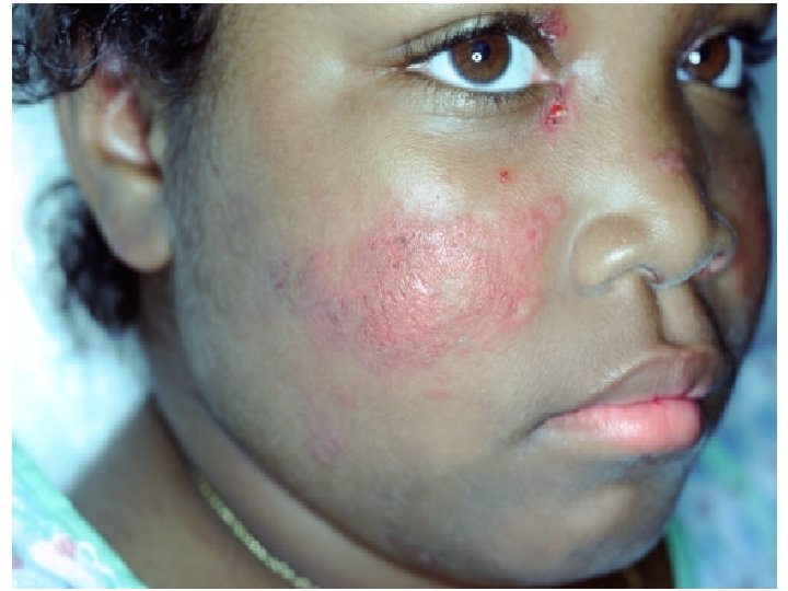
• Malar rash: This is a "butterfly-shaped" red rash over the cheeks below the eyes and across the bridge of the nose. It may be a flat or a raised rash. The rashes are made worse by sun exposure.
• Maculopapular eruption
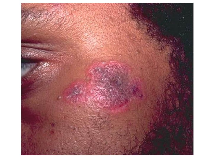
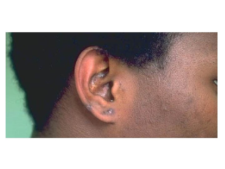
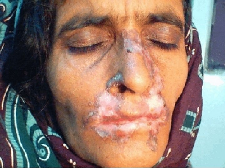
• Discoid lupus These are red, raised patches with scaling of the overlying skin.
Seal’s facial scars are the result of discoid lupus
SLE • Drug induced lupus has been seen with hydralizine, procainamide, isoniazid and Dpenicillamine*; all usually remit when the offending drug is discontinued.
• Vasculitic skin lesin
• Alopecia
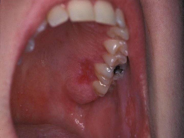
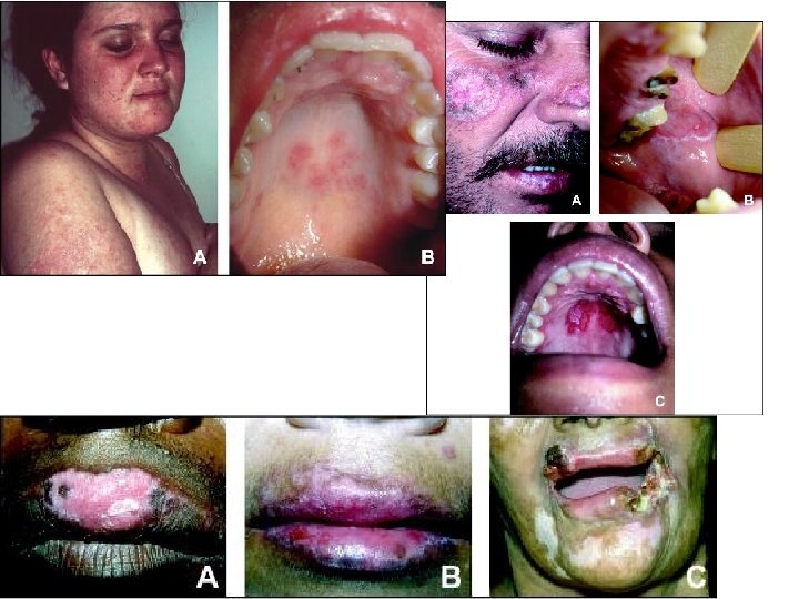
• Oral ulcer: Painless sores in the nose or mouth need to be observed and documented by a doctor.
• Ulcerated leukocytoclastic vasculitis in SLE
53 yo BF with severe generalized weakness, weight loss, and chronic psychosis Alopecia Malar rash Arthritis Psychosis
Musculoskeletal system • The arthritis of lupus is usually found on both sides of the body and does not cause deformity of the joints. Swelling and tenderness must be present. • The most frequently involved joints are those of the hand, knees, and wrists. • People with lupus can suffer from a certain type of low blood flow injury to a joint causing death of the bone in the joint. • The muscle involvement was reported in 3050% of lupus patients
• Avacular necrosis of bone. It may be caused by prednisone therapy
Kidney system • Haematuria • Proteinure (>0. 5 g protein/d or 3+ ) • Cast
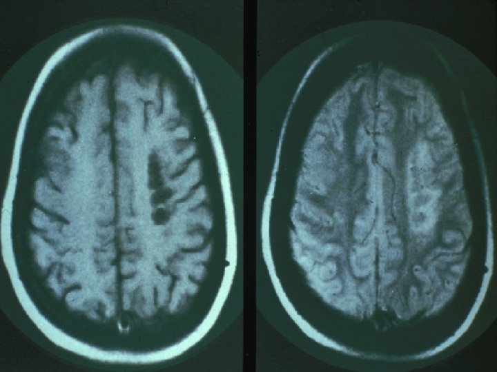
Nervous system • The brain , nerve problems and psychiatric syndromes are common in lupus affecting up to two-thirds of people. • Potential disorders include seizures, nerve paralysis, severe depression, and even psychosis. • Spinal cord involvement in lupus is rare and occurs primarily when there is clot formation in a critical vessel that supplies blood to the spinal cord.
Hematological abnormalities • Red blood cells a normochromic, normocytic anemia is frequently found in SLE. They appears to be related to chronic inflammation, drugrelated haemorrhage. haemolytic anemia as detected by the Coombs’ test is the feature of SLE. on rare occasion, a serum antibody may be produced which impairs red cell production.
• Platelets. thrombocytopenia (<100*109/L) appears to be mediated by anti-platelet antibodies or/and anti-phospholipid antibodies.
• White blood cell leucopenia (<4. 0*109/L), its cause is probably a combination of destruction of white cells by autoantibodies, decreased marrow production, increased or marginal splenic pooling, and complement activation. it should also noted that the immunosuppressive drugs used in the treatment of SLE may cause a marked leucopenia.
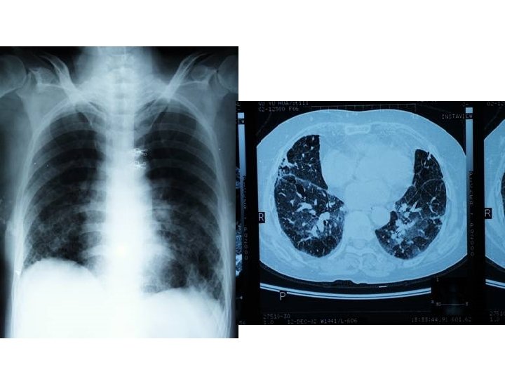
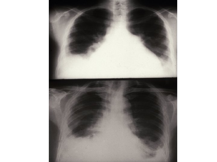
Pulmonary manifestations • Pleurisy it is the most common manifestation of pulmonary involvement of SLE. The volume of pleural effusions usually is small to moderate and maybe unilateral or bilateral. Large pleural effusion are uncommon. It usually exudative in character. Pleural effusions may also occur in SLE patients with nephrotic syndrome, infection, cardiac failure.
• Lung 1) acute lupus pneumonitis: fever, dyspnea, cough with scanty sputum, hemoptysis, tachypnea and pleuritic chest pain. 2) pulmonary hemorrhage 3) chronic diffuse interstitial lung disease. the diagnosis should not be made until infectious processes such as viral pneumonia, tuberculosis, and other bacterial, fungal and pneumocystis carinii infection have been completely excluded.
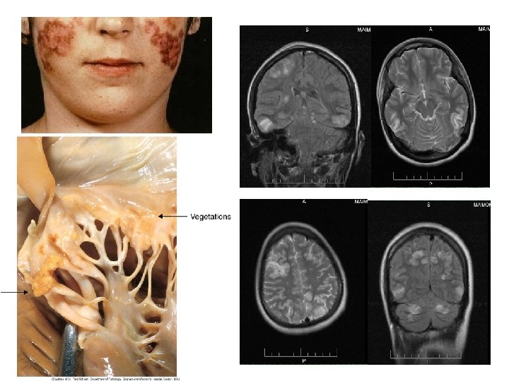
Cardiovascular manifestations • Pericarditis is the most common cardiac manifestation of SLE. • Myocarditis (the clinical features of lupus myocarditis resembles that of viral myocarditis) • Libman-Sacks endocarditis and valvular disease • Hypertension, cardiac failure
• Pericarditis
• SLE can be associated with endocarditis. Shown here is Libman-Sacks endocarditis in which there are many flat, reddish-tan vegetations spreading over the mitral valve and chordae. Transesophageal image of a mitral valve with masses characteristic of Libman. Sacks endocarditis.
Gastrointestinal and hepatic manifestation • Esophagitis, dysphagia, nausea, vomiting: (drug related in most cases) • Chronic intestinal pseudo-obstruction, mesenteric vasculitis, protein-losing enteropathy • Pancreatitis • Lupus hepatitis
Eyes • The eyes are rarely involved in lupus except for the retina. People with lupus often have to be screened by an ophthalmologist if they are taking the antimalarial drugs chloroquine or hydroxychloroquine
Secondary sjogren’s syndrome • Dry eyes • Dry mouth exocrine glands were infiltrated with lymphocytes
Secondary Antiphospholipid syndrome • Antiphospholipid syndrome (APS) is characterized by recurrent arterial and /or venous thrombosis, fetal loss and thrombocytopenia. High titer of Antiphospholipid antibody can be found in APS patients.
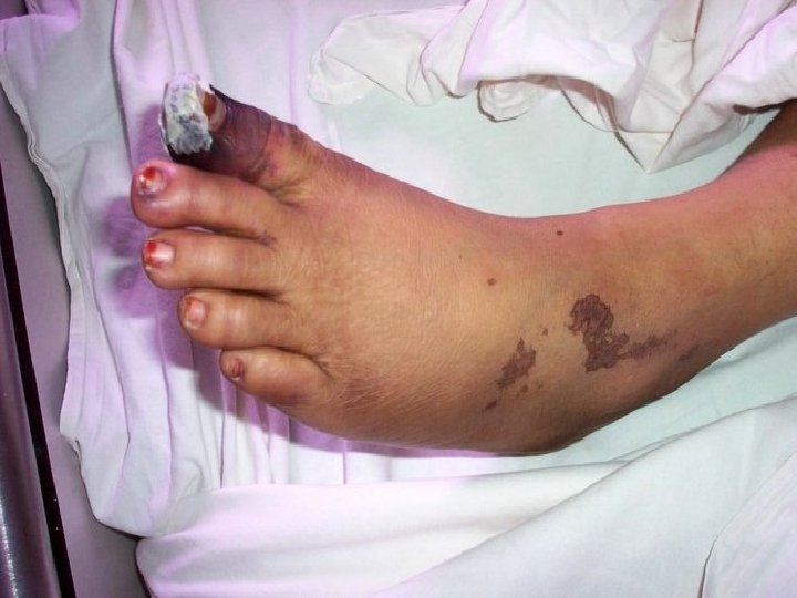
(A) Cutaneous necrosis of the legs. Fiehn C et al. Ann Rheum Dis 2001; 60: 908 -912 © 2001 by BMJ Publishing Group Ltd and European League Against Rheumatism
• Deep venous thrombosis (blood clot). Notice the contrast between the involved left leg and the normal right leg. Redness, swelling, and warmth combined with discomfort in the involved leg are cardinal manifestations of a deep venous thrombosis.
Laboratory investigation
Autoantibodies in SLE • Antibodies to cell nucleus component ANA, anti-ds. DNA, antibodies to extracellular nuclear antigen (ENA, anti-Sm, anti-RNP, anti-Jo 1) • Antibodies to cytoplasmic antigens anti-SSA, anti-SSB • Cell-specific autoantibodies lymphocytotoxic antibodies, anti-neurone antibodies, anti-erythrocyte antibodies, antiplatelet antibodies • Antibodies to serum components antiphospholipid antibody anticoagulants antiglobulin (rheumatoid factor)
Anti-nuclear antibodies • The lupus erythematosus (LE) cell it has been superseded by the ANA and anti-ds. DNA techniques. • ANA is a screening test anti-Sm, anti-ds. DNA antibodies are lupus specific antoantibodies.
• This homogenous pattern of diffuse bright green staining of nuclei seen by immunofluorescence microscopy with a Hep 2 cell substrate is called homogenous, and is the most common pattern with autoimmune diseases overall.
• This rim (peripheral ) pattern of linear bright green staining around the peripheral of nuclei seen by immunofluorescence microscopy with a Hep 2 cell substrate. • ds. DNA
• Nucleolar pattern
• Speckled pattern Scl 70, SSA, SSB, Sm
• These little Crithidia organisms have a small kinetoplast between the nucleus and the flagella which glows bright green under immunofluorescence microscopy, and is indicative of anti-native DNA antibody that is very specific for SLE.
• Immu-blotting method to detect anti -Sm, RNP, SSA, SSB, Jo 1, Scl 70 and ribosomal P.
Antibodies in SLE • Antinuclear Antibody (ANA) • Anti-Native DNA Antibody (Anti-n. DNA) • Anti-Smith (Anti-Sm) • Anti-Ribonucleoprotein Antibody (Anti-RNP) • Anti-RO/SSA; Anti-LA/SSB
Antinuclear Antibody (ANA) • Most useful in SLE • Sensitive but not specific for SLE • Also seen in drug-induced lupus, RA, scleroderma, chronic hepatitis • Since non-specific for SLE other more specific antibodies were sought
ANA: peripheral pattern • If ANA titer is > 1: 160, and there is a peripheral pattern, it strongly suggests SLE
Anti-n. DNA Antibody • More specific than ANA • Antibodies to “native” double-stranded DNA, a specific nuclear constituent that functions as an autoantigen • Occurs in ~70% of patients with SLE • Is for SLE
Anti-n. DNA Antibody • Used to confirm SLE in someone suspected of having SLE who has a positive ANA • Virtually all patients with a positive Antin. DNA antibody have a positive ANA • Therefore, don’t order if patient has a negative ANA
Anti-Sm Antibody • Antibody to Smith (Sm) antigen • Specific for SLE • Present in only 30% of patients with SLE (therefore not sensitive for SLE)
Lupus band test • Immunofluorescence of skin with antibody to Ig. G demonstrates a band-like deposition of immune complexes that is bright green at the dermal epidermal junction in this skin biopsy taken from an area with a visible rash. With SLE such deposition can be found in skin uninvolved by a rash, whereas with DLE the immune complexes are found only in involved skin.
Vasculitis • Vasculitis in arteries throughout the body can account for signs and symptoms from a variety of organ involvements. Seen here is an artery with extensive vasculitis with chronic inflammatory cells.
• SLE is associated with a peculiar periarteriolar fibrosis in the spleen, as shown here.
Kidney biopsy • WHO classification of lupus nephritis is based on light, immunofluorescence, and electron microscopic findings.
WHO classification of lupus nephritis immunofluorence Pattern mesangial peripheral electron microscopy mesangial subendothelial subepithelial Ⅰnormal 0 0 + 0 0 ⅡA mesangial deposit + 0 0 ⅡB mesangial hypercellularity + 0 ++ + ++ + + 0 Ⅲ focal segmental GN + Ⅳ diffuse GN + Ⅴ membranous GN ++
Semiquantitative assessment of activity and chronicity • Active indicators cellular proliferation, necrosis, karyorrhexis, cellular crescents, wire loops, hyaline thrombi, leukocytic infiltration, interstitial infiltration. • Chronicity indicators glomerular sclerosis, fibrous crescents, interstitial fibrosis, tubular atrophy Indicators are scored on a scale of 0 to 3, with necrosis, karyorrhexis, and cellular crescents weighted two times. The maximum of activity is 24, and the maximum of chronicity is 12.
Diagnosis
Criteria for diagnosing lupus • The diagnosis of lupus is a clinical one made by observing symptoms. Lab tests provide only a part of the picture. The American College of Rheumatology has designated 11 criteria for diagnosis. To receive the diagnosis of lupus, a person must have 4 or more of these criteria:
Criteria of the ARA for the classification of SLE 1. Malar rash: Fixed erythema over malar areas, sparing nasolabial folds 2. Discoid rash: Erythematous raised patches with keratotic scaling and follicular plugging 3. Photosensitivity: Skin rash after exposure to sunlight, history or physical exam 4. Oral ulcers: Oral or nasopharyngeal, painless, by physical exam 5. Arthritis: Tenderness, swelling, effusion in 2 or more peripheral joints 6. Serositis: A) pleuritis or B) pericarditis 7. Renal disorder A) proteinuria>0. 5 g/24 hour or 3+ or B) cellular casts 8. Neurological disorder: A) seizures or B) psychiatric disorder (having excluded other causes, e. g. drigs) 9. Haematological disorder: A) haemolytic anaemia or B) leucopenia or C) thrombocytopenia 10. Immunologic disorder: A) positive LE cells or B) raised anti-native DNA antibdy binding or C) anti-Sm antibody or D) false positive serological test for syphilis. 11. Positive antinuclear antibody:
Management and treatment
1. Monitoring the lupus patients • It cannot be emphasized too strongly that lupus is a disease requiring regular and careful follow-up. • Important initial advice should be given about avoiding UV light, infections, extreme stress or fatigue • Laboratory test—blood test, ESR, C 3, IC, liver function tests and antids. DNA.
2. Grading clinical activity • The highly variable nature of the syndrome • Evaluation of lupus activity is the base or beginning of therapy. • Non-life-threatening features such as arthralgia, skin rash, RP, alopecia • Severe complication such as renal, cerebral and heart involvement.
SLE disease activity index (SLEDAI) Clinical feature seizure , psychosis , organ brain syndrome visual disturbance, cranial nerve disorder lupus headache, cerebrovascular accidents, vasculitis arthritis myositis urinary casts, hematuria, proteinure, pyuria rash, alopecia, mucosal ulcers, pleurisy, pericarditis low complement, increased DNA binding fever thrombocytopenia, leucopenia score 8 8 4 4 4 2 2 2 1 1
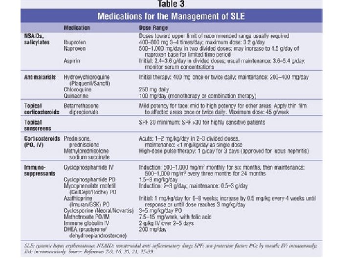
3. Clinical therapy • There are four main groups drugs useful in the treatment of lupus: the non-steroid antiinflammatory drugs, anti-malarials, corticosteroid and cytotoxic drugs. • How to treat lupus is a kind of art. Which and the dosage of drugs will be used to treat the patient depend on lupus activity.
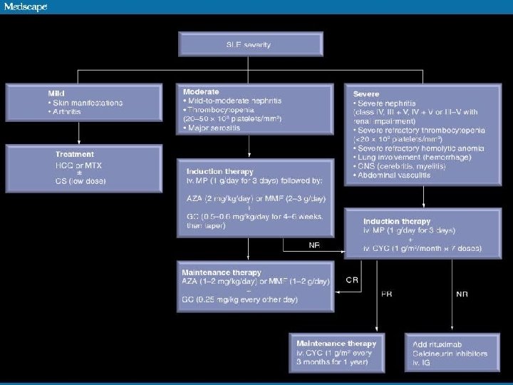
Treatment principles • Depends on disease severity • Fever, skin, musculoskeletal and serositis = milder disease • CNS and renal involvement – aggressive Rx • Emergencies: - severe CNS involvement - systemic vasculitis - profound thrombocytopenia (TTP-like syndrome) - rapidly progressive nephritis - diffuse alveolar hemorrhage
Medications used • • Steroids Cyclophosphamide Azathioprine Mycophenolate Chloroquine (Rituximab) Plasma exchange/ IVIG NSAIDS
Disease-modifying antirheumatic drugs • Disease-modifying antirheumatic drugs (DMARDs) are used preventively to reduce the incidence of flares, the process of the disease, and lower the need for steroid use; when flares occur, they are treated with corticosteroids. • DMARDs commonly in use are antimalarials and immunosuppressants (e. g. methotrexate and azathioprine). • Hydroxychloroquine is an FDA-approved antimalarial used for constitutional, cutaneous, and articular manifestations, • whereas cyclophosphamide is used for severe glomerulonephritis or other organ-damaging complications.
Steroids in Lupus • Steroid responsive • Steroid non-responsive • • • Dermatitis (local) Polyarthritis Serositis Vasculitis Hematological Glomerulonephritis (most) • Myelopathies Thrombosis Chronic renal damage Hypertension Steroid-induced psychosis • Infection
Preventive care • Medication-related (steroid) complications (Ca, vit D, bisphonates) • Aggressive BP and lipid control • Immunization (complement deficient) • Stress-dose steroid protocols for patients on maintenance corticosteroids (surgery/ infection) • Avoid UV exposure • Avoid estrogen therapies • Avoid sulfa-containing medications • Pregnancy planning
1. Mildly active lupus Category I arthritis, arthralgia, myalgia, fatigue, mild mucocutaneous involvement, low-grade fever, mild serositis, lupus headache combination of NSAID and / or antimalarials(chloroquine, hydroxychloroquine). The drug of choice is hydroxychloroquine (200 mg BD for 3 months and then 200 mg daily). Prednisolone remain the drugs of first choice to control lupus activity. Low dosage <=10 mg/d can be used therapy (Prednisolone 0. 3 -0. 5 mg/kg/day)
1. Category II (Moderate SLE) high-grade fever, toxaemia, severe mucocutaneous manifestations, marked photosensitivity, moderate to severe serositis, lupus pneumonitis, mild to moderate myocarditis, mesangioproliferative or minimal change lupus nephritis, haemolytic anaemia and thrombocytopenia prednisolone 1 mg/kg orally per day High dose of steroid must be continued till disease activity is well controlled that usually takes up to 6 weeks when it should be tapered off slowly over 6 to 12 months. In a toxic appearing patient, the administration of intravenous pulse methylprednisolone (15 mg/kg, max. 1 g) over an hour for 3 or 5 consecutive days may achieverapid control of lupus activity.
1. Category III (Severe SLE) organ/life-threatening features such as : focal/diffuse proliferative glomerulonephritis with or without azotaemia/hypertension lupus cerebritis with recurrent seizures, acute confusional state, coma; systemic necrotizing vasculitis such as one causing peripheral gangrene, GI bleeding or mononeuritis ultiplex A combination therapy consisting of high-dose daily oral prednisolone (40 -60 mg/day) and intravenous cyclophosphamide pulses (0. 75 gm/m 2, maximum of 1 g, over 1 hour) is recommended. The cyclophosphamide pulses are given once a month for 6 months by which time usually remission is achieved and then a aintenance pulse is administered every 3 months for a total of 2 years of cytotoxic therapy. Prednisolone is tapered off or reduced to a very low dose i. e. 5 -7. 5 mg per day by 6 months.
1. Category IV (SLE with miscellaneous features) antiphospholipid syndrome (recurrent DVT, CVAs, recurrent foetal loss etc. ), pure membranous lupus nephritis, chronic sclerosing lupus nephritis, seizures without other evidence of lupus activity, behavioural disorders without other serious manifestations, resistant thrombocytopenia or haemolytic anaemia Immunosuppressive therapy does not play any significant role in these conditions. Treatment of antiphospholipid syndrome. Heparin and warfarin should be started simultaneously so as to allow an overlap of about 5 days. INR should be adjusted between 3 and 4 on long-term warfarin therapy. The duration of warfarin therapy is life-long in patients with recurrent venous thrombosis. For refractory thrombocytopenia, danazol may be useful.
regimen for induction of remission (the first 6 months), which is then maintained with azathioprine 22. 5 mg/kg/day for about 2 years.
Biological agents Abatacept Abetimus Blocks CD 28 -mediated costimulation Blocks the production of antids. DNA antibodies Anakinra (IL-1 receptor antagonist) Blocks IL-1 signalling Atacicept (TACI-Ig) Soluble TACI receptor that binds to BLy. S and APRIL Reduction in B cells and immunoglobulin levels Infliximab (anti-TNF) Blocks TNF Rituximab (anti-CD 20 m. Ab) Depletion of B cells Tocilizumab (anti-IL-6 receptor m. Ab) Blocks IL-6
Treatment of SLE Arthritis, arthralgias, myalgias: 1. NSAIDS, 2. anti-malarials (eg. Plaquenil), 3. Steroids-injections, 4. oral methotrexate • Photosensitivity, dermatitis 1. 2. 3. • • 1. 2. • avoid Sun exposure topical steroids Plaquenil Weight loss and fatigue steroids Abortion, fetal loss ASA immunosuppression Thrombosis • 1. 2. 3. • Glomerulonephritis steroids pulse cytotoxics mycophenylate mofetil CNS disease anti-coagulants for thrombosis Steroids and cytotoxics for vasculitis Infarction (secondary to vasculitis) 1. steroids 2. cytotoxics 3. prostacyclin • Cytopenias steroids
Management of proliferative (class III–IV) lupus nephritis. 39 Systemic lupus erythematosus patients with proliferative nephritis may be stratified into those with moderate-to-severe versus severe disease based on impairment of renal function (increase in serum creatinine ≥ 30% and/or proteinuria ≥ 3. 0 g/day) and/or presence of adverse renal biopsy histological findings (crescents and/or fibrinoid necrosis >25% of glomeruli, chronicity index >4 or chronicity index >3 and activity index >10). Bertsias G K et al. Ann Rheum Dis 2010; 69: 1603 -1611 © 2010 by BMJ Publishing Group Ltd and European League Against Rheumatism
Management of renal lupus based on our own current practice. Ioannou Y , Isenberg D A Postgrad Med J 2002; 78: 599 -606 Copyright © The Fellowship of Postgraduate Medicine. All rights reserved.
Otherapy • • Plasma exchange Intravenous Immunoglobulin Stem cell transplantation Immune therapy ( anti-IL 10, anti-CD 20, and immune tolerance therapy)
SLE and pregnancy • SLE has been stable for more than 1 year. • Prednisone is no more than 10 mg/d, and cytotoxic drug has been stopped for more than 6 moth. SLE patients can plan to have a baby.
Case #1 • 30 yo female with polyarthritis, fever, malaise, and malar rash
Lab Work-up • CBC • Direct Coomb’s • Platelet Count • Urinalysis • • Anemia in 60 -70% (usu. normo/normo; hemolytic in 10%); WBC in 50% • Thrombocytopenia in 15 -30% • UA: RBCs; +/-casts; proteinuria
Which of the following is the probable diagnosis: A. Dermatomyositis B. Scleroderma C. Rheumatoid arthritis D. Polyarteritis nodosa E. SLE
Case #1 • 30 yo female with polyarthr itis(1), fever, malaise, and malar rash(2)
Systemic Lupus Erythematosus Lab Work-up • CBC • Direct Coomb’s • Platelet Count • Urinalysis • • Anemia (3) in 60 -70% (usu. normo/normo; hemolytic in 10%); WBC in 50% • Thrombocytopenia • (3) in 15 -30% • UA: RBCs; +/-casts; proteinuria(4)
Which of the following is the probable diagnosis: A. Dermatomyositis B. Scleroderma C. Rheumatoid arthritis D. Polyarteritis nodosa E. SLE • ANSWER: E
Case 2: History • A 36 -year-old female is seen for migratory arthritis of 6 months’ duration. She also reports some fatigue and a photosensitive skin rash. ROS notes: • • • Patchy hair loss 4 months ago that regrew Aphthous-like mouth ulcers every 4 to 6 weeks A diagnosis of “walking pneumonia” made last month based on symptoms of pleuritic chest pain
Case 2: Objective Findings • Pain with mild synovitis over the MCPs and PIPs • Rash over her face, legs, and trunk • Hgb = 12. 1; ESR = 33 • UA = 3+ protein • ANA = 1: 640 titer
Case 2: Question • With this clinical history, what is the most important thing to do now? A. Start an NSAID for the joint pain B. Start hydroxychloroquine to treat the rash and prevent recurrent pleurisy C. Fully evaluate her renal status and initiate appropriate therapy D. Start prednisone at 80 mg qd
Case 2: Answer • C. Fully evaluate her renal status • • Don’t Wait Aggressively evaluate renal status if the urinalysis is abnormal in SLE patients
Case 2: History • A 36 -year-old female is seen for migratory arthritis months’ duration. She also reports some fatigue and a photosensitive skin rash. (2) (1) of 6 ROS notes: • Patchy hair loss 4 months ago that regrew • Aphthous-like • A diagnosis of “walking pneumonia” made last month based on symptoms of mouth ulcers(3) every 4 to 6 weeks pleuritic (4)chest pain • UA = 3+ protein(5) • ANA = 1: 640 titer(6)
Prognosis • Benign to rapidly progressive • Better for isolated skin + musculoskeletal disease vs renal and CNS • Death rate 3 X age-comparable general population Mortality Ø Ø Nephritis (most within 5 yrs of symptoms) Infectious (active SLE + Rx – most common) CVS disease (50 X more MI than other woman) Malignancy (chronic inflammation + Rx)
Summary • Autoimmune disorder • Multiple manifestations • Aggressive investigation and treatment • Continued surveillance
AMERICAN COLLEGE OF RHEUMATOLOGY (ACR) CRITERIA FOR DIAGNOSIS OF SLE • • Serositis (Pleurisy, pericarditis) Oral ulcers Arthritis Photosensitivity Blood disorders (Leukopenia, thrombocytopenia) Renal involvement Antinuclear antibodies (ANA) Immunologic phenomena [false-positive Rapid Plasma Reagin (RPR)] • Neurologic disorder • Malar rash • Discoid rash
Course and prognosis • An episodic course is characteristic, with exacerbations and complete remissions that may last for long periods. These remissions may occur even in patients with renal disease. • A chronic course is occasionally seen. Earlier estimates of the mortality in SLE were exaggerated; 10 -year survival rate is about 90%. In most cases the pattern of the disease becomes established in the first 10 years; if serious problems have not developed in this time, they are unlikely to do so. The arthritis is usually intermittent. Chronic progressive destruction of joints as seen in RA and OA occurs rarely, but a few patients develop deformities such as ulnar deviation.
Summary - Key symptoms • • Arthralgia Alopecia Discoid lesions Fever Malar rash Oral ulcer Photosensitivity Weight loss
Summary - Key signs • A patient must have 4 or more of the following 11 criteria to be classified as having SLE - Malar rash - Discoid rash - Photosensitivity - Oral ulcer - Arthritis - Serositis - Renal disease - Neurologic disease - Hematologic disorders - Immunologic abnormalities - Positive antinuclear antibodies (ANA)
Summary - Key tests • • • ANA Anti-ds. DNA Anti-Sm Complement levels (especially C 3) False-positive test for syphilis
Summary – Key treatment • Corticosteroids: Solu-Medrol, prednisone • Immunosuppressive drugs: methotrexate (MTX), azathioprine • Antimalariais: hydroxychloroquine • NSAIDs
Thank you very much!