SYPHILIS Causative Organism Treponema pallidum Characteristics of Treponema


 Ø Can be seen")
 Treponema Pathogenic Non-pathogenic Non-venereal")
 Blood transfusion Via")


 Tabes dorsalis b) General paralysis of insane")

 g) h) i) Deafness Bone sclerosis, Arthritis Juvenile general paralysis of insane Damage")


 2 - Serological tests for Syphilis for all stages A.")

 Stage of disease VDRL TPHA No Past or")
 (Continued) Stage of disease VDRL TPHA FTA-ABS §Biological")
 (Continued) Treatment of syphilis : * Penicillin is")
 Borrelia Treponema Borrelia recurrentis Leptospira Borrelia vincenti Pathogenic genera of spirochaetes")
 (Continued) Borrelia vincenti : Gram –ve irregular spiral bacteria Culture :")

- Slides: 36
SYPHILIS
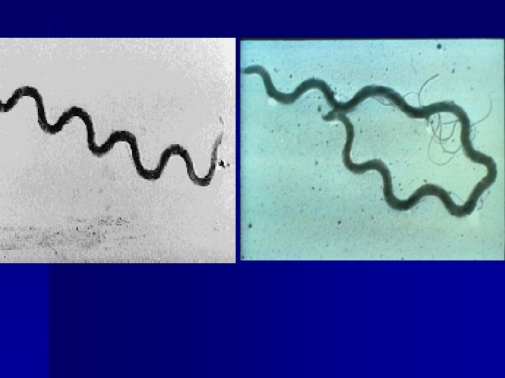
Causative Organism – Treponema pallidum Characteristics of Treponema pallidum Ø Ø Ø Spirochaetes or spiral organisms, they are motile, slender. Do not grow in artificial medium (some treponema are part of the normal oral flora e. g. T. denticulc) Cannot be seen by light microscopy because they are very thin (0. 15 μm), long 5 -15 μm. Note: Does not stain with gram stain
Causative Organism – Treponema pallidum Characteristics of Treponema pallidum (Continued) Ø Can be seen by § Dark field microscopy, by § Phase contract technique Ø Can be stained by § Silver impregnation § Fluorescent antibody technique Ø Ø Sensitive to penicillin They can be propagated by inoculation in rabbits in testes and anterior chamber of eye.
Causative Organism – Treponema pallidum Characteristics of Treponema pallidum (Continued) Treponema Pathogenic Non-pathogenic Non-venereal disease by direct contact T. pallidum Syphilus Oral commensals T. Denticula T. macrodentium T. microdentium T. Pertenue T. carateum T. pallidum A sexually transmitted disease Yaws Pinta Bejal
Mode of Transmission Ø Ø Direct sexual contact (90 – 96%) Blood transfusion Via placenta from infected pregnant mother faetus causes congenital syphilis. Contact accidental contact E. g. Medical personnel. Source of T. pallidum: Primary and secondary syphilis lesions.
Causative Organism: Treponema pallidum is the causative organism of syphilis. Syphilis can be Acquired Congenital Clinical Features of Syphilis / Symptoms and signs of Acquired syphil Syphilis is a sexually transmitted disease / a venereal disease Incubation Period: 10 – 90 days (average – 21 days) Ø
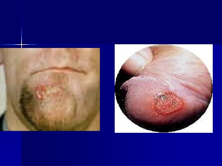
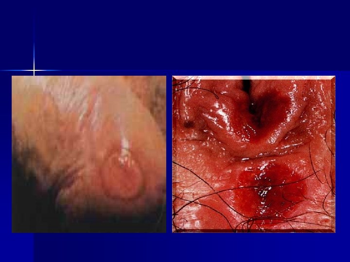
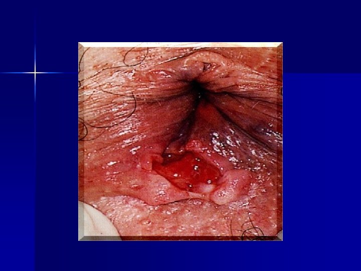
3 Stage of Syphilis: 1 - Primary syphilis: Primary chancre develops after 2 -10 weeks a well defined indurated painless ulcer mainly on the genitalia (90%) and extra-genital on Lips (5 -10%). In female, chancre occurs in the cervix. The chancre is painless and exudate is formed in the centre. This fluid is highly infectious and examination by dark field microscopes shows Spirochaetes. There is regional lymphadenopathy. § Primary chancre heals spontaneously without treatment within 38 weeks. § Primary syphilis is highly infectious. § Serological tests for Syphilis are positive in 80% cases.
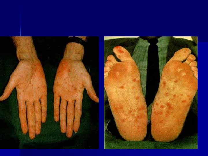
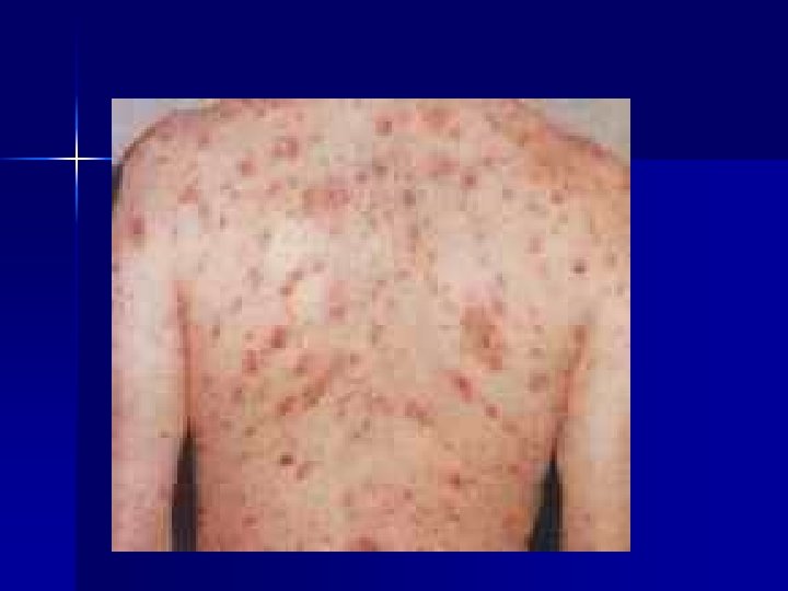
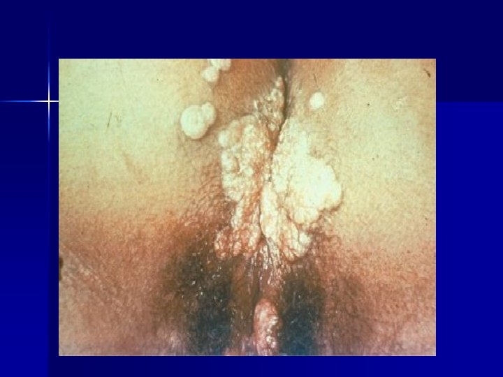
2 - Secondary Syphilis: After 6 -8 weeks of primary chancre: § Desseminated secondary stage develops. Muco-cutaneous lesion occurs e. g. Skin rash, mucasal ulcers, condylomata on genitalia, Lymphadenopathy, fever headache malaise, alopecia. § Secondary syphilis is highly infectious § Snal – truck mucosal ulcers in the mouth Hepatitis, glomerulonephritis, periostitis, iridocyclitis, choroidoretinitis, arthritis. § Serological tests for syphilis becomes almost uniformly positive. Secondary Stage may follows by the following: a) b) c) d) Cured spontaneously Early latent Latent Tertiary stage
Latent Stage: Ø Ø After the secondary syphilis symptoms subsides, the disease enters a latent stage. After about 2 years, the syphilis is NOT normally infectious, except from mother to the foetus.
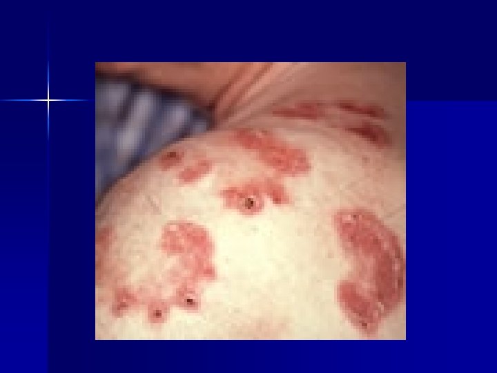
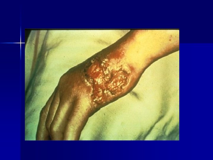
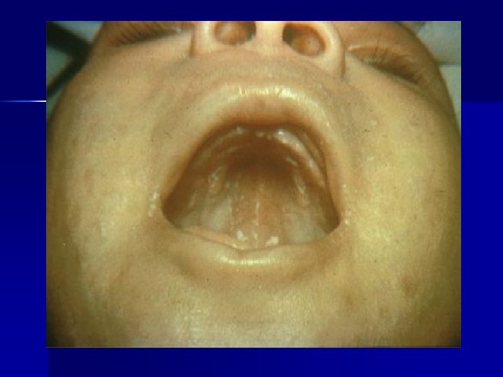
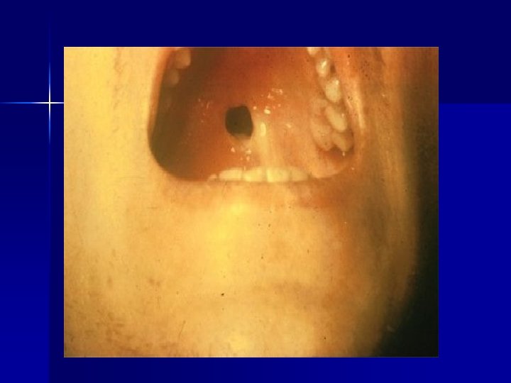
3 - Tertiary Syphilis: After 2 -20 yrs, tertiary stage develops produces §Gummatous Lesions in perforation of the palate (Roof of the mouth) which interferes with speech §Skin §Bone §Joints Charcoat’s joints § Cardiovascular System E. g. (a) Aortic aneurism (b) Aortic valve incompetence
Central Nervous System: Neurosyphilis E. g. a) Tabes dorsalis b) General paralysis of insane c) ØTertiary stage is not infectious. Meningovascular sympilis
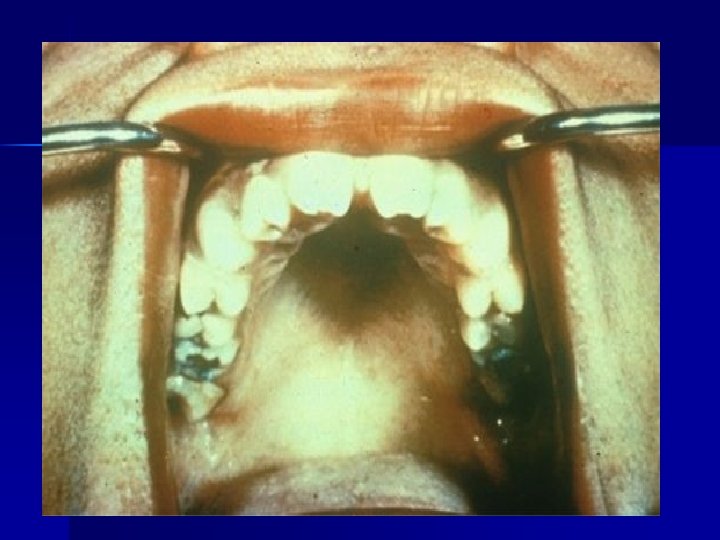
Ø Ø Ø Congenital Syphilis: most distressing and dangerous form of Syphilis. Early Congenital syphilis: a) Skin: rash maculopapular rash b) Mucosal Lesion: Mucocutaneous lesions c) Hepatospleenomegaly d) Lymphadenopathy Late Congenital Syphilis: a) b) c) d) d) Hutchinson’s teeth (Notching of the incisor teeth) Mulberry molars, Moon’s molars Sadle nose Sabre shin (tibia) Interstitial keralitis, blindness
f) g) h) i) Deafness Bone sclerosis, Arthritis Juvenile general paralysis of insane Damage of Mental development and other neurological symptoms. j) Stillbirths
Laboratory of Syphilis 1 - Dark ground microscopy to demonstrate, Spirochaetes T. pallidum in fluid or exudate from lesions of primary and secondary syphilis. a) Primary Syphilis exudate from chancre b) Secondary syphilis mucous path exudate taken for dark ground microscopy. n Direct immunofluorescent microscopy can be used.
Laboratory of Syphilis (Continued) 2 - Serological tests for Syphilis for all stages A. Non-specific Tests B. Specific Tests (for non-treponemal) (for non-treponemal or reagin antibody) (Ag used is cardiolipin) 1 - VDRL (Venereal Disease Research Laboratory) 2 - RPR (Rapid Plasma reagin) 3 - WR (Wasserman Reaction) (Ag used is Treponemal antigen) 1 - FTA – ABS Test (Florescent Treponemal Antibody Absorption) 2 - TPHA (Treponema pallidum Haemagglutination. 3 - TPI (Treponema pallidum immobilization) Congenital Syphilis Baby’s blood Ig. M –FTA-ABS TEST
Serological Tests for Syphilis (With Interpretation) Stage of disease VDRL TPHA No Past or Present Infec. §No Past /present Infection §Primary (Early) §Primary (Late) Or §Secondary + Tertiary §Latent §Treated syphilis (had infection before) FTA-ABS Or (Active Syphilis) in secondary syphilis.
Serological Tests for Syphilis (With Interpretation) (Continued) Stage of disease VDRL TPHA FTA-ABS §Biological false positive (No Infection by T. pallidum) §Congenital syphilis* * Early primary syphilis – FTA – ABS * After successful treatment – VDRL Note: FTA – ABS after successful treatment remains positive for life Positive Negative But FTA – ABS + TPHA Remain positive * V. D. R. L. is used to see efficacy (effect) of treatment. After successful treatment V. D. R. L. becomes negative.
Serological Tests for Syphilis (With Interpretation) (Continued) Treatment of syphilis : * Penicillin is the drug of choice Primary: * Penicillin for 15 days. Secondary and Tertiary syphilis : Penicillin for 21 days usually followed by 10 injection at weekly intervals. Note: Spirochetes are spiral motile bacteria. Their motility is due to contractile axial fibers run along the bacterial cell.
Spirochetes (spiral bacteria) Borrelia Treponema Borrelia recurrentis Leptospira Borrelia vincenti Pathogenic genera of spirochaetes are : * Borrelia, * Leptaspira, * Treponema Borrelia recurrentis Source : Rodents Disease : • Epidemic Louse borne relapsing fever • Endemic Louse borne relapsing fever Treatment: Tetracycline
Spirochetes (Spiral bacteria) (Continued) Borrelia vincenti : Gram –ve irregular spiral bacteria Culture : Strict anaerobic bacteria, difficult to culture. * Serum enriched media used Anaerobic culture Laboratory diagnosis : Mainly by Microscopic Examination of Gram stained smear only Disease : Borrelia vincenti and Fusobacterium species together produce: • Vincent’s angina (Pharynigitis) or Acute necrotizing ulcerative gingivitis • Gingivo - Stomatitis • Sore Throat Treatment : • Penicillin or Metronidazole • Oral hygiene