Synthetic Function Plasma proteins albumin globulins cholesterol triglycerides
, cholesterol, triglycerides and lipoproteins § Detoxification")
§ Synthetic Function § Plasma proteins (albumin, globulins), cholesterol, triglycerides and lipoproteins § Detoxification and excretion § Ammonia to urea (urea cycle), bilirubin, cholesterol, drug metabolites § Storage Function § Vitamins A, D, E, K and B 12 § Production of bile salts § Helps in digestion
 § Hepatitis § Icterus §")
§ Hepatocellular disease § Cholestasis (obstruction of bile flow) § Hepatitis § Icterus § Liver cancer § Steatosis (fatty liver)

§ Noninvasive methods for screening of liver dysfunction § Help in identifying general types of disorder § Assess severity and allow prediction of outcome § Disease and treatment follow up
 2. Uptake and excretion of")
1. Protein synthesis (albumin, α- and β-globulins, clotting factors) 2. Uptake and excretion of bilirubin and bile acids 3. Uptake of ammonia and conversion of ammonia to urea (BUN) 4. Glucose homeostasis and glycogen storage

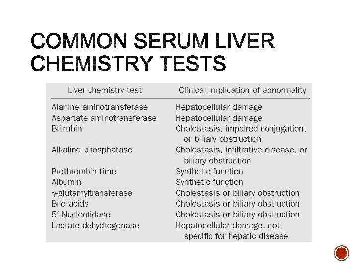
Broadly classified as: 1. Tests to detect hepatic injury: • Mild or severe; acute or chronic • Nature of liver injury (hepatocellular or cholestasis) 2. Tests to assess hepatic function

Group I: Markers of liver dysfunction ▫ ▫ ▫ Serum bilirubin: total and conjugated Urine: bile salts and urobilinogen Total protein, serum albumin and albumin/globulin ratio
 Aspartate aminotransferase (AST)")
Group II: Markers of hepatocellular injury ▫ ▫ Alanine aminotransferase (ALT) Aspartate aminotransferase (AST)
 g-glutamyltransferase (GGT)")
Group III: Markers of cholestasis ▫ ▫ Alkaline phosphatase (ALP) g-glutamyltransferase (GGT)

§ Normal LFT values do not always indicate absence of liver disease § Liver a has very large reserve capacity § Asymptomatics may have abnormal LFT results § Diagnosis should be based on clinical examination

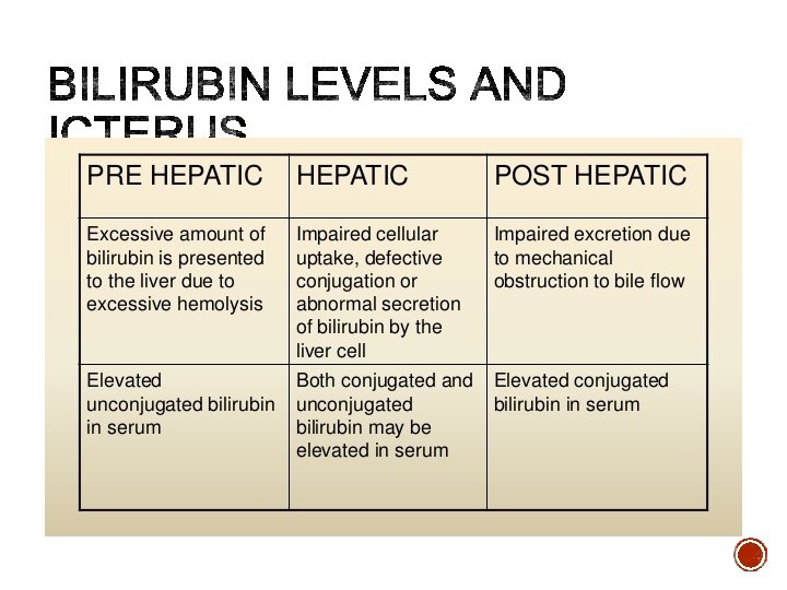
§ A byproduct of red blood cell breakdown § It is the yellowish pigment observed in icterus § High bilirubin levels are observed in: § Gallbladder stones § acute and chronic hepatitis
 § <0. 3 mg/d. L")
§ Normal § 0. 1 -0. 6 mg/dl (dogs) § <0. 3 mg/d. L (cats) § Conjugated (direct): § 0. 0 – 0. 4 mg/d. L (dogs) § 0. 0 – 0. 1 mg/d. L (cats)
")
Hepatocellular disease may cause increased concentrations of both unconjugated bilirubin and conjugated bilirubin. (1) The presence of unconjugated bilirubin is due to decreased bilirubin uptake and conjugation by damaged hepatocytes. (2) The presence of conjugated bilirubin is due to intrahepatic cholestasis from hepatocyte swelling. Post-hepatic biliary obstruction is expected to increase conjugated bilirubin concentration, but hepatic injury secondary to cholestasis also may result in increased concentration of unconjugated bilirubin.

Dogs and cats with pre-hepatic or post-hepatic hyperbilirubinemia often have a mixture of conjugated and unconjugated bilirubin. Evaluation of other laboratory parameters may provide more information about the cause of hyperbilirubinemia than does the percentage of conjugated vs. unconjugated bilirubin. (1) Anemia suggests hemolytic disease, particularly with evidence of a hemolytic anemia (spherocytes, positive Coombs’ test, Heinz bodies, etc. ). (2) Increased GGT or ALP activity in conjunction with hyperbilirubinemia suggests cholestatic disease

§ Most UBG is metabolized in the large intestine but a fraction is excreted in urine § Normally bile salts are NOT present in urine § Obstruction in the biliary passages causes: § Leakage of bile salts into circulation § Excretion in urine

§ The most abundant protein synthesized by the liver § Normal serum levels: 3. 5 – 5 g/d. L § Synthesis depends on the extent of functioning liver cell mass § Longer half-life: 20 days § Its levels decrease in all chronic liver diseases

§ Normal serum levels: 2 – 3. 5 g/d. L § a and b-globulins mainly synthesized by the liver § They constitute immunoglobulins (g-globulins-antibodies) § High serum g-globulins are observed in chronic hepatitis and cirrhosis:

§ Normal A/G ratio: 0. 5 -1. 7 § Globulin levels increase in hypoalbuminemia as a compensation

§ Prothrombin: synthesized by the liver, a marker of liver function § Half-life: 6 hrs. (indicates the present function of the liver) § PT is prolonged only when liver loses more than 80% of its reserve capacity § Vitamin K deficiency also causes prolonged PT § Intake of vitamin K does not affect PT in liver disease

§ Also present in erythrocytes and myocytes § Normal range: 10– 40 U/L § A marker of hepatocellular damage § High serum levels are observed in: § Chronic hepatitis, cirrhosis and liver cancer
: 10 -60 § High serum")
§ More liver-specific than AST § Normal range (U/L): 10 -60 § High serum levels in acute hepatitis (300 -1000 U/L) § Mild to moderate increases in ALT activity may occur with anticonvulsants, corticosteroids(dog), and thiacetarsemide. This is most likely due to mild hepatocellular injury rather than induction.

§ Appears in plasma many days before clinical signs appear § A normal value does not always indicate absence of liver damage § Obese but otherwise normal individuals may have elevated ALT levels

§ The liver of all animals contains high SDH activity. Increases in serum SDH activity generallyare considered liver specific in all species studied.

§ SDH is the enzyme of choice to detect hepatocellular injury in horses, sheep, goats, and cattle, because it is more specific for hepatic disease than AST and ALT in these species. § (1) AST is not a liver-specific enzyme. § (2) Hepatic ALT activity is too low to serve as a marker of hepatocellular injury in these species.

§ A non-specific marker of liver disease § Produced by bone osteoblasts (for bone calcification) § Present on hepatocyte membrane § Normal range: 40 – 150 U/L § Modearte elevation observed in: § Infective hepatitis and hepatocellular carcinoma

§ High levels are observed in: § Extrahepatic obstruction and intrahepatic cholestasis § Very high levels are observed in: § Bone diseases
 Cholestatic diseases (2) Bone lysis")
§ Increased serum ALP activity may occur in: (1) Cholestatic diseases (2) Bone lysis or remodeling (e. g. , bone tumors, young, growing animals) (3) Corticosteroid treatment or Cushing’s disease (dogs) (4) Phenobarbital treatment (likely due to hepatic injury/cholestasis) (5) Hepatic nodular hyperplasia (dogs) (6) Colic in horses (7) Feline hepatic lipidosis (8) Hyperthyroidism in cats

§ Used for glutathione synthesis § Normal range: <10 U/L § Moderate elevation observed in: § Infective hepatitis § GGT is increased in fatty liver
- Slides: 30