Synthesis and Secretion of Glucagon Glucagon a polypeptide


 of protein")


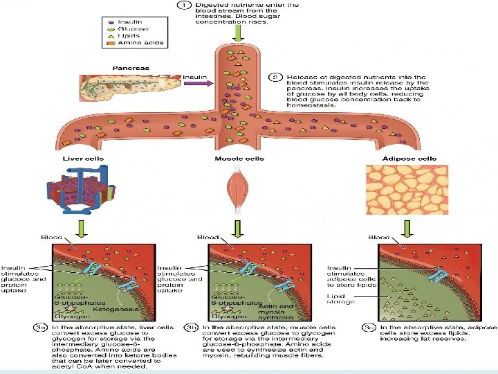
 • Fate of Carbohydrates: digested to monosaccharide’s glucose is")


 fast. The circled numbers")


, but in greatly reduced")


- Slides: 28
• Synthesis and Secretion of Glucagon • Glucagon, a polypeptide hormone, is synthesized in the α-cells of pancreas • The preproglucagon (160 aa) is produced on the RER and is converted to proglucagon as it enters the ER lumen. • Proteolytic cleavage at various sites produces the mature 29–amino acid glucagon. • Glucagon is rapidly metabolized, primarily in the liver and kidneys. • Glucagon plasma half-life is only about 3 to 5 minutes. • Increasing levels of glucose and insulin inhibit glucagon release. • Glucagon levels may increase, stimulating gluconeogenesis in the absence of dietary glucose. • The relative amounts of insulin and glucagon in the blood after a mixed meal are dependent on the composition of the meal, because glucose stimulates insulin release, and amino acids stimulate glucagon release.
29 aa
Structure of insulin receptor • The active insulin receptor consists of 1 - two identical α chains protruding from the outer face of the plasma membrane 2 - two transmembrane β subunits with their carboxyl termini protruding into the cytosol. 3 - The α chains contain the insulin binding domain, 4 - the intracellular domains of the β chains contain the protein kinase activity that transfers a phosphoryl group from ATP to the hydroxyl group of Tyr residues in specific target proteins.
Signalling through the insulin receptor 1 - Binding of insulin to the α chains activates the Tyr kinase activity of the β chains, 2 - Each αβ monomer phosphorylates three critical Tyr residues near the carboxyl terminus of the β chain of its partner in the dimer. 3 - This autophosphorylation opens up the active site so that the enzyme can phosphorylate Tyr residues of other target proteins. 4 - One of these target proteins is insulin receptor substrate IRS. 5 - Once IRS phosphorylated on its Tyr residues, it becomes a multiple binding sites for different proteins with SH 2 domains for a complex of proteins that carry the message from the insulin receptor to end targets in the cytosol and nucleus, through a long series of intermediate proteins. 6 - phosphorylated–Tyr residue in IRS is bound by the SH 2 domain of the protein Grb 2.
7 - A number of signalling proteins contain SH 2 domains, all of which bind P –Tyr residues in a protein partner. 8 - Grb 2 also contains a second protein-binding domain, SH 3 that binds to regions rich in Proline residues. Grb 2 binds to a proline-rich region of Sos. When bound to Grb 2, Sos binds Ras causing a large conformational change in Ras that permits release of GDP and binding of GTP and thus activating Ras. 9 - Ras can activate a protein kinase, Raf-1 the first of three protein kinases—Raf-1, MEK, and ERK—that form a cascade in which each kinase activates the next by phosphorylation. The protein kinase ERK is activated by phosphorylation of both a Threonine and a Tyr residue. When activated, it mediates some of the biological effects of insulin by entering the nucleus and phosphorylating proteins such as Elk 1, which modulates the transcription of about 100 insulin-regulated genes.
Signalling through the insulin receptor
• Signal transduction by glucagon • Glucagon, binds G proteins, • Activates adenylyl cyclase, increasing c. AMP. • c. AMP activates protein kinase A, which changes the activity of enzymes by phosphorylating key regulatory enzymes in the pathways of carbohydrate and fat metabolism at specific serine residues. • Some enzymes are activated and others are inhibited by this change in phosphorylation state. • In general, the major actions of glucagon occur in liver, adipose tissue, and certain cells of the kidney that contain glucagon receptors. • Glucagon-stimulated phosphorylation of enzymes simultaneously activates glycogen degradation, inhibits glycogen synthesis, and inhibits glycolysis in the liver.
Protein kinase A activation by c. AMP When the regulatory subunits (R) of protein kinase A bind the allosteric activator, c. AMP, they dissociate from the enzyme, thereby releasing active catalytic subunits (C).
adipose triacylglycerol lipase
• Signal Transduction by Norepinephrine and Cortisol Epinephrine, • The stress of a low blood glucose level mediates the release of the major insulin counter regulatory hormones through neuronal signals. • Hypoglycemia is one of the stress signals that stimulates the release of cortisol, epinephrine, and norepinephrine. • • Signal transduction by the glucocorticoid cortisol involves hormone binding to intracellular (cytosolic) receptors • Neuronal signals also play a minor role in the release of glucagon.
The Fed State (Absorptive state) • Fate of Carbohydrates: digested to monosaccharide’s glucose is oxidized by various tissues for energy, enters biosynthetic pathways, and is stored as glycogen, mainly in liver and muscle. • Glucose is also converted to triacylglycerols. The liver packages triacylglycerols, made from glucose or from fatty acids obtained from the blood, into very low-density lipoproteins (VLDL) and releases them into the blood. The fatty acids of the VLDL are mainly stored as triacylglycerols in adipose tissue, but some may be used to meet the energy needs of cells. • Fate of Proteins: digested to amino acids, amino acids are converted to proteins or used to make various nitrogen-containing compounds such as neurotransmitters and heme. • The carbon skeleton may also be oxidized for energy directly, or converted to glucose. • Fate of Fats: (mainly triacylglycerols) are digested packaged in chylomicrons, and secreted by way of the lymph into the blood. • The fatty acids of the chylomicron triacylglycerols are stored mainly as triacylglycerols in adipose cells.
The fed state. The circled numbers indicate the approximate order in which the processes occur. TG triacylglycerols; FA: fatty acid; AA amino acid; RBC red blood cell; VLDL very-low-density lipoprotein; I insulin; +: stimulated by.
Some Facts • The most important metabolic fuels are glucose and fatty acids. • In normal circumstances, glucose is the only fuel by the brain. • Glucose is also preferentially used by muscle during the initial stages of exercise. • Liver glycogen can supply glucose for no longer than 18 h. • Gluconeogenesis provides glucose over longer periods. • The caloric value of fats (9 Kcal/g) is higher than that of either carbohydrate or protein (4 Kcal/g). • The body has a virtually unlimited capacity for the accumulation of fats as adipose tissue triglycerides. • Fatty acids can support the body’s energy needs over prolonged periods of time. In extreme circumstances, humans can fast for as long as 60 -90 days and obese persons longer.
The fasting state • Blood glucose levels peak approximately 1 hour after eating and then decrease as tissues oxidize glucose or convert it to storage forms of fuel. • By 2 hours after a meal, the level returns to the fasting range (between 80 and 100 mg/d. L). • This decrease in blood glucose causes the pancreas to decrease its secretion of insulin, and the serum insulin level decreases. • The liver responds to this hormonal signal by starting to degrade its glycogen stores and release glucose into the blood. If we eat another meal within a few hours, we return to the fed state. • However, if we continue to fast for a 12 -hour period, we enter the basal state (also known as the postabsorptive state). • A person is generally considered to be in the basal state after an overnight fast, when no food has been eaten since dinner the previous evening. By this time, the serum insulin level is low and glucagon is rising.
• A. Blood Glucose and the Role of the Liver during Fasting • Glucose is the major fuel for brain and neural tissue, • Most neurons lack enzymes required for oxidation of fatty acids, but can use ketone bodies to a limited extent. • Glucose is the sole fuel for red blood cells. • Red blood cells lack mitochondria, which contain the enzymes of fatty acids and ketone bodies oxidation • Liver glycogen stores are limited. 1 - Although liver glycogen levels may increase to 200 to 300 g after a meal, only approximately 80 g remain after an overnight fast. 2 - Liver produce glucose through gluconeogenesis and glycogenolysis. • In gluconeogenesis, lactate, glycerol, and amino acids are used as carbon sources to synthesize glucose. 3 - Lactate is a product of glycolysis in red blood cells and exercising muscle, • Glycerol is obtained from lipolysis of adipose triacylglycerols, • Amino acids are generated by the breakdown of protein. • Most of the amino acid is supplied from degradation of muscle protein.
• Because the nitrogen of the amino acids can form ammonia, which is toxic to the body, the liver converts this nitrogen to urea. • Urea has two amino groups for just one carbon (NH 2 -CONH 2). It is a very soluble, nontoxic compound that can be readily excreted by the kidneys and thus is an efficient means for disposing of excess ammonia. • As fasting progresses, gluconeogenesis becomes increasingly more important as a source of blood glucose. • After a day or so of fasting, liver glycogen stores are depleted and gluconeogenesis is the only source of blood glucose.
• B. Role of Adipose Tissue During Fasting • Adipose triacylglycerols are the major source of energy during fasting. • Fatty acids are partially oxidized in the liver to 4 -carbon products called ketone bodies. • Ketone bodies are subsequently oxidized as a fuel by other tissues. • Adipose triacylglycerols are mobilized by a process known as lipolysis. They are converted to fatty acids and glycerol, which enter the blood. • Fatty acids cannot provide carbon for gluconeogenesis. • only the small glycerol portion travels to the liver to enter the gluconeogenic pathway. • Fatty acids serve as a fuel for muscle, kidney, and most other tissues. • They are oxidized to acetyl Co. A, and subsequently to CO 2 and H 2 O in the TCA cycle,
• Most of the fatty acids that enter the liver are converted to ketone bodies rather than being completely oxidized to CO 2. • The process of conversion of fatty acids to acetyl Co. A produces a considerable amount of energy (ATP), which drives the reactions of the liver under these conditions. • • The acetyl Co. A is converted to the ketone bodies acetoacetate and β-hydroxybutyrate, which are released into the blood. • The liver lacks an enzyme required for ketone body oxidation. However, ketone bodies can be further oxidized by most other cells with mitochondria, such as muscle and kidney. • In these tissues, acetoacetate and β-hydroxybutyrate are converted to acetyl Co. A and then oxidized in the TCA cycle, with subsequent generation of ATP.
Basal state. This state occurs after an overnight (12 -hour) fast. The circled numbers serve as a guide indicating the approximate order in which the processes begin to occur. (KB: ketone bodies)
Prolonged fasting • Metabolic changes during prolonged fasting • If the pattern of fuel utilization that occurs during a brief fast were to persist for an extended period, the body’s protein would be quite rapidly consumed to the point at which critical functions would be compromised. • Fortunately, metabolic changes occur during prolonged fasting that conserve (spare) muscle protein by causing muscle protein turnover to decrease.
A- Role of Liver During Prolonged Fasting • After 3 to 5 days of fasting the body enters the starved state, • Muscle decreases its use of ketone bodies and depends mainly on fatty acids for its fuel. • The liver, however, continues to convert fatty acids to ketone bodies. • The result is that the concentration of ketone bodies rises in the blood. • The brain begins to take up these ketone bodies from the blood and to oxidize them for energy. Therefore, the brain needs less glucose than it did after an overnight fast. • Glucose is still required for red blood cells, and the brain continues to use a limited amount of glucose, which it oxidizes for energy and for the synthesis of neurotransmitters. • Less glucose is used by the body, and, therefore, the liver needs to produce less glucose per hour during prolonged fasting than during shorter periods of fasting.
• Because the stores of glycogen in the liver are depleted by approximately 30 hours of fasting, gluconeogenesis is the only process by which the liver can supply glucose to the blood if fasting continues. • The amino acid pool, produced by the breakdown of protein, continues to serve as a major source of carbon for gluconeogenesis. • A fraction of this amino acid pool is also being used for biosynthetic functions (e. g. , synthesis of heme and neurotransmitters). • However, as a result of the decreased rate of gluconeogenesis during prolonged fasting, protein is “spared”; less protein is degraded to supply amino acids for gluconeogenesis. • While converting amino acid carbon to glucose in gluconeogenesis, the liver also converts the nitrogen of these amino acids to urea. • Consequently, because glucose production decreases during prolonged fasting compared with early fasting, urea production also decreases.
• B- Role of Adipose Tissue During Prolonged Fasting • Adipose tissue continues to break down its triacylglycerol stores, providing fatty acids and glycerol to the blood. • These fatty acids serve as the major source of fuel for the body. • The glycerol is converted to glucose, whereas the fatty acids are oxidized to CO 2 and H 2 O by tissues such as muscle. • In the liver, fatty acids are converted to ketone bodies that are oxidized by many tissues, including the brain. • A number of factors determine how long we can fast and still survive. 1 - The amount of adipose tissue is one factor, because adipose tissue supplies the body with its major source of fuel. However, 2 - Body protein levels can also determine the length of time we can fast.
• Glucose is still used during prolonged fasting (starvation), but in greatly reduced amounts. • Protein still degraded for glucose production and can become so depleted that the heart, kidney, and other vital tissues stop functioning, or we can develop an infection and not have adequate reserves to mount an immune response. • In addition to fuel problems, we are also deprived of the vitamin and mineral precursors of coenzymes and other compounds necessary for tissue function. • Because of either a lack of ATP or a decreased intake of electrolytes, the electrolyte composition of the blood or cells could become incompatible with life. Ultimately, we die of starvation.
Starved state. Dashed lines indicate processes that have decreased, and the heavy solid line indicates a process that has increased relative to the fasting state.
Metabolism during absorptive state
Metabolism during postabsorptive state