
Syndromes of Orthopaedic Importance Meghan Imrie MD Core




- Slides: 64
Syndromes of Orthopaedic Importance Meghan Imrie, MD Core Course Curriculum January 27, 2016
Syndromes overview n “orthopaedic importance” – What ABOS/AAOS/POSNA expects you to know – What the patient’s parent in peds clinic that day wants you to know – What you actually need to know for practice
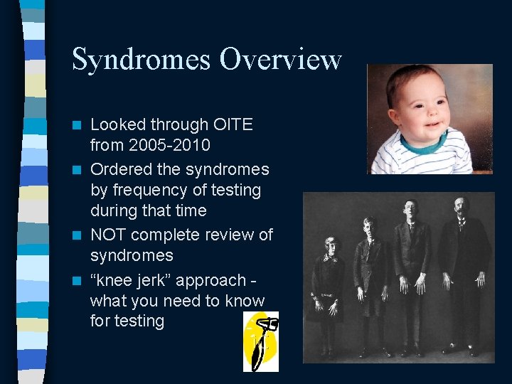
Syndromes Overview Looked through OITE from 2005 -2010 n Ordered the syndromes by frequency of testing during that time n NOT complete review of syndromes n “knee jerk” approach what you need to know for testing n
Syndromes overview n A few practical tips – Take close family history – Make an excuse to leave the room and consult book or internet – Re-evaluate patient for further features learned from above – If any question, call or refer to genetics
Syndromes overview n – Prader-Willi – Beckwith. Wiedemann – Ehler’s Danlos – Gaucher’s – Osteopetrosis – Duchenne’s MD Based on 2005 -2010 OITEs: – Osteogenesis imperfecta – Neurofibromatosis – Charcot Marie Tooth – Arthrogryposis – Myelomeningocele – Marfan’s – Down’s n Will also discuss – MPSs – Loeys-Dietz – Rett’s
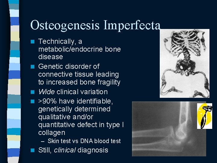
Osteogenesis Imperfecta Technically, a metabolic/endocrine bone disease n Genetic disorder of connective tissue leading to increased bone fragility n Wide clinical variation n >90% have identifiable, genetically determined qualitative and/or quantitative defect in type I collagen n – Skin test vs DNA blood test n Still, clinical diagnosis
Osteogenesis Imperfecta n Pathophysiology – (forgive the simplification, Lane!) – Type I collagen • major structural connective tissue protein of skeletal system • Composed of 3 strands of collagen protein – 2 alpha 1 strands and 1 alpha 2 strand • COL 1 A 1 encodes pro-alpha 1; COL 1 A 2 encodes proalpha 2 – Quantitative defect: often heterozygous with one copy not producing any – Qualitative defect: error in substitution or deletion leading to abnormal, less effectual collagen
Osteogenesis Imperfecta Wide clinical variation n Non-accidental trauma vs fracture from mild OI n – Difficult distinction if no family history, blue sclerae, etc – Involve CPS if any question With increased genetic knowledge, classification increasingly difficult/muddled n Some thought to simplified phenotypic classification: mild, moderate, severe, lethal n Sillence classification n – Most commonly used
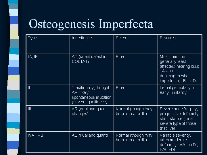
Osteogenesis Imperfecta Type Inheritance Sclerae Features IA, IB AD (quant defect in COL 1 A 1) Blue Most common, generally least affected; hearing loss; 1 A - no dentinogenesis imperfecta; 1 B - + DI II Traditionally, thought AR; likely spontaneous mutation (severe, qualitative) Blue Lethal perinatally or early in infancy III AR (qual and quant changes) Normal (though may be bluish at birth) Severe bone fragility, progressive deformity, short stature (most severe type of those that live) IVA, IVB AD (qual and quant) Normal (though may be bluish at birth) Variable severity, often moderate deformity; IVA, no DI; IVB, +DI
Osteogenesis Imperfecta n General treatment principles – Bones heal fairly predictably, but do not remodel reliably • Our usual “as long as they are in the same room” kid rules therefore don’t always apply • Under age 2, can usually treat more or less regularly – For minimally displaced, angulated fractures, immobilize for as short a duration as possible • Until child no longer symptomatic • To prevent further osteopenia/fracture risk – Don’t use plates • Intramedullary devices best – Rush rods, telescoping rods
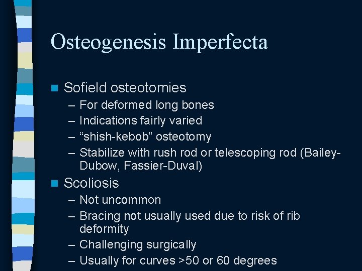
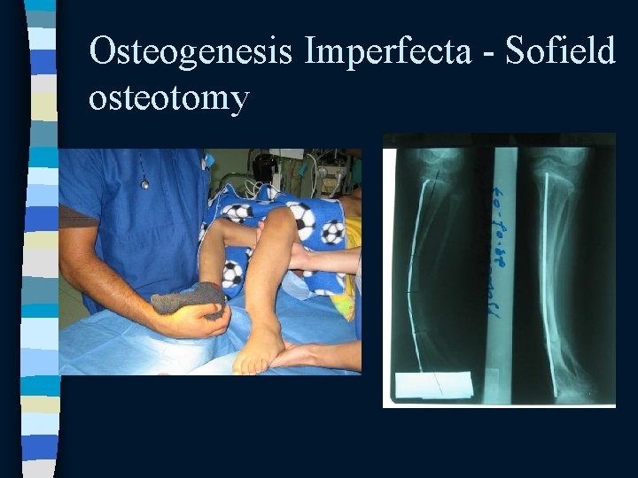
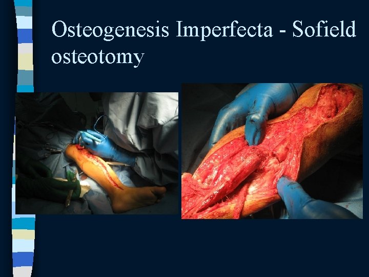
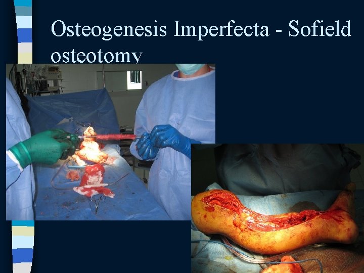
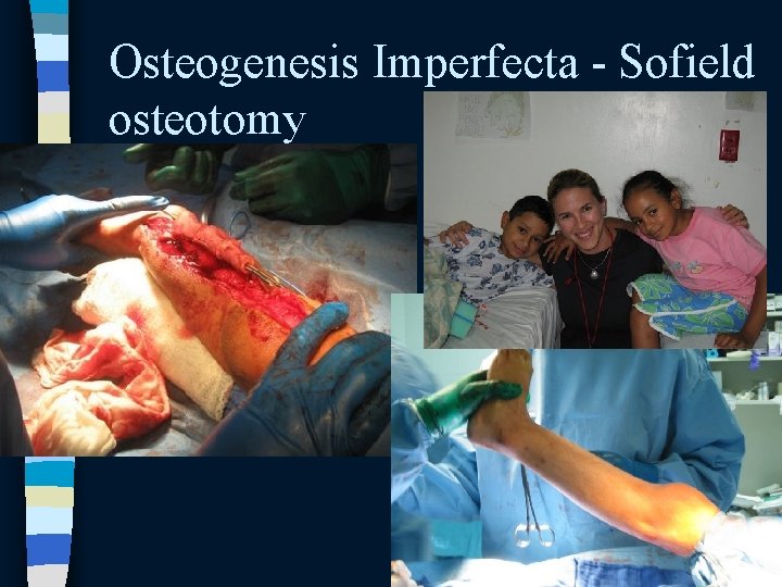
Osteogenesis Imperfecta n Sofield osteotomies – – n For deformed long bones Indications fairly varied “shish-kebob” osteotomy Stabilize with rush rod or telescoping rod (Bailey. Dubow, Fassier-Duval) Scoliosis – Not uncommon – Bracing not usually used due to risk of rib deformity – Challenging surgically – Usually for curves >50 or 60 degrees
Osteogenesis Imperfecta - Sofield osteotomy
Osteogenesis Imperfecta - Sofield osteotomy
Osteogenesis Imperfecta - Sofield osteotomy
Osteogenesis Imperfecta - Sofield osteotomy
OI - possibly tested material n Basilar impression/invagination – Due to soft bone of foramen magnum/microfractures of skull base – Leads to direct brainstem compression – Most common in type IVB – Sxs: headache, dysarthria, dysphagia, spasticity, increasing contractures – Plain films not very helpful • Need 3 -D imaging (MRI probably best) – Rx: often, anterior decompression/PSF (neurosurg) n Bisphonate use in OI – Depending on area of practice, still controversial – IV pamidronate currently “gold standard” • Numerous studies show increase in BMD by DEXA (? clinical implication? ) • Some studies report – – Decreased fxs Decreased pain Better mobility Increased height – Placebo trial vs po alendronate: no change in fx rate – Bottom (biased) line: don’t forget the half-life of these meds…
OI - summary n “brittle bone” disease – Due to quantitative and/or qualitative defects in type I collagen – COL 1 A 1 and COL 1 A 2 culprits – Varied clinical spectrum • Mild OI and NAT can be difficult to distinguish – Olecranon fracture in adolescent on OITE = probably has OI – Usual peds fracture principles don’t necessarily apply • Remodeling unpredictable • Don’t plate – Sofield osteotomy work horse for deformities – Be on look out for basilar invagination (especially in adults) – Bisphonates may be tested: pamidronate gold standard, increase DEXA definitely, probably reduce fracture rate and pain • But we don’t currently recommend them…
Syndromes overview n Based on 2005 -2010 OITEs: – Osteogenesis imperfecta – Neurofibromatosis – Charcot Marie Tooth – Arthrogryposis – Myelomeningocele – Marfan’s – Down’s – – – n Prader-Willi Beckwith-Wiedemann Ehler’s Danlos Gaucher’s Osteopetrosis Duchenne’s MD Will also discuss – MPSs – Loeys-Dietz – Rett’s
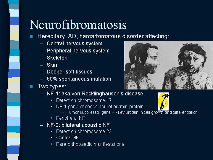
Neurofibromatosis n Hereditary, AD, hamartomatous disorder affecting: – – – n Central nervous system Peripheral nervous system Skeleton Skin Deeper soft tissues 50% spontaneous mutation Two types: – NF-1: aka von Recklinghausen’s disease • Defect on chromosome 17 • NF-1 gene encodes neurofibromin protein – Tumor suppressor gene --> key protein in cell growth and differentiation • Peripheral NF – NF-2: bilateral acoustic NF • Defect on chromosome 22 • Central NF • Rare orthopaedic manifestations
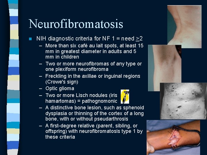
Neurofibromatosis n NIH diagnostic criteria for NF 1 = need >2 – More than six café au lait spots, at least 15 mm in greatest diameter in adults and 5 mm in children – Two or more neurofibromas of any type or one plexiform neurofibroma – Freckling in the axillae or inguinal regions (Crowe's sign) – Optic glioma – Two or more Lisch nodules (iris hamartomas) = pathognomonic – A distinctive bone lesion, such as sphenoid dysplasia or thinning of the cortex of a long bone, with or without pseudarthrosis – A first-degree relative (parent, sibling, or offspring) with neurofibromatosis type 1 by these criteria
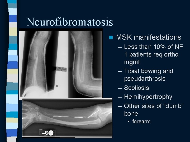
Neurofibromatosis n MSK manifestations – Less than 10% of NF 1 patients req ortho mgmt – Tibial bowing and pseudarthrosis – Scoliosis – Hemihypertrophy – Other sites of “dumb” bone • forearm
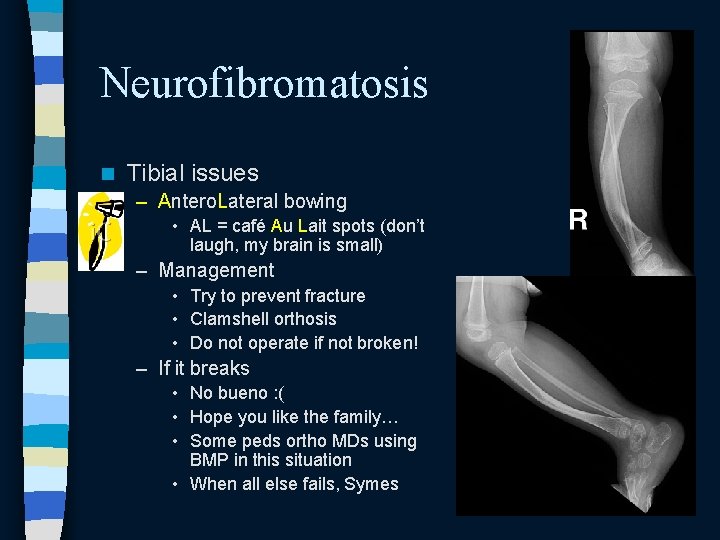
Neurofibromatosis n Tibial issues – Antero. Lateral bowing • AL = café Au Lait spots (don’t laugh, my brain is small) – Management • Try to prevent fracture • Clamshell orthosis • Do not operate if not broken! – If it breaks • No bueno : ( • Hope you like the family… • Some peds ortho MDs using BMP in this situation • When all else fails, Symes
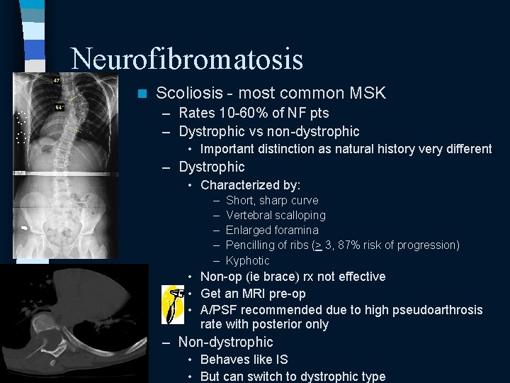
Neurofibromatosis n Scoliosis - most common MSK – Rates 10 -60% of NF pts – Dystrophic vs non-dystrophic • Important distinction as natural history very different – Dystrophic • Characterized by: – – – Short, sharp curve Vertebral scalloping Enlarged foramina Pencilling of ribs (> 3, 87% risk of progression) Kyphotic • Non-op (ie brace) rx not effective • Get an MRI pre-op • A/PSF recommended due to high pseudoarthrosis rate with posterior only – Non-dystrophic • Behaves like IS • But can switch to dystrophic type
Neurofibromatosis n Hemihypertrophy – Rare – Debulking unsatisfactory – Try epiphysiodesis for LLD n Other areas of “dumb” bone – Forearm – Has been tested on OITE (on diagnosis, not on treatment)
Neurofibromatosis - summary n AD disease of CNS, PNS, skin, skeleton – Café au lait spots: Coast of California NF 1 (chromosome 17, neurofibromin protein) has MSK manifestations n Most common genetic disorder caused by mutation in single gene n Dumb bone problems n – Anterolateral tibial bowing: prevent fracture, don’t do osteotomy! – Scoliosis: dystrophic curves worse, need MRI and A/PSF – Dumb bone can be anywhere • Forearm seems favored by OITE
Syndromes overview n – – – Based on 2005 -2010 OITEs: – Osteogenesis imperfecta – Neurofibromatosis – Charcot Marie Tooth – Arthrogryposis – Myelomeningocele – Marfan’s – Down’s n Prader-Willi Beckwith-Wiedemann Ehler’s Danlos Gaucher’s Osteopetrosis Duchenne’s MD Will also discuss – MPSs – Loeys-Dietz – Rett’s
Charcot Marie Tooth Disease One of hereditary motor sensory neuropathies (HMSN) n Motor sensory demyelinating or axonal neuropathy n – But motor much more involved than sensory n 2 main types with two more recent forms identified – CMT 1: • manifests earlier • AD • More common – CMT 2: • manifests in 3 rd decade • AD or AR • DTRs preserved – CMTX: • Second most common form • X-linked dominant – Orthopaedic manifestations similar
Charcot Marie Tooth Diagnosis confirmed by DNA testing n Numerous genetic abnormalities – CMT 1: duplication in chr 17, coding for peripheral myelin protein 22 (PMP 22) – CMT X (x-linked CMT): mutations in connexin 32 gene n Little correlation between genotype and phenotype n
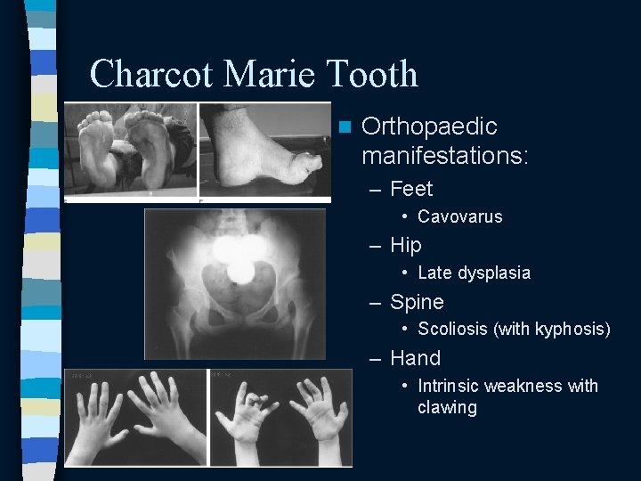
Charcot Marie Tooth n Orthopaedic manifestations: – Feet • Cavovarus – Hip • Late dysplasia – Spine • Scoliosis (with kyphosis) – Hand • Intrinsic weakness with clawing
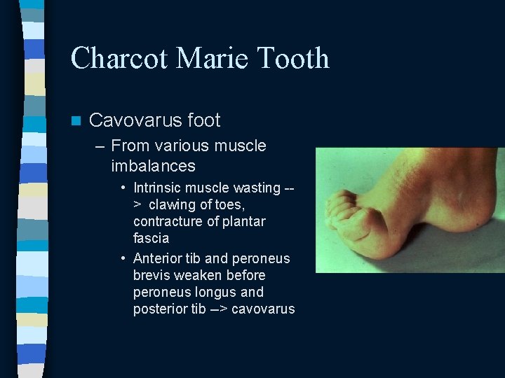
Charcot Marie Tooth n Cavovarus foot – From various muscle imbalances • Intrinsic muscle wasting -> clawing of toes, contracture of plantar fascia • Anterior tib and peroneus brevis weaken before peroneus longus and posterior tib --> cavovarus
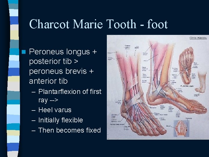
Charcot Marie Tooth - foot n Peroneus longus + posterior tib > peroneus brevis + anterior tib – Plantarflexion of first ray --> – Heel varus – Initially flexible – Then becomes fixed
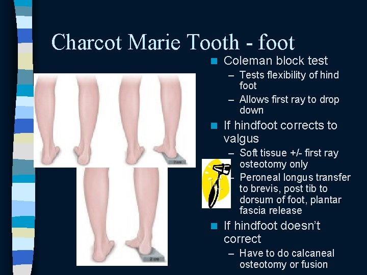
Charcot Marie Tooth - foot n Coleman block test – Tests flexibility of hind foot – Allows first ray to drop down n If hindfoot corrects to valgus – Soft tissue +/- first ray osteotomy only – Peroneal longus transfer to brevis, post tib to dorsum of foot, plantar fascia release n If hindfoot doesn’t correct – Have to do calcaneal osteotomy or fusion
Charcot Marie Tooth - summary n Diagnosis confirmed with DNA test – CMT 1: chr 17, PMP 22 – CMT X: x linked, connexin n Ortho manifestations – Feet • Cavovarus since peroneus longus stays stronger longer than anterior tib • If flexible (heel corrects on Coleman block), no calcaneal osteotomy or triple needed – Hip: Late dysplasia – Hand: Intrinsic wasting – Spine: scoliosis
Syndromes overview n – – – Based on 2005 -2010 OITEs: – Osteogenesis imperfecta – Neurofibromatosis – Charcot Marie Tooth – Arthrogryposis – Myelomeningocele – Marfan’s – Down’s n Prader-Willi Beckwith-Wiedemann Ehler’s Danlos Gaucher’s Osteopetrosis Duchenne’s MD Will also discuss – MPSs – Loeys-Dietz – Rett’s
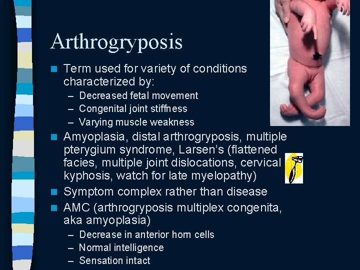
Arthrogryposis n Term used for variety of conditions characterized by: – Decreased fetal movement – Congenital joint stiffness – Varying muscle weakness Amyoplasia, distal arthrogryposis, multiple pterygium syndrome, Larsen’s (flattened facies, multiple joint dislocations, cervical kyphosis, watch for late myelopathy) n Symptom complex rather than disease n AMC (arthrogryposis multiplex congenita, aka amyoplasia) n – Decrease in anterior horn cells – Normal intelligence – Sensation intact
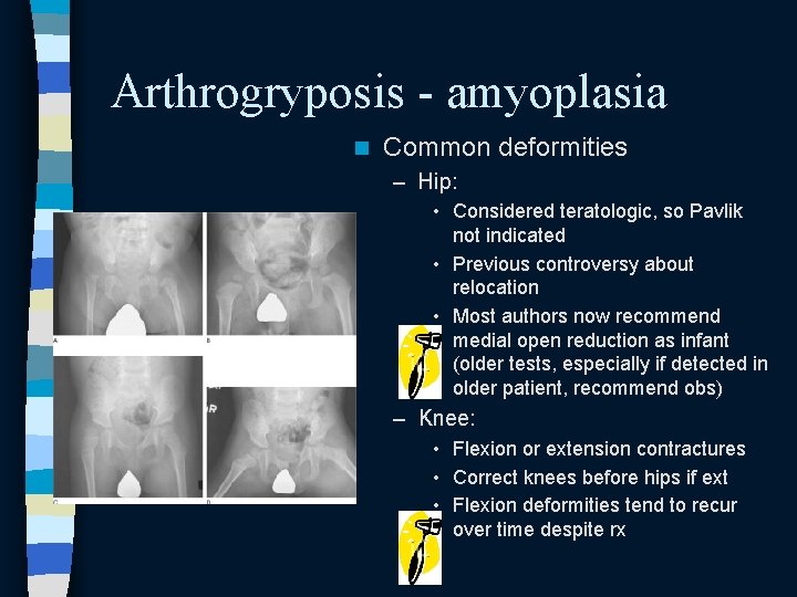
Arthrogryposis - amyoplasia n Common deformities – Hip: • Considered teratologic, so Pavlik not indicated • Previous controversy about relocation • Most authors now recommend medial open reduction as infant (older tests, especially if detected in older patient, recommend obs) – Knee: • Flexion or extension contractures • Correct knees before hips if ext • Flexion deformities tend to recur over time despite rx
Arthrogryposis - amyoplasia n Common deformities cont’d – Foot • Rigid clubfoot or vertical talus • For TEV, some authors report success with Ponseti method (prob won’t be tested) • Treat initially with PMR • Recurrence common – Treat with bony procedure like talectomy or triple arthrodesis • CVT requires surgery – Spine • Often C shaped, neuromuscular-esque • Bracing ineffective
Syndromes overview n – – – Based on 2005 -2010 OITEs: – Osteogenesis imperfecta – Neurofibromatosis – Charcot Marie Tooth – Arthrogryposis – Myelomeningocele – Marfan’s – Down’s n Prader-Willi Beckwith-Wiedemann Ehler’s Danlos Gaucher’s Osteopetrosis Duchenne’s MD Will also discuss – MPSs – Loeys-Dietz – Rett’s
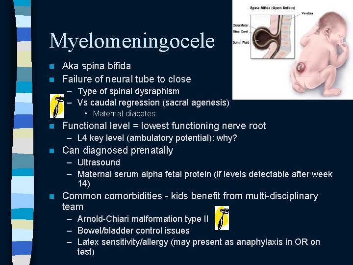
Myelomeningocele Aka spina bifida n Failure of neural tube to close n – Type of spinal dysraphism – Vs caudal regression (sacral agenesis) • Maternal diabetes n Functional level = lowest functioning nerve root – L 4 key level (ambulatory potential): why? n Can diagnosed prenatally – Ultrasound – Maternal serum alpha fetal protein (if levels detectable after week 14) n Common comorbidities - kids benefit from multi-disciplinary team – Arnold-Chiari malformation type II – Bowel/bladder control issues – Latex sensitivity/allergy (may present as anaphylaxis in OR on test)
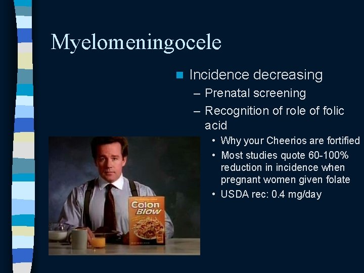
Myelomeningocele n Incidence decreasing – Prenatal screening – Recognition of role of folic acid • Why your Cheerios are fortified • Most studies quote 60 -100% reduction in incidence when pregnant women given folate • USDA rec: 0. 4 mg/day
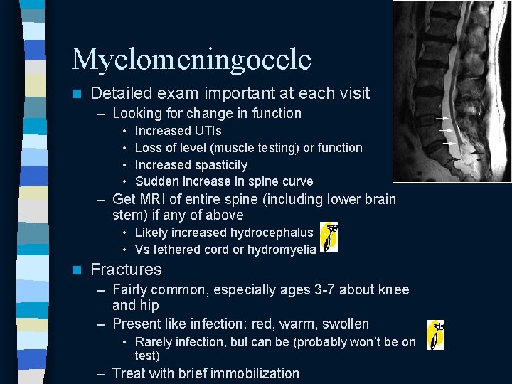
Myelomeningocele n Detailed exam important at each visit – Looking for change in function • • Increased UTIs Loss of level (muscle testing) or function Increased spasticity Sudden increase in spine curve – Get MRI of entire spine (including lower brain stem) if any of above • Likely increased hydrocephalus • Vs tethered cord or hydromyelia n Fractures – Fairly common, especially ages 3 -7 about knee and hip – Present like infection: red, warm, swollen • Rarely infection, but can be (probably won’t be on test) – Treat with brief immobilization
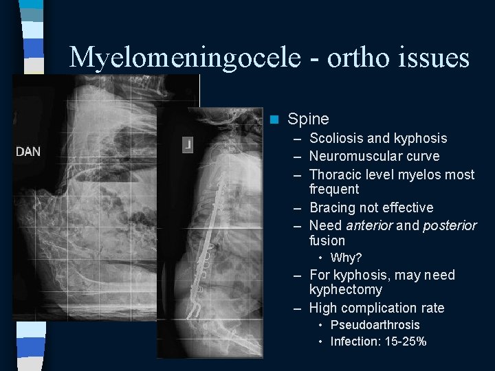
Myelomeningocele - ortho issues n Spine – Scoliosis and kyphosis – Neuromuscular curve – Thoracic level myelos most frequent – Bracing not effective – Need anterior and posterior fusion • Why? – For kyphosis, may need kyphectomy – High complication rate • Pseudoarthrosis • Infection: 15 -25%
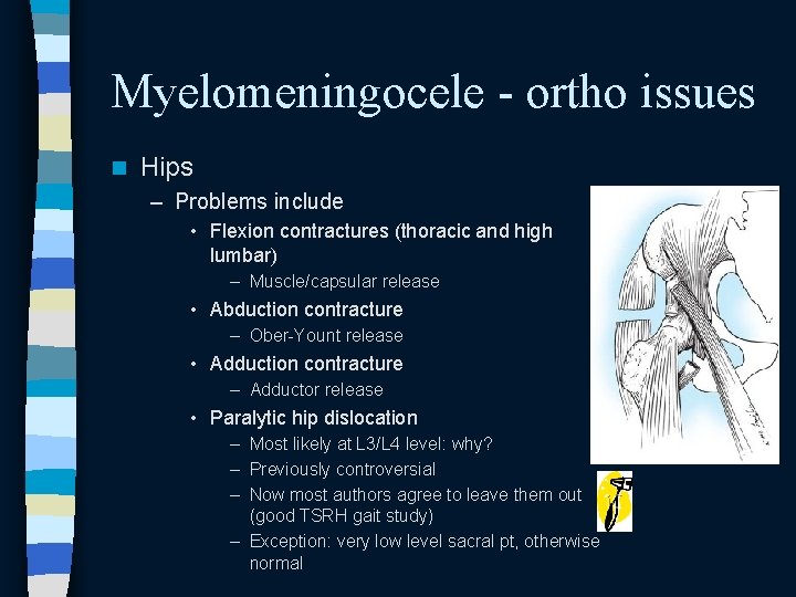
Myelomeningocele - ortho issues n Hips – Problems include • Flexion contractures (thoracic and high lumbar) – Muscle/capsular release • Abduction contracture – Ober-Yount release • Adduction contracture – Adductor release • Paralytic hip dislocation – Most likely at L 3/L 4 level: why? – Previously controversial – Now most authors agree to leave them out (good TSRH gait study) – Exception: very low level sacral pt, otherwise normal
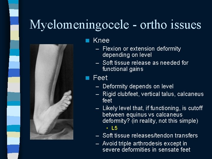
Myelomeningocele - ortho issues n Knee – Flexion or extension deformity depending on level – Soft tissue release as needed for functional gains n Feet – Deformity depends on level – Rigid clubfeet, vertical talus, calcaneus feet – Likely level that, if functioning, is cutoff between equinus vs calcaneus deformity? (in reality, not this simple) • L 5 – Soft tissue releases/tendon transfers – Avoid triple arthrodesis except in severe deformities in sensate feet
Myelomeningocele - summary n n n n Folic acid prevention Sacral agenesis (on same spectrum): maternal diabetes Latex allergy = anaphylaxis (crashing patient) in surgery Change in function/scoli curve --> get MRI of entire spine --> increased hydrocephalus (no bueno, call neurosurg) or tethered cord Non-op limb that looks like it might have osteo = likely fracture Scoli surgery = go anterior and posterior Leave dislocated hips out (unless patient looks totally normal and you are told has sacral myelo)
Syndromes overview n – – – Based on 2005 -2010 OITEs: – Osteogenesis imperfecta – Neurofibromatosis – Charcot Marie Tooth – Arthrogryposis – Myelomeningocele – Marfan’s – Down’s n Prader-Willi Beckwith-Wiedemann Ehler’s Danlos Gaucher’s Osteopetrosis Duchenne’s MD Will also discuss – MPSs – Loeys-Dietz – Rett’s
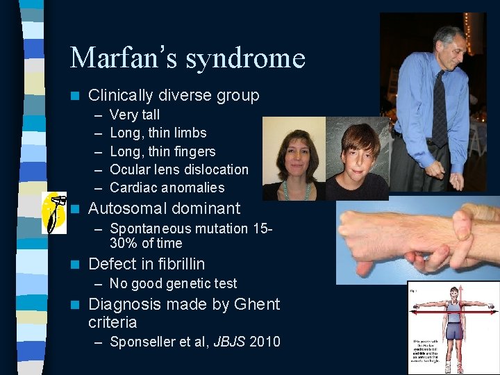
Marfan’s syndrome n Clinically diverse group – – – n Very tall Long, thin limbs Long, thin fingers Ocular lens dislocation Cardiac anomalies Autosomal dominant – Spontaneous mutation 1530% of time n Defect in fibrillin – No good genetic test n Diagnosis made by Ghent criteria – Sponseller et al, JBJS 2010
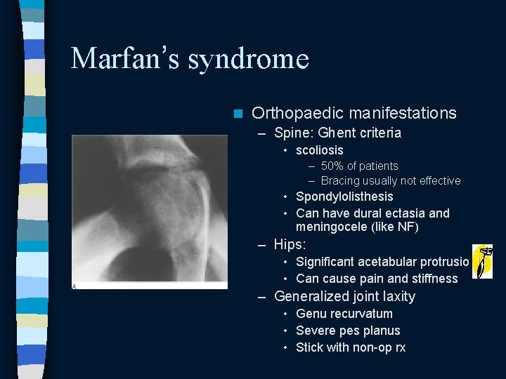
Marfan’s syndrome n Orthopaedic manifestations – Spine: Ghent criteria • scoliosis – 50% of patients – Bracing usually not effective • Spondylolisthesis • Can have dural ectasia and meningocele (like NF) – Hips: • Significant acetabular protrusio • Can cause pain and stiffness – Generalized joint laxity • Genu recurvatum • Severe pes planus • Stick with non-op rx
Syndromes overview n – – – Based on 2005 -2010 OITEs: – Osteogenesis imperfecta – Neurofibromatosis – Charcot Marie Tooth – Arthrogryposis – Myelomeningocele – Marfan’s – Down’s n Prader-Willi Beckwith-Wiedemann Ehler’s Danlos Gaucher’s Osteopetrosis Duchenne’s MD Will also discuss – MPSs – Loeys-Dietz – Rett’s
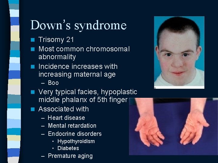
Down’s syndrome Trisomy 21 n Most common chromosomal abnormality n Incidence increases with increasing maternal age n – Boo Very typical facies, hypoplastic middle phalanx of 5 th finger n Associated with n – Heart disease – Mental retardation – Endocrine disorders • Hypothyroidism • Diabetes – Premature aging
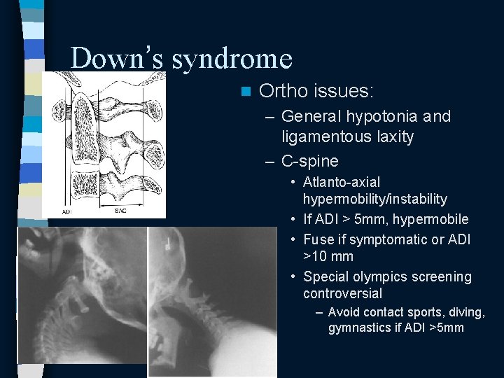
Down’s syndrome n Ortho issues: – General hypotonia and ligamentous laxity – C-spine • Atlanto-axial hypermobility/instability • If ADI > 5 mm, hypermobile • Fuse if symptomatic or ADI >10 mm • Special olympics screening controversial – Avoid contact sports, diving, gymnastics if ADI >5 mm
Down’s syndrome n Ortho issues cont’d – Hip • SCFE – Check thyroid! – Unstable/stable doesn’t apply » They’ll walk on unstable slips • Dislocation – Not congenital » Often late and dynamic – Rx challenge » Req OR, + likely osteotomy – Knees • Patellofemoral instability – Very debilitating – If symptomatic, realignment procedure – If asymptomatic, detected late, and patient can extend knee, don’t operate
Syndromes overview n – – – Based on 2005 -2010 OITEs: – Osteogenesis imperfecta – Neurofibromatosis – Charcot Marie Tooth – Arthrogryposis – Myelomeningocele – Marfan’s – Down’s n Prader-Willi Beckwith-Wiedemann Ehler’s Danlos Gaucher’s Osteopetrosis Duchenne’s MD Will also discuss – MPSs – Loeys-Dietz – Rett’s
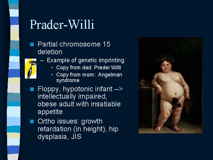
Prader-Willi n Partial chromosome 15 deletion – Example of genetic imprinting • Copy from dad: Prader Willi • Copy from mom: Angelman syndrome Floppy, hypotonic infant --> intellectually impaired, obese adult with insatiable appetite n Ortho issues: growth retardation (in height), hip dysplasia, JIS n
Beckwith-Wiedemann syndrome On differential of hemihypertrophy n Characterized by: n – – n Large tongue Omphalocele Organomegaly Exophthalmos Watch for – Neonatal hypoglycemia (can be fatal if undetected) – Intra-abdominal tumors • 40% chance of malignancy • Wilms, hepatoblastoma
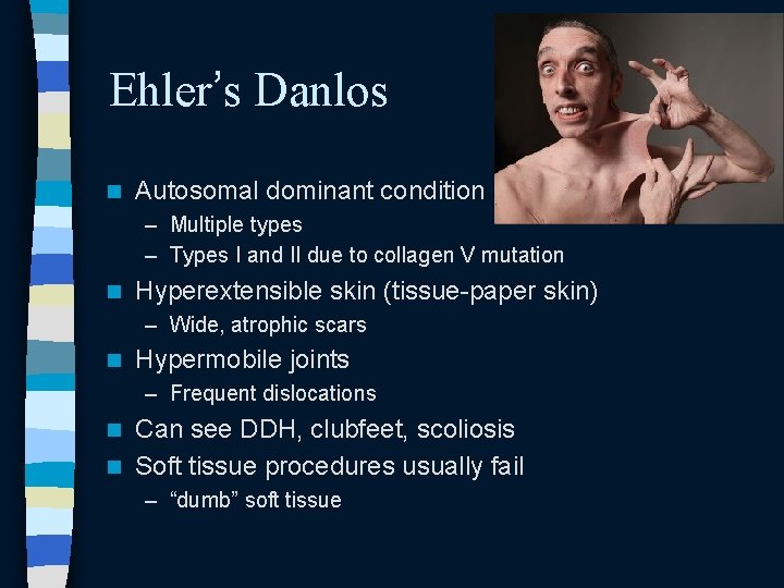
Ehler’s Danlos n Autosomal dominant condition – Multiple types – Types I and II due to collagen V mutation n Hyperextensible skin (tissue-paper skin) – Wide, atrophic scars n Hypermobile joints – Frequent dislocations Can see DDH, clubfeet, scoliosis n Soft tissue procedures usually fail n – “dumb” soft tissue
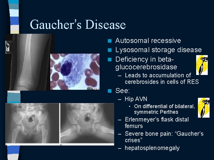
Gaucher’s Disease Autosomal recessive n Lysosomal storage disease n Deficiency in betaglucocerebrosidase n – Leads to accumulation of cerebrosides in cells of RES n See: – Hip AVN • On differential of bilateral, symmetric Perthes – Erlenmeyer’s flask distal femurs – Severe bone pain: “Gaucher’s crises” – hepatosplenomegaly
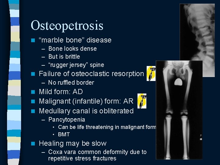
Osteopetrosis n “marble bone” disease – Bone looks dense – But is brittle – “rugger jersey” spine n Failure of osteoclastic resorption – No ruffled border Mild form: AD n Malignant (infantile) form: AR n Medullary canal is obliterated n – Pancytopenia • Can be life threatening in malignant form • BMT n Healing may be slow – Coxa vara common deformity due to repetitive stress fractures
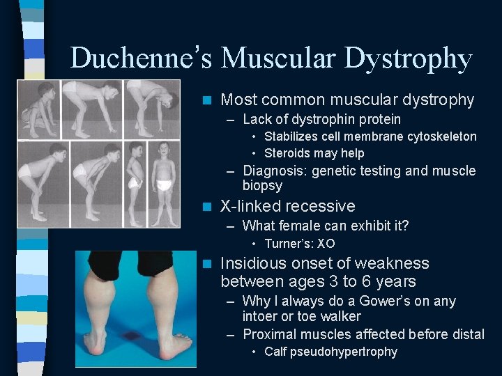
Duchenne’s Muscular Dystrophy n Most common muscular dystrophy – Lack of dystrophin protein • Stabilizes cell membrane cytoskeleton • Steroids may help – Diagnosis: genetic testing and muscle biopsy n X-linked recessive – What female can exhibit it? • Turner’s: XO n Insidious onset of weakness between ages 3 to 6 years – Why I always do a Gower’s on any intoer or toe walker – Proximal muscles affected before distal • Calf pseudohypertrophy
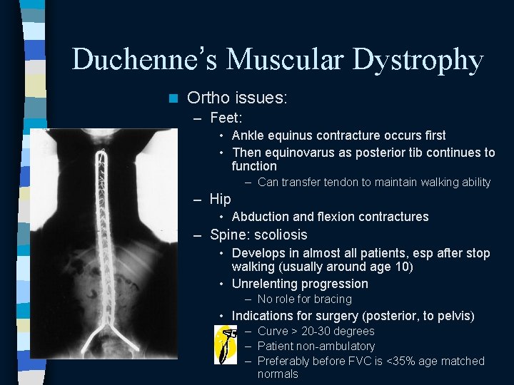
Duchenne’s Muscular Dystrophy n Ortho issues: – Feet: • Ankle equinus contracture occurs first • Then equinovarus as posterior tib continues to function – Can transfer tendon to maintain walking ability – Hip • Abduction and flexion contractures – Spine: scoliosis • Develops in almost all patients, esp after stop walking (usually around age 10) • Unrelenting progression – No role for bracing • Indications for surgery (posterior, to pelvis) – Curve > 20 -30 degrees – Patient non-ambulatory – Preferably before FVC is <35% age matched normals
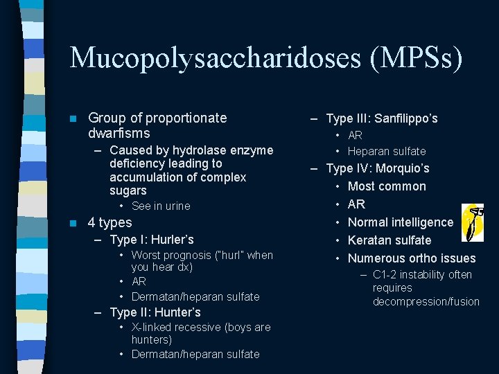
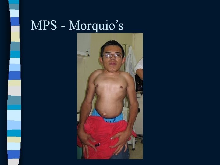
Mucopolysaccharidoses (MPSs) n Group of proportionate dwarfisms – Caused by hydrolase enzyme deficiency leading to accumulation of complex sugars • See in urine n 4 types – Type I: Hurler’s • Worst prognosis (“hurl” when you hear dx) • AR • Dermatan/heparan sulfate – Type II: Hunter’s • X-linked recessive (boys are hunters) • Dermatan/heparan sulfate – Type III: Sanfilippo’s • AR • Heparan sulfate – Type IV: Morquio’s • Most common • AR • Normal intelligence • Keratan sulfate • Numerous ortho issues – C 1 -2 instability often requires decompression/fusion
MPS - Morquio’s
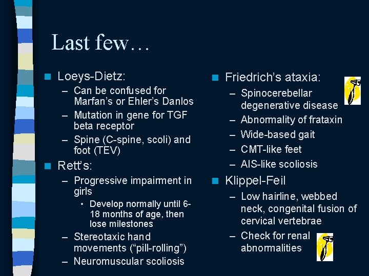
Last few… n Loeys-Dietz: n – Can be confused for Marfan’s or Ehler’s Danlos – Mutation in gene for TGF beta receptor – Spine (C-spine, scoli) and foot (TEV) n – Spinocerebellar degenerative disease – Abnormality of frataxin – Wide-based gait – CMT-like feet – AIS-like scoliosis Rett’s: – Progressive impairment in girls • Develop normally until 618 months of age, then lose milestones – Stereotaxic hand movements (“pill-rolling”) – Neuromuscular scoliosis Friedrich’s ataxia: n Klippel-Feil – Low hairline, webbed neck, congenital fusion of cervical vertebrae – Check for renal abnormalities
That’s it!