Syncope Learning objectives Gain organised knowledge in the

Syncope

Learning objectives • Gain organised knowledge in the subject area syncope • Be able to correctly interpret clinical findings in patients with syncope • Know and apply the relevant evidence and/or guidelines • Be aware of common cognitive biases in the diagnosis and management of syncope
 Syncope is a symptom, (not a diagnosis) which has 5 essential elements:")
Syncope (‘interrupt’) Syncope is a symptom, (not a diagnosis) which has 5 essential elements: • • loss of consciousness loss of voluntary muscle tone ( fall) relatively rapid onset recovery is spontaneous, complete and usually prompt The underlying mechanism is transient global cerebral hypoperfusion

Syncope is common England data: syncope and collapse

Collapse ? cause transient loss of consciousness Due to acute illness Syncope Seizure Hypoglycaemia Intoxication etc

Collapse ? cause transient loss of consciousness Due to acute illness 1. 2. 3. 4. Syncope Neurally-mediated Orthostatic hypotension Cardiac arrhythmia Structural Seizure Hypoglycaemia Intoxication etc TLOC alone is never a TIA

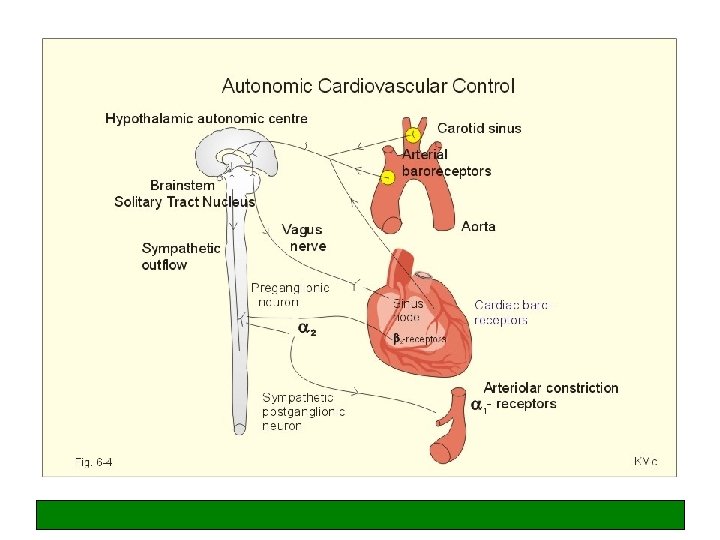
‘Initial evaluation’ • • • History from patient History from any available eye-witnesses! Cardiovascular (and neurological) examination 12 -lead ECG Lying and standing BP

Scenario 1 A 60 -year-old man was admitted to AMU following a blackout. He said he was walking along the street when he experienced brief dizziness then found himself on the floor. This has never happened before. There were no available eye-witnesses. His past medical history included type 2 diabetes and hypertension for which he was taking metformin, ramipril and bendroflumethiazide. On examination there was no abnormality to find. Bloods, lying and standing BP and 12 -lead ECG were normal.

Scenario 2 A 20 -year-old woman was admitted following a collapse. Eyewitnesses reported she felt unwell (pale and nauseated) while standing in the pub and decided to go outside for some fresh air. Before reaching the exit she collapsed and was observed to jerk all four limbs. She recovered quickly and an ambulance was called. The patient described feeling like her vision was closing in, and palpitations, before blacking out. She has had previous collapses at work (in a café kitchen) which were similar. There was no family history of collapses or sudden death. On examination, she was back to normal but complaining of feeling ‘washed out’. Clinical examination, lying and standing BP and 12 -lead ECG was normal.

Scenario 3 An 18 -year-old man was admitted for a routine arthroscopy of the knee. In the anaesthetic room he was observed to become asystolic for 10 seconds during cannula insertion. The procedure was postponed while the anaesthetist sought more information. The patient’s mother stated that the patient and his father were ‘fainters’ and this was often triggered by unpleasant stimuli. The anaesthetist phoned the medical registrar to ask, ‘Can you get vasovagal syncope while lying down? ’

Common pitfalls • • ‘…and all four limbs jerked’ ‘…and was incontinent of urine’ ‘…was talking nonsense’ ‘…and felt really tired afterwards’ ‘…had palpitations before collapsing’ ‘…he injured himself badly’ ‘…he went rigid as we dragged him out of the restaurant’

Syncope vs seizure SYNCOPE MORE LIKELY • Posture • Pale, nausea/ vomiting, sweaty, palpitations • Short duration jerky movements, start after LOC • Quick recovery • Fatigue for several hours afterwards common • Incontinence of urine SEIZURE MORE LIKELY • Aura / blue face • Prolonged tonic-clonic movements, coincide with LOC • Automatisms, tongue biting • Prolonged confusion, headache or drowsy* • At night • Faecal incontinence

Any questions at this point?

Scenario 4 A 65 -year-old man was admitted following a collapse. He described half a dozen previous collapses with no warning. All were while in the standing position, but one was while driving – this was while he was reversing in to a parking space. One had occurred while crossing the road. He had a past medical history of type 2 diabetes on metformin. He was normally active and independent. Examination of the cardiovascular system and lying and standing BP was normal. Bloods and a 12 -lead ECG were normal.

Scenario 5 An 85 -year-old woman was found lying on the floor in her nursing home. She had a scalp laceration and an ambulance was called. Her past medical history included paroxysmal atrial fibrillation, hypertension and dementia. She was taking warfarin and bisoprolol 2. 5 mg od. On examination she had normal vital signs and was back to her usual self. Blood results were normal apart from an INR of 2. 5. A 12 -lead ECG showed sinus rhythm and a CT scan of the head was normal apart from some atrophy in keeping with the patient’s age and moderate small vessel disease.

Syncope in general: pitfalls in older people • • ‘…and slumped to one side (sitting)’ ‘…I don’t know, I must have tripped’ ‘…collapsed without warning’ ‘…I don’t go dizzy, I just feel queer’ Unwitnessed Lying and standing BPs are normal More than one diagnosis

Scenario 6 An 80 -year-old woman was admitted following a collapse. Her husband described how they were out shopping when she started to feel ‘drained’ and went to sit down, but before she could do so, she collapsed to the floor. She recovered and was back to normal after a few minutes. Previous collapses had been in similar circumstances – once while in the supermarket, and another while queuing at the Post Office. Her past medical history included type 2 diabetes, hypertension and asthma. There was nothing abnormal to find on examination, postural BP, bloods or 12 -lead ECG.
 VVS 120 OH 60")
BP responses in different types of syncope BP (mm. Hg) VVS 120 OH 60 Elderly dysautonomic pattern BP after standing Time (mins)

Scenario 7 A 65 -year old man was admitted following a collapse. This had never happened before. Eyewitnesses described him waiting at the bus stop, then looking pale and sweaty and feeling unwell briefly before falling to the ground. He made a quick recovery. His past medical history included a previous MI. He was normally fit and well and did not experience angina. Cardiovascular exam, lying and standing BP, and blood results were normal. A 12 -lead ECG showed sinus rhythm and anterior Q waves.

Common cognitive biases in the diagnosis and management of syncope • Not taking the time to get an eye-witness account or perform a proper ‘initial evaluation’, including lying and standing BP • Knowledge gaps application of the wrong heuristic

Any questions at this point?

Causes of TLOC in patients referred to syncope clinics 50% 2% 30% in elderly 20% 3%
 History, physical examination, ECG, lying & standing BP Certain or suspected")
Syncope (ESC Guidelines) History, physical examination, ECG, lying & standing BP Certain or suspected diagnosis Initial evaluation Unexplained syncope Structural heart disease or abnormal ECG No structural heart disease and normal ECG Evaluate/confirm disease/disorder No Diagnosis made Cardiac evaluation + - Frequent or severe NMS evaluation + Treatment Single/rare No further evaluation - Treatment Re-appraisal

Vasodepressor VVS

Cardio-inhibitory CSH

Summary of NICE Guidelines
 Box 3. Red flags Refer within 24 hours for specialist cardiovascular")
Syncope (NICE guidelines) Box 3. Red flags Refer within 24 hours for specialist cardiovascular assessment (by the most appropriate local service) anyone with TLOC who also has any of the following: –an ECG abnormality –heart failure (history or physical signs) –TLOC during exertion –family history of sudden cardiac death in people aged younger than 40 years and/or an inherited cardiac condition –new or unexplained breathlessness –a heart murmur • Consider referring within 24 hours anyone aged older than 65 years who has experienced TLOC without prodromal symptoms No Uncomplicated faint (vasovagal syncope – 3 Ps), situational syncope or orthostatic hypotension? If so – give advice and treat. Yes • Refer for specialist cardiovascular assessment by the most appropriate local service within 24 hours (AMU) • If the person presents to the ambulance service, take them to the Emergency Department
 Reassess: –history, including any previous events –medical history,")
Specialist cardiovascular assessment and diagnosis (NICE) Reassess: –history, including any previous events –medical history, and any family history of cardiac disease –drug therapy at the time of TLOC and any subsequent changes • Conduct a clinical examination and measure lying and standing BP • Repeat 12 -lead ECG and examine previous ECGs Assign to suspected cause of syncope and offer further testing as directed below, or other tests as clinically appropriate Suspected structural heart disease cause Suspected cardiac arrhythmic cause Suspected neurally mediated cause* Unexplained cause
 Because")
NICE contd Suspected structural heart disease cause Investigate appropriately (for example, cardiac imaging) Because other mechanisms for syncope are possible in this group, also consider investigating for a cardiac arrhythmic (see opposite), and for orthostatic hypotension or for neurally mediated syncope Suspected cardiac arrhythmic cause • Offer an ambulatory ECG as a first-line investigation –choose type of ambulatory ECG based on person’s history (and in particular, frequency) of TLOC • Do not offer a tilt test as a first-line investigation 24/48 T if several times a week EER if every 1 -2 weeks IER if less than once a fortnight

NICE contd Unexplained cause *Suspected neurally mediated cause Vasovagal syncope suspected Is the person 60 years or older? Carotid sinus syncope suspected Yes Do not offer a tilt test to people who have a diagnosis of vasovagal syncope on initial assessment No • Offer carotid sinus massage • Carry out this test in a controlled environment, with ECG recording and resuscitation equipment available ** Only consider a tilt test if the person has recurrent episodes of TLOC that adversely affect their quality of life, or represent a high risk of injury, to assess whether the syncope is accompanied by a severe cardioinhibitory response (usually asystole) • Offer an ambulatory ECG – choose type of ambulatory ECG based on history and frequency of TLOC • Do not offer a tilt test before the ambulatory ECG Syncope due to marked bradycardia/asystole and/or marked hypotension reproduced? Yes Diagnose carotid sinus syncope No Negative carotid sinus massage test (normal or asymptomatic non-significant bradycardia and/or hypotension)
")
Implantable event recorder (Reveal ® Device)

What about tilt testing? ESC • • • VVS is not always diagnose-able on the history Tilt testing discriminates well for VVS between symptomatic patients and asymptomatic controls (specificity 90% p. HUT ) No ‘gold standard’ to compare Well known that different hemodynamic picture occurs at different times in same patient - so not used to tailor treatment Tilt testing is safe/well tolerated Indicated in unexplained syncope in absence of heart disease NICE • • • Tilt test studies are mainly (heterogeneous) case control studies Pre-test probability of neurally mediated syncope is high in patients without structural heart disease, even if test is negative Analysed evidence for tilt testing vs IER (A 1. as standard) in diagnosis of cardio-inhibitory NM syncope (A 2. as would benefit from pacing) and found more cost-effective to use IER
 1. Recurrent unexplained (or single serious) syncope in absence")
Indications for tilt testing (ESC) 1. Recurrent unexplained (or single serious) syncope in absence of heart disease 2. Recurrent unexplained (or single serious) syncope in absence of heart disease, after cardiac causes of syncope have been excluded 3. Assessing recurrent pre-syncope (incl POTS) 4. After an aetiology of syncope has been established, but where demonstration of susceptibility to neurallymediated syncope would alter therapeutic approach 5. Differentiating syncope with myoclonic jerks from epilepsy (also PNES and psychogenic pseudo-syncope) 6. Evaluating patients with recurrent unexplained ‘falls’

Any questions at this point?

What tests should I do in syncope? • FBC, U&E, CRP*, glucose • 12 -lead ECG • Patients may need investigating for postural hypotension… • Do not do the following: – Troponin if no chest pain / ECG changes – CT brain – Heart tests in people with normal hearts!

When to admit a patient with syncope • Suspected or known significant heart disease • ECG abnormalities suggesting an arrhythmia • Syncope during exercise • Syncope occurring in supine position. . . • Syncope causing severe injury • Family history of sudden death • Sudden onset palpitations in the absence of heart disease • Frequent recurrent episodes. . ? • Old and needs ‘sorting out’
")
Prognostic stratification Poor prognosis • Structural heart disease (independent of the cause of syncope) Excellent prognosis • Young, healthy, normal 12 -lead ECG • Neurally-mediated syncope • Orthostatic hypotension • Unexplained syncope after thorough evaluation

Treatment of non-cardiac syncope • • • Patient education General measures Reduce / stop exacerbating medication Medication for syncope Dual chamber PPM for certain patients (rare)
 Disorder Group 1 Group 2 (bus, lorry) and taxi VVS")
UK driving regulations (DVLA) Disorder Group 1 Group 2 (bus, lorry) and taxi VVS / situational - - Cough syncope Cannot drive until controlled Unexplained – low risk recurrence Can drive after 4 weeks Can drive after 3 months Unexplained – high risk recurrence Can drive after 4 weeks if treated, otherwise 6 months Can drive after 3 months if treated, otherwise 1 year LOC with seizure markers Cannot drive for 1 year Cannot drive for 5 years LOC and no pointers at all Cannot drive for 6 months Cannot drive for 1 year

Any questions at this point?

Read strategically! www. internalmedicineteaching. org
- Slides: 43