Synaptic Transmission and Cellular Signaling an Overview Department


: Cm = QAm/V (Q is the")

: directly measure the release of oxidizable neurotransmitter,")


 observed")


 noted the high specificity and potency of biological")

")
")

- Slides: 30
Synaptic Transmission and Cellular Signaling: an Overview Department of Pharmacology Jin-Chung Chen, Ph. D. Room 664; x 5282
Synaptic Transmission: historic view 1. Histologist Golgi and Ramon Cajal: neuronal connection via “Synapse” 2. Oliver, Shafer, Langley and Elliot (1890): Chemical transmission 3. Experimental biology by Otto Loewi: electrical stimulation on vagus n. decrease heart contraction
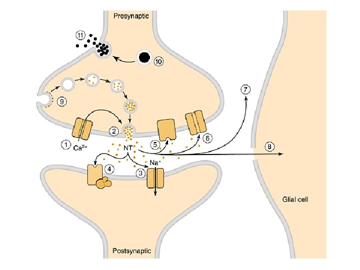
Criteria for chemical transmission 1. Synthesis of the neurotransmitter in the presynaptic nerve terminals 2. Storage of the neurotransmitter in secretory vesicles. 3. Regulated release of neurotransmitter in the synaptic space between the pre- and postsynaptic neurons. 4. Presence of receptors on the postsynaptic membrane mimics the effect of nerve stimulation 5. A means for “termination” of the action of the released neurotransmitter.
Eukaryotic neurotransmitter release 1. Classic neurotransmitter: ACh, DA, NE, EPI and 5 -HT are low-molecular-weight substance that have no other physiological function 2. They store in “synaptic vesicles”: acidic interior (p. H = 5. 5), maintained by a vacuolar-type, proton-translocating ATPase driven by H+ pump and specific vesicular transporter
3. Peptides and proteins can also be released from the n. terminals. They utilize the ER, Golgi and trans-Golgi network in the cell body (not the terminal) and transport into the nerve terminal by axonal transprot. 4. Constitutive secretion and endocytosis in yeast and mammalian cells are similar in presynaptic events of synaptic transmission
Methods to study exocytosis 1. Membrane capacity (Cm): Cm = QAm/V (Q is the charge across the membrane) specific capacitance is mainly determined by the thickness and dielectric constant of the phospholipid bilayer. Therefore, the increase in plasma membrane area due to exocytosis is proportional to the increase in Cm. Patch clamp: micropipet has tight seal to allow noise, high sensitivity, electric measurement
Electrochemical: amperometric Electrophysiological : capacity
2. Electrochemical detection (carbon fiber amperometric electrode): directly measure the release of oxidizable neurotransmitter, such as catecholamines and serotonin. 3. Synaptosome: homogenize brain tissues to shear off the nerve terminals. Each of the synaptosome is about 0. 5 – 1. 0 m in diameter, contains hundreds of synaptic vesicles, trace mitochondria and postsynaptic membrane. It can function for several hours.
Synaptosomal preparation Superfusion apparatus
4. Tissue culture: adrenal medullary chromaffin cells have the same precursor cells as postganglionic sympathetic neurons 5. Cell culture: collagenase digestion of bovine adrenal glands or using PC 12 cells to study the neurochemical nature of neurotransmitter release.
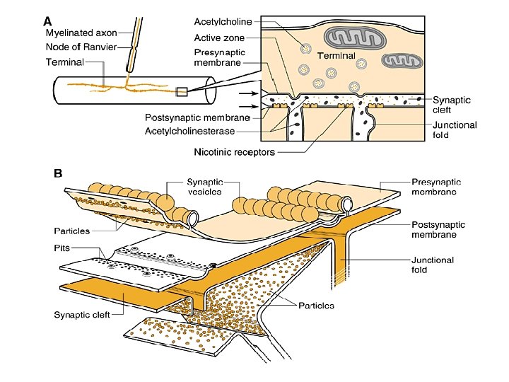
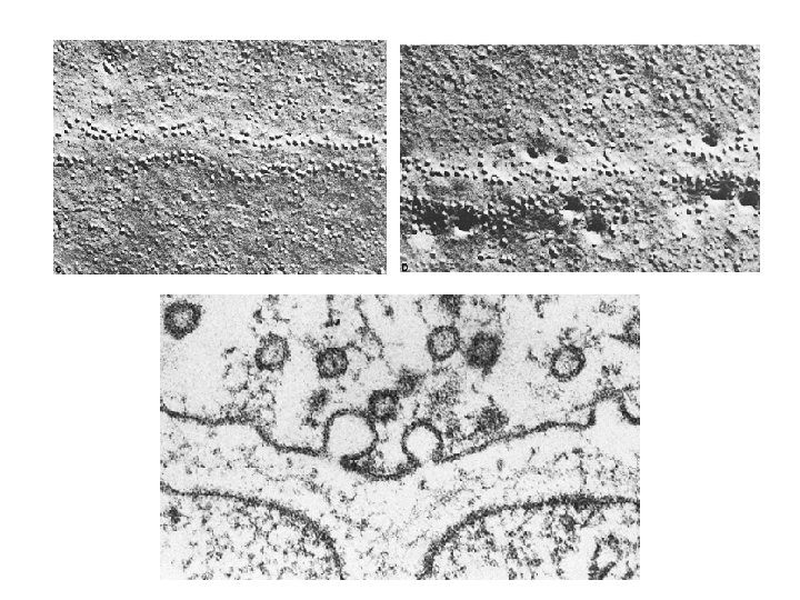
Neuromuscular junction: ACh release and postsynaptic effect 1. There approximately 300 active zones per NMJ; 500, 000 vesicles in all of the active zones at one NMJ. It is estimated a vesicle contains 20, 000 ACh molecules. 2. The specialized postsynaptic membrane structure consists a high density of nicotinic ACh receptors (n. ACh. Rs) 3. Basal lamina matrix proteins are important for the formation and maintenance of the NMJ and are concentrated in the cleft; enriched with ACh. E.
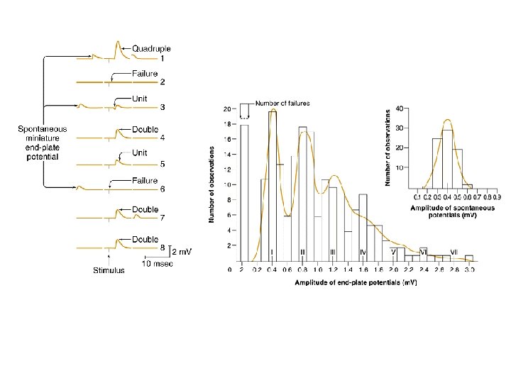
Quantum release : mechanism of release as Exocytosis 1. Fatt and Katz (1952) observed motor neuron stimulation results in spontaneous potentials of approximately 1 m. V at the motor end plate (miniature end-plate potentials [MEPPs]). 2. Measurement of quantum release: amplitude fixed; but number variated.
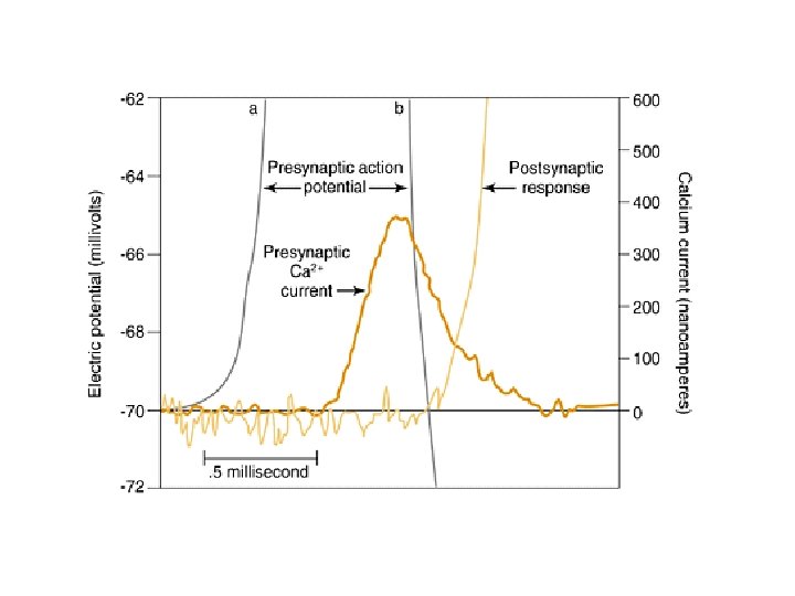
Calcium is necessary for transmission at the NMJ and other synapses 1. In most cases in the CNS and PNS, chemical transmission does not occur unless Ca 2+ is present. 2. EPPs could not be elicited if the calcium pulse immediately followed the depolarization; but should be beforehand (pulse as short as 1 msec preceded the depolarization). 3. Extracellular calcium: 2 m. M vs. intracellular 0. 1 M; the requirement to activate calcium channels: 50 -100 M. 4. Origin of calcium could be extracellular (thru voltagedependent Ca 2+ channel) or intracellular: GPCR-coupled PI-PLC activation release IP 3, thus Ca 2+ release from ER
Top trace illustrates the postsynaptic membrane potential
Pre-synaptic event and time scale 1. The time between calcium influx and exocytosis in the n. terminal is very short (0. 5 -1. 0 msec at NMJ; 200 sec at squid axon; 60 sec in CNS neuron) 2. In such short time (due to docking and fusion of synaptic vesicle), synaptic vesicle can not move significantly (calcium diffuse only 850 Å) 3. The supply of synaptic vesicles in the terminal in limited. Rapid recycled from the synaptic membrane with plasma membrane via clathrin-mediated endocytosis is essential (endocytosis is occurred away from active zones).
Calcium-dependent proteins are required for vesicle docking
The use of fluorescent dye FM 1 -43 to label the membrane and visualize endocytosis and exocytosis at the NMJ: 1. Complete cycle of exocytosis and endocytosis app. 1 min 2. A single n. impulse release 0. 1% of the recycling pool. 3. Endocytosis follows app. 20 sec. after exocytosis.
Endocytosis and clathrin cage
Fast synaptic transmission vs. peptide/protein release at the nerve terminals 1. Fast transmission: catecholamines, amino acid transmitters; can be synthesized within the n. terminals (synaptic vesicle) or transported rapidly across the membrane (uptake carrier). 2. In contrast, peptide/protein are inserted in the secretory granules, then transport down to n. terminals (no carrier) 3. Peptidergic granules are far less numbers than vesicles involved in fast exocytosis and are not localized at active zone (maybe function to maintain the long-lasting effect on postsynaptic neuron)
Function of ATP in presynaptic secretion ? 1. ATP usually is released along with transmitter in the vesicle; and important in maintaining the “exocytosis mechanism”. 2. ATP is necessary for the function of NSF (an ATPase) to dissociate complexes of the SNARE protein VAMP. 3. Maintain PIP 2 and PIP by phosphorylation of phosphatidylinositol 4 -kinaes and PIP kinase (PIP 2 binds to synaptotagmin and rabphilin 3 a and regulate cytoskeleton: profilin, gelsolin, scinderin and myosin).
Cellular signaling mechanisms Langley (1900 s) noted the high specificity and potency of biological response and postulated the existence of “receptor” or “acceptor”. Three phases of receptor-mediated signaling: 1. Polar molecule forms a complex with cell-surface receptor 2. Activated receptor-ligand complex elicits an increase of 2 nd messenger, or channel open/close 3. Signal cascades
Cell-surface receptors utilize four distinct molecular mechanisms for transmembrane signaling
Signal transduction cascade mediated by GPCR (G-protein coupled receptor)
Signal transduction cascade mediated by RTK (receptor tyrosine kinase)
Cross-talk between GPCR, ligand-gated ion channels and receptor tyrosine kinase