Synapse and its types n n n The


")


 receive stimuli from internal viscera n Monitor a variety of")


 n n Junction of tendon & muscle Monitors force")
- Slides: 49
Synapse and its types
n n n The Synapse Site at which neurons communicate Signals pass across synapse in one direction Types of cells in synapse n Presynaptic neuron - conducts impulse toward the synapse n Postsynaptic neuron - conducts impulse away from the synapse n Average postsynaptic neuron has up to 10, 000 synapses n Some in cerebellum have up to 100, 000 synapses Two major types of synapses n Electrical - not common in nervous system n Chemical - most common type
n Types of Chemical Synapses n Axodendritic n n n Axosomatic n n Between axon terminals of presynaptic neuron and dendrite of postsynaptic neuron Most common type of synapse Between axon of pre- and soma (cell body) of post-synaptic neuron Axoaxonic n n Between two axons Not common
n Types of Neural Synapses
n n n n Presynaptic bulb has secretory vesicles that contain neurotransmitter chemical (NT) NT must pass across the synaptic cleft, space that separates pre- and postsynaptic membranes Postsynaptic membrane contains receptors specific for each type of NT Binding of NT to its receptor causes ion channels to open or close
n Chemical Synapse
Lecture 2
n n Receptors are specialized cells for detecting particular changes in the environment.
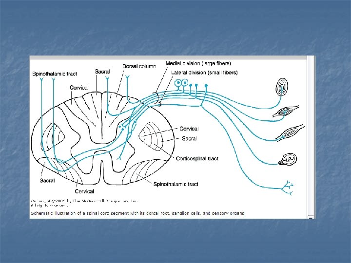
n Exteroceptors include receptors affected mainly by the external environment: Meissner's corpuscles, Merkel's corpuscles, and hair cells for touch; Krause's endbulbs for cold; Ruffini's corpuscles for warmth; and free nerve endings for pain.
n Receptors are not absolutely specific for a given sensation; strong stimuli can cause various sensations, even pain, even though the inciting stimuli are not necessarily painful.
n Proprioceptors receive impulses mainly from pacinian corpuscles, joint receptors, muscle spindles, and Golgi tendon organs
n Each efferent fiber from a receptor relays stimuli that originate in a receptive field and gives rise to a component of an afferent sensory system. Each individual receptor fires either completely or not at all when stimulated. The greater the intensity of a stimulus, the more endorgans that are stimulated, the higher the rate of discharge is, and the longer the duration of effect is.
n Adaptation denotes the diminution in rate of discharge of some receptors on repeated or continuous stimulation of constant intensity; the sensation of sitting in a chair or walking on even ground is suppressed.
n today s
Adaptation of Sensory Receptors n Change in sensitivity to long-lasting stimuli n decrease in responsiveness of a receptor bad smells disappear n very hot water starts to feel only warm n
n Receptors vary in their ability to adapt n Rapidly adapting receptors (smell, pressure, touch) n n adapt quickly; specialized for signaling stimulus changes Slowly adapting receptors (pain, body position) n continuation of nerve impulses as long as stimulus persists
Sensory Receptors and their Classification n Specialized cell or cell process that monitors specific conditions Arriving information is a sensation Awareness of a sensation is a perception
Peripheral Sensory Receptors n Structures that pick up sensory stimuli n Initiate signals in sensory axons
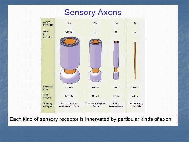
Peripheral Sensory Receptors n Two main categories of sensory receptors n Special nerve endings of sensory neurons n n Monitor general sensory information Independent receptor cells – specialized epithelial cells or small neurons n Monitor most types of special sensory information
Peripheral Sensory Receptors n Sensory receptors also classified according to: Location n Type of stimulus detected n Structure n
Classification by Location n Exteroceptors – sensitive to stimuli arising from outside the body Located at or near body surfaces n Include receptors for touch, pressure, pain, and temperature n
n Interoceptors – (visceroceptors) receive stimuli from internal viscera n Monitor a variety of stimuli
n Proprioceptors – monitor degree of stretch n Located in musculoskeletal organs
Classification by Modality n n n Mechanoreceptors – respond to mechanical forces Thermoreceptors – respond to temperature changes Chemoreceptors – respond to chemicals in solution Photoreceptors – respond to light – located in the eye Nociceptors – respond to harmful stimuli that result in pain
n Elecromagnetic receptors Thermo and photo Rods and cones
Chemoreceptors n Chemoreceptors are located in Carotid bodies n Aortic bodies n Special senses of taste and smell n Respiratory area of medulla n
Classification by Structure n General sensory receptors n n Widely distributed Nerve endings of sensory neurons monitor: n n n Touch, pressure, vibration, stretch Pain, temperature, proprioception Divided into two groups Non encapsulated Free nerve endings Merkels disc Hair follicle receptors n
Encapsulated nerve endings Meisners corposcles Pacinian corposcles Ruffinis corposcles n
Encapsulated nerve endings
Meissner’s Corpuscle
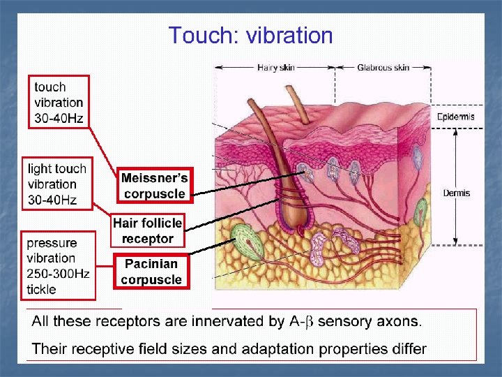
Meissner’s Corpuscle n n n Dermal papilla of palms and soles Ovoid in shape Stack of modified flattened schwaan cells Capsule is continous with endoneurium Rapidly adapting Two point tactile discrimination
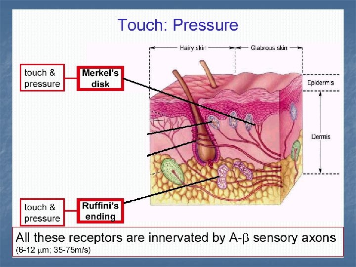
Pacinian corpuscle n n Onion-like connective tissue capsule enclosing a dendrite Found in subcutaneous tissues , ligaments, joint capsules Rapidly adapting high-frequency vibration
Ruffini Corpuscle n n n Found deep in dermis of hairy skin Detect heavy touch, continuous touch, & pressure Slowly adapting
Non encapsulated Free nerve endings n Epithelial cells of skin, cornea, dermis fascia n Pain, crude touch, pressure
• Free nerve endings found around follicles, detects movement of hair
Merkel’s Disc n n Flattened dendrites touching cells of stratum basale Used in discriminative touch
n Hairless skin fingertips
CUTANEOUS RECEPTORS n End organ of Ruffini crude touch n Merkel discs n Root hair plexus discriminative touch hair movement
Tactile Receptors in the Skin
Proprioceptors Monitor stretch in locomotory organs n Three types of proprioceptors n
Two Types of Proprioceptors n Muscle spindles – measure the changing length of a muscle n n Imbedded in the perimysium between muscle fascicles Golgi tendon organs – located near the muscle-tendon junction n Monitor tension within tendons
Anatomy of the muscle spindle and Golgi tendon organ Efferent motor fiber to spindle Secondary sensory endings (type II fiber) Primary sensory endings (type Ia fiber) Muscle spindle Connective tissue capsule Capsule a Efferent motor fiber to extrafusal muscle fibers Extrafusal muscle fiber Intrafusal muscle fibers Sensory fiber Tendon Golgi tendon organ
MUSCEL SPINDLE FIBERS n n n Monitor changes in length of skeletal muscle Degree of muscle stretch Aid in coordination & efficiency of muscle contraction
TENDON ORGANS (GOLGI TENDON ORGANS) n n Junction of tendon & muscle Monitors force of muscle contraction Detect tension applied to tendon Protects tendon & muscle from excessive tension