Swim Bladder an important ARO in fishes Core

Swim Bladder – an important ARO in fishes Core Course No. ZOOA P 2 T Group A, Unit: 1, Topic No. 6

Introduction to Swim Bladder In most of the fishes a characteristic sac like structure is present between the gut and the kidneys. This structure is called by various names, viz. , swim bladder, or gas bladder, or air bladder. In our present discussion, the name of the bladder is followed as the swim bladder to avoid confusion. The swim bladder occupies the same position as the lungs of higher verte brates and is regarded as homologous to the lungs. It differs from the lungs of higher forms mainly in origin and blood supply. The swim bladder is present in almost all the bony fishes and functions usually as a hydrostatic organ. Starting as a very insignificant cellular extension from the gut, the swim bladder in fishes leads the whole group through an evo lutionary channel.

Its Origin…… The swim bladder arises from the dorsal wall of the gut and gets the blood supply usually from the dorsal aorta, while the vertebrate lung origi nates from the ventral wall of the pharynx and receives blood from the sixth aortic arch.

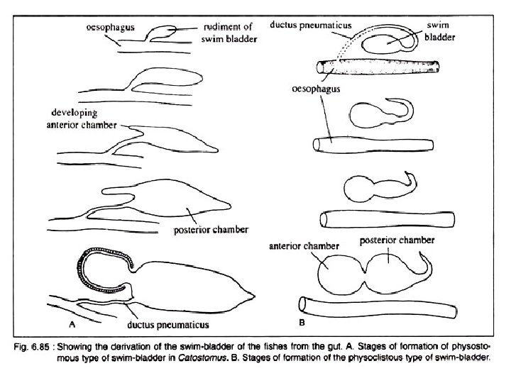
Development of Swim Bladder Opinions differ as regards the development of swim bladder in fishes. In teleosts, it originates as an unpaired dorsal or dorsolateral diverticulum of the oesophagus. It starts as a small pouch budded off from the oesophagus. The diverti culum with an opening in the oesophagus becomes subsequently divided into two halves. Of these two, the left one often atrophies except in a few primitive forms. The right half becomes well developed and take a median position. In dipnoans and Polypteridae, the swim bladder is modified into the ‘lungs’ and originates as the down growths from the floor of the pharynx.


These out growths have been rotated around the right side of the alimentary canal to occupy the dorsal position. As a con sequence of shifting of the position, the original right ‘lung’ becomes the left one. Spengel advocates the view that the swim bladder in fishes originates from the posterior pair of the gill pouches, but definite embryological evi dence in support of this idea is lacking.

Swim Bladders in different groups of fishes

Basic Structure of Swim Bladder The swim bladder in fishes varies greatly in struc ture, size and shape. a. It is essentially a tough sac like structure with an overlying capillary network. b. Beneath the capillary system there is a connective tissue layer called tunica externa. c. Below this layer lies the tunica interna consisting primarily of smooth muscle fibres and epithelial gas gland. d. The swim bladder lies below the kidneys, between the gonads and above the gut. e. The connection with the oesophagus may be retained through out life or may be lost in the adult.
 and Morean (1876) have shown that the")
Gas Composition of Swim Bladder Biot (1807) and Morean (1876) have shown that the gas secreted by the swim bladder is mostly oxygen. Nitrogen, and little quantity of carbon dioxide are also present. Generally the gas composition varies in different species. In salmonids, the maximum amount of gas in the swim bladder is Nitrogen. Again in many species the composition includes mostly a mix ture of oxygen and carbon dioxide. Jean Baptiste Biot
 between the swim bladder")
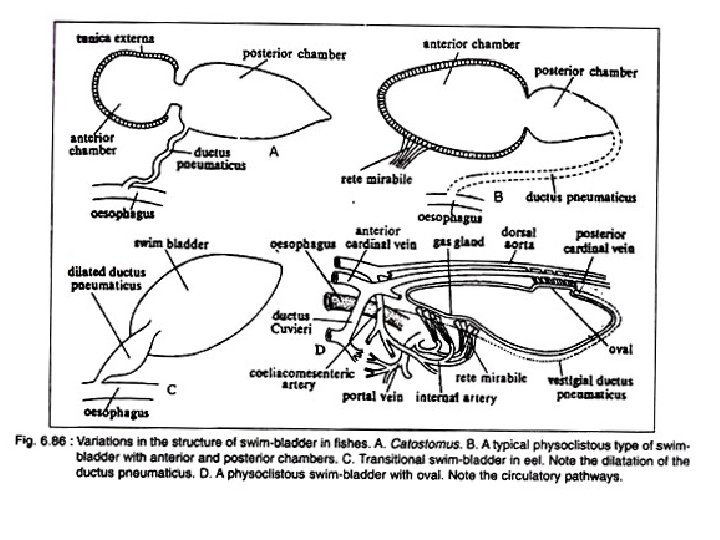
Types Depending on the presence of the duct (ductus pneumaticus) between the swim bladder and the oesophagus, the swim bladder in fishes can be divided into two broad categories: Physostomous [Gk. physi = a bladder; stomata, mouth] and Physoclistous types [Gk. clistic = enclosed]. Depending on the condition of the swim bladder, the teleosts are classified by older taxonomists into two groups Physostomi and Physoclisti. A transi tional condition is observed in eels.

A. Physostomous Condition: The swim bladder develops from the oesopha gus. When the ductus pneumaticus is present between the swim bladder and the oesopha gus, the swim bladder is called physostomous type (Fig. 6. 85 A). A vessel emerging from the coeliacomesenteric artery supplies the swim bladder and the blood from it is conveyed to the heart through a vein joining the hepatic portal vein. This condition is observed in bony ganoid fishes, the dipnoans and soft rayed teleosts.
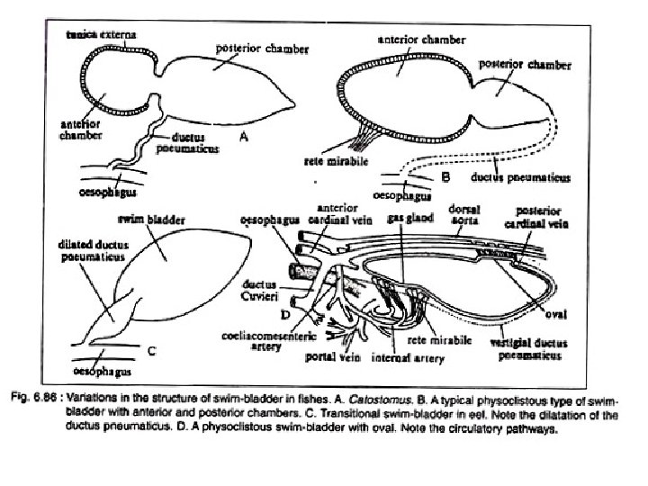

B. Physoclistous Condition: In this condition the ductus pneumaticus is either closed or atrophied (Fig. 6. 85 B). This type of swim bladder is observed in spiny rayed fishes. In this type of swim bladder, there lies an anteroventral secretory gas gland (containing retia mirabilia) and a posterodorsal gas absorbing region called the oval. The oval develops out of the degenerating ductus pneu maticus. The rete mirabilis of the gas gland, the oval and the walls of the bladder are sup plied by the coeliacomesenteric artery and also by arteries from the dorsal aorta. But the blood from the different parts of the swim bladder is returned by two routes. The blood from the gas gland is returned to the heart by the hepatic portal vein, while from the rest of the bladder by the posterior cardinal veins. The bladder, specially the gas gland, gets the lateral branches from the vagus, while the oval is innervated by sympathetic nerves.

Modifications of Physostomous Condition The typical physostomous pattern becomes modified in different fishes and the basic trends are: (1) The formation of paired sacs and (2) The gradual acquisition of two chambers— an anterior and a posterior. The swim bladder in Polypterus (bichir) (Fig. 6. 87 A, B) represents the primitive condi tion. It is a bilobed sac with two unequally developed lobes. The left lobe is shorter and the right lobe is longer. The bilobed sac opens on the floor of the pharynx through a slit like glottis. The glottis is provided with muscular sphincter. The internal lining of the bladder is smooth and partly ciliated.
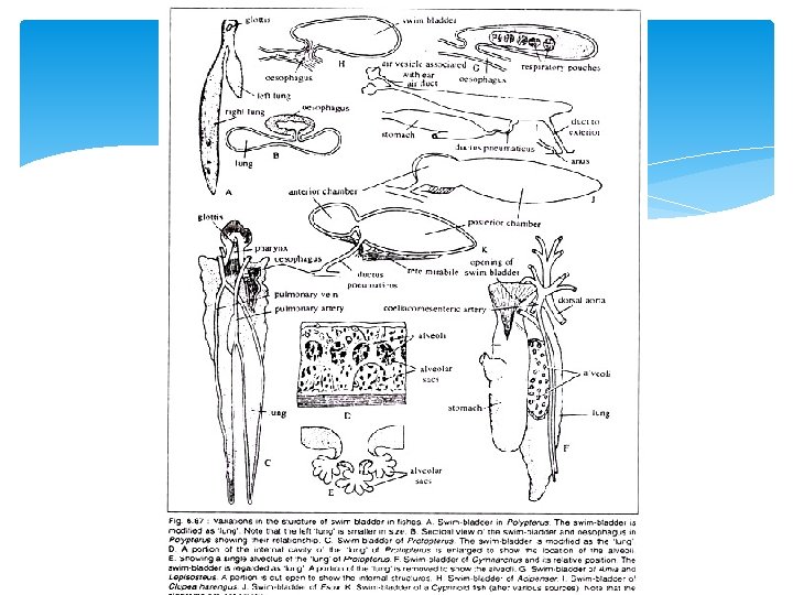

The lack of alveolar sacculations and the pre sence of muscular walls are the two noted feature in the swim bladder of Polypterus. The walls of the bladder are highly vascular and are lined by two layers of striated muscle fibres. The bladder is supplied by a pair of pulmonary arteries arising from the last pair of epibranchial arteries and the corres ponding veins enter into the hepatic vein below the sinus venosus. In the dipnoans, the swim bladder is called the lung and the inner walls are produced into numerous alveoli. The swim bladder resem bles the tetrapod lungs both structurally as well as functionally. In Neoceratodus it is single lobed, while in Protopterus and Lepidosiren it is bilobed (Fig. 6. 87 C, D, E).

Histological Modifications in swim Bladder In some physostomous and all physoclistous fishes this process of gas transference is done directly from the blood stream. Inside the bladder there is an oxygen producing device and an oxygen absorbing device. The swim bladder is a vascular structure but the degree of vascularization varies in different teleosts. In some species of the families Clupeidae and Salmonidae the capillaries are uniformly pre sent all over the swim bladder, but in most cases these highly vascular interlacing and tightly packed capillaries form a mass called rete mirabilis. The anterior chamber of swim bladder shows the tendency to become diffe rentiated into oxygen producing area called red body. The oxygen is produced by the reduction of the oxyhaemoglobin in the ery throcytes when brought into close contact with the secreting epithelial cells of the gas gland. The red body consists of internal oxygen secreting cells (gas gland) and sup plied by the blood vessels from the retia Mirabella (sing, rete mirabilis).

It forms a com plicated structure where the arterial and venous capillaries communicate only after reaching the gas gland. The most primitive condition is observed in Pickerel where the gland is covered by thick glandular epithelium which is thrown into a number of folds. In eels and some other fishes, the red bodies are non glandular in nature but serve the same physio logical function. The red gland is supplied with blood from the coeliac artery and is returned to the portal vein. The activity of the red gland is controlled by the vagus nerve. In the fishes with functional ductus pneumaticus the gas glands are absent but in eels this func tion is taken up by the red gland.

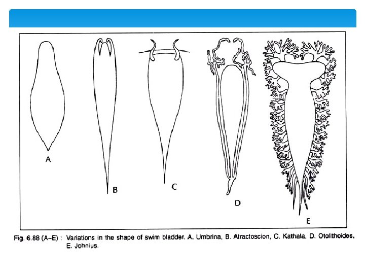
Variable shape & Size of Swim Bladder The swim bladder varies extensively in shape and size. In Umbrina (Fig. 6. 88 A), it is oval shaped and without any appendage. In Atractoscion (Fig. 6. 88 B), it gives off only one pair of simple diverticula that extends from the anterior side. In Kathala (Fig. 6. 88 C), the swim bladder develops a pair of appendage extending in front of transverse septum into head. In Otolithoides (Fig. 6. 88 D), the appendages attached to posterior end of bladder and at least the main part lying parallel to the blad der. In. Corvina lobata, many such branched diverticula develop from the lateral walls of the swim bladder. In Johnius (Fig. 6. 88 E), it is hammer shaped with 12 to 15 pairs arbores cent appendages, the first branching in the head and the posterior tip are highly pointed.

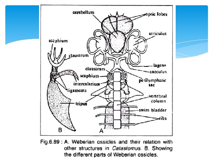
Weberian Ossicles The perilymphatic sac and the anterior end of the swim bladder are connected by a series of four ossicles (Fig. 6. 89), which are articulated as a conducting chain. Of the four, the tripus, intercalarium and scaphium actu ally form the chain, while the fourth one, claustrum lies dorsal to the scaphium and lies in the wall of posterior prolongation of the perilymphatic sac. The function of these ossi cles is controversial. It is regarded that the Weberian ossicles either help to intensify sound vibrations and convey these waves to the internal ear of help to understand the state of tension of air pressure in the bladder and transit changes of such pressure to the peri lymph to set up a reflex action. There are various views regarding the actual process of derivation of these ossicles.
 De Beer (1937) and Watson (1939) regarded that these are detached or modified")
1) De Beer (1937) and Watson (1939) regarded that these are detached or modified processes of the first three anterior vertebrae. As regards the actual mode of origin of the four ossicles there are differences of opinion. 2) The claustrum is regarded to be modified interspinous ossicle or modified spine of first vertebra or modified neural arch of first vertebra or modified intercalated cartilage or modified neural process of first cartilage. 3) The scaphium is considered to be the modified neural arch of the first ver tebra or modified rib of the first vertebra or derived from the neural arch of the first vertebra and also from the mesenchyme. 4) The intercalarium is derived from the neural arch and trans verse process of the second vertebra or from the neural arch of the second vertebra and also from the ossified ligament or from the neural arch of the second vertebra only. 5) The tripus is formed from the rib of the third vertebra and the ossified ligament or from the trans verse process of the third vertebra along with ossified wall of the swim bladder or from the transverse process of the third vertebra and the ribs of third and fourth vertebrae.

Functions of Swim Bladder: Hydrostatic Organ It is primarily a hydro static organ and helps to keep the weight of the body equal to the volume of the water, the fish displaces. It also serves to equilibrate the body in relation to the surrounding medium by increasing or decreasing the volume of gas content. In the physostomous fishes the expul sionof the gas from the swim bladder is caused by way of the ductus pneumaticus, but in the physoclistous fishes where the ductus pneumaticus is absent the superfluous gas is removed by diffusion.

Swim Bladder acts as Adjustable Float The swim bladder also acts as an adjustable float to enable the fishes to swim at any depth with the least effort. When a fish likes to sink, the specific gravity of the body is increased. When it ascends the swim bladder is distended and the specific gravity is diminished. By such adjustment, a fish can maintain equilibrium at any level.

Swim Bladder Maintains Proper Centre of Gravity: The swim bladder helps to maintain the proper centre of gravity by shifting the contained gas from one part of it to the other and this facilitates in exhibiting a variety of movement. Swim Bladder helps in Respiration: The respiratory function of the swim bladder is quite significant. In many fish es living in water in which oxygen content is considerably low, the oxygen produced in the bladder may serve as a source of oxygen. In a few fishes, specially in the dipnoans, the swim bladder becomes modified into the ‘lung’. The ‘lung’ is capable of taking atmospheric air. Swim Bladder as Resonator: The swim bladder is regarded to act as a resonator. It intensifies the vibra tions of sound and transmits these to the ear through the Weberian ossicles.

Production of sound The swim bladder helps in the production of sound. Many fishes, Doras, Platystoma, Malapterurus, Trigla can produce grunting or hissing or drumming sound. The circulation of the contained air inside the swim bladder causes the vibration of the incomplete septa. The sound is pro duced as the consequence of vibration of the incomplete septa present on the inner wall of the swim bladder. The vibrations are caused by the movement of the contained air of the swim bladder. Sound may also be produced by the compression of the extrinsic and intrinsic musculature of the swim bladder. Polypterus, Protopterus and Lepidosiren can produce sound by compression and forceful expulsion of the contained gas in the swim bladder. In Cynoscion male, the musculus sonorificus probably helps in compression.
 – Ganguly, Sinha, Adhikari")
Ref: Biology of Animals (Vol 2) – Ganguly, Sinha, Adhikari
- Slides: 31