Survival curve radiation dose cell survival fraction Reproductive


 = Width of shoulder =semi-threshold dose (almost the threshold dose) n")


, nuclear membrane Chromosome, specifically the")
 http: //www-micro. msb. le. ac. uk/3035/kalmakoff/baculohostinteract. html")


 Loss reproductive integrity")


 from exchangetype chromosomal aberration. log-linear plot with broad shoulder Characterized")


 gene")


 = 300 c. Gy = 3 Gy")
 has a value close to 300 c.")


- Slides: 49
Survival curve = radiation dose & cell survival fraction
Reproductive Integrity Cell survival cell death For differentiated cells that do not proliferate e. g. , nerve, muscle lose of specific function (death) For proliferating cells e. g. , hematopoietic stem cells, culture cells lose for sustained proliferation (death) lose of reproductive integrity (reproductive death)
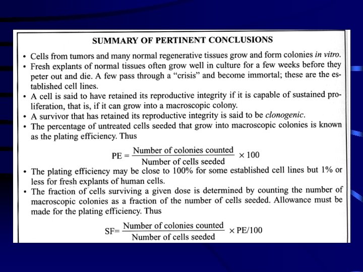
Definition of survival for radiobiology Retain reproductive integrity Proliferate indefinitely to produce colony (a large clone = colony = clonogenic)
The in vitro survival curve Cell culture Established cell lines Tissue trypsin cell culture in vitro Dose-survival curve
PE = plating efficiency
Serial dilution
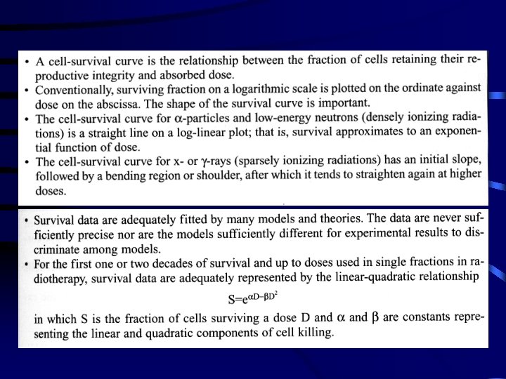
The shape of the survival curve Width of shoulder = Dq or n =semi-threshold dose log Linear –quadratic function
Multi-target model Initial slope single-event killing = D 1 final slope multiple-event killing = D 0
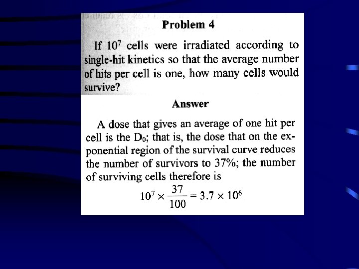
D 1 & D 0 = the dose required to reduce the fraction of surviving cells to 37% of it previous value. D 0 (straight at high dose) = the average dose required to deliver one inactivating event (one hit) per cell. = the dose required to reduce survival from 0. 1 to 0. 037 or 0. 01 to 0. 0037. = D 0 (37% survival) dose required to reduce survival to e-1 (0. 37)
Dq (quasithreshold dose) = Width of shoulder =semi-threshold dose (almost the threshold dose) n = extrapolation number Logen = Dq/D 0 D 0/0. 37(e-1) = Dq/n D 0/e-1 = Dq/n Threshold dose = n= e-1 x. Dq/D 0 ln n = -Dq/D 0 the dose below which there is no effect.
Linear –quadratic model Two components to cell killing by radiation One that is proportional to dose One that is proportional to square of dose (dual-radiation action; two separate breaks) S = e-a. D-b. D 2
S= 2 -a. D-b. D e S = the fraction of surviving a dose D a, b = constant When a. D = b. D 2 D = a/b Linear & quandratic contribution equally to cell killing at same dose
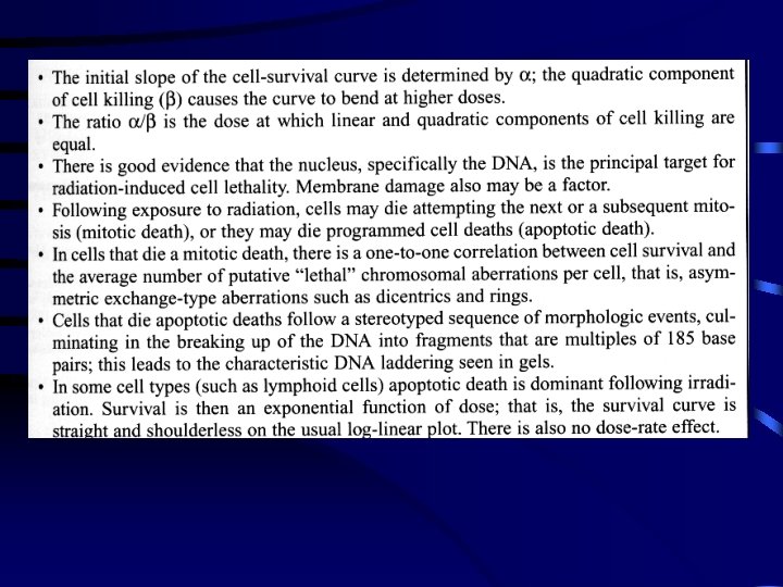
The mechanism of cell killing Target : DNA (nucleus), nuclear membrane Chromosome, specifically the DNA, as the primary target for radiation-induced lethality.
Apoptotic and mitotic death: AD (falling off) http: //www-micro. msb. le. ac. uk/3035/kalmakoff/baculohostinteract. html
http: //www. niaaa. nih. gov/publications/arh 25 -3/image 01. gif
http: //www. copewithcytokines. de/cope. cgi? 000638
http: //www. ucihs. uci. edu/anatomy/histo/corenotes/celldeath 2004. pdf
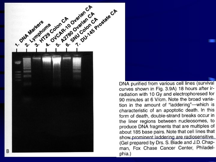
The detection of this DNA ladder is still currently used to distinguish at the molecular level apoptosis from necrosis. • Apoptosis: Dsb in linker DNA between nucleosome 185 bp [“ladder” in Gel] • nercosis [“smear” in Gel] Fig. 3 -9
Radiation-induced apoptosis is highly cell type dependent. Hemopoietic cells & lymphoid cells prone to rapid radiation-induced apoptosis. Most tumors mitotic cell death & apoptosis. or mitotic cell death only.
Apoptotic and mitotic death: MD • The most common form of cell death from radiation is mitotic death (MD). Cell die in attempting to divide because of damaged chromosomes. Review Cell death by mitotic catastrophe: a molecular definition http: //www. nature. com/cgi-taf/Dyna. Page. taf? file=/onc/journal/v 23/n 16/abs/1207528 a. html&dynoptions=doi 1097246946
Asymmetric exchange-type chromosome aberrations (i. e. , dicentrics and rings ) Loss reproductive integrity Unable to proliferate death Asymmetric exchange-type chromosome aberrations represent the principle mechanism for radiation-induced mitotic death in mammalian cells.
No apoptosis Cell surviving & cell without visible aberration correlation
Exchange type aberrations require two chromosome breaks. The probability of an interaction between the two breaks is related to D (low dose) or D 2 (high dose).
Chromosome aberrations in human lymphocytes Ch 2
Survival curves for various mammalian cells in culture First in vitro survival curve All mammalian cells, normal & malignant, exhibit similar x-ray survival curve (initial shoulder but size vary) The Do of X-ray survival curves for most culture cells range from 1 to 2 Gy (100 -200 rad or c. Gy). (page 41)
radiosensitivity
Survival-curve shape and mechanism of cell death Most cells fall between apopototic & mitotic death Note! Shoulder
Mitotic death results (principally) from exchangetype chromosomal aberration. log-linear plot with broad shoulder Characterized by subsequently dose-rate effect (page 74). Apoptotic death result unknow mechanism. straight line on log-linear plot. Characterized by expotential function of dose. little or no dose-rate effect.
S= 2 -a. D-b. D e Linear –quadratic model apoptotic death S= a. A mitotic death 2 -(a +a )D-b D M A M e = cell killing from apoptotic death (vary linear) a. M = cell killing from mitotic death (vary linear) b. M = cell killing from mitotic death (vary square)
Oncogenes and radioresistance Transfection of activated oncogenes to culture cells Increase radioresistance
Genetic control of radiosensitivity ATM (AT-mutated) gene
Intrinsic Radiosensitivity and predictive assay Courtenary assay Semisolid agar gel with growth factor Nonclonogenic assay Cell growth in multi-wells plate, e. g. MTT assay or chapter 15
補充 Surviving cell number is then determined indirectly by MTT dye reduction. (Fig 23. 4) The amount of MTT-formazan produced can be determined spectrophotometrically once and solublilized it in a suitable solvent.
The effective survival curve for a multifraction regimen are most often used in clinical radiotherapy What is multifraction regimen? Dilute dose to fraction at time intervals sublethal damage & time for repair Shoulder The effective survival curve
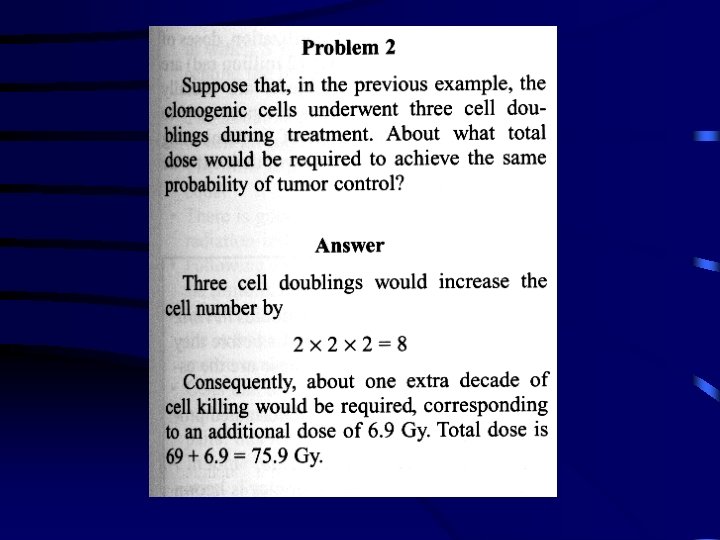
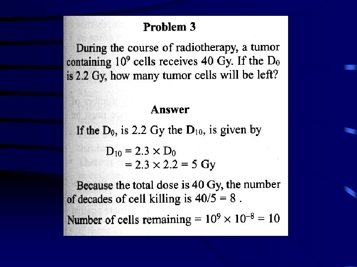
For human, effective D 0 (37% survival) = 300 c. Gy = 3 Gy D 10 (the dose required to kill 90% = 10 % survival) = one decade of cell killing = 2. 3 X D 0 • Natural log 10 = 2. 3 • equal Slope Ps: equal Slope Logen = Dq/D 0 (F 3. 3 & page 37)
The Do of effecitive survival curve (slope) has a value close to 300 c. Gy for cells of human origin. This is an avarage value and can differ significantly for different tumors.
Calculations of tumor cell kill 109
The radiosensitivity of mammalian cells compared with microorganisms