Surgical Treatment of Sinusitis Dr Vishal Sharma Maxillary

")
")


 2. Pain & swelling of cheek (breach")
 • Region of antral puncture in inferior meatus perforated with Tilley's")


 + superior part of")


 2. Anterior ethmoidectomy 3. Middle")


 Major epistaxis Orbital hematoma Diplopia Blindness or ed visual acuity Internal")

- Slides: 69
Surgical Treatment of Sinusitis Dr. Vishal Sharma
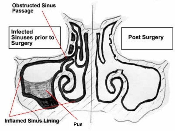
Maxillary Sinusitis Surgical Treatment Methods: 1. Antral Washout 2. Intra-nasal Inferior meatal antrostomy (INA) 3. Caldwell - Luc surgery 4. Middle meatal antrostomy 5. Functional Endoscopic Sinus Surgery (FESS)
Antral Washout (proof puncture, antral lavage)
Indications Diagnosis & treatment of chronic maxillary sinusitis not responding to conservative medications Cytology/culture sensitivity of antral contents Contraindications Age < 3 yrs Hypoplastic maxilla with thick bony walls Acute maxillary sinusitis untreated by antibiotics Trauma to maxillary sinus or Fracture of orbital floor Drainage of maxillary antral hematoma
Tilley Lichwitz Antrum Puncture Trocar & Cannula
Higginson Syringe
Trocar directed towards I/L tragus
Hole made 1. 25 cm behind anterior end of inferior turbinate
Antral irrigation
Anesthesia: L. A. for adults. G. A. for children & uncooperative pt. Position: Sitting / supine. Technique: Puncture lateral wall of inferior meatus with Tilley-Litchwitz antral trocar & cannula, just anterior to turbinate genu, trocar directed towards tragus of ipsilateral ear, with gentle boring action. Advance till it hits posterior wall, then withdraw slightly. Remove trocar & wash sinus with saline at 370 C with pt leaning forwards & saying k k. Wash till clear fluid comes. Remove cannula.
Complications 1. Hemorrhage ( Lateral Sphenopalatine artery) 2. Pain & swelling of cheek (breach of anterior wall) 3. Orbital damage (perforation of orbital floor) 4. Perforation of posterior wall (maxillary artery injury) 5. Vasovagal attack 6. Fatal air embolism
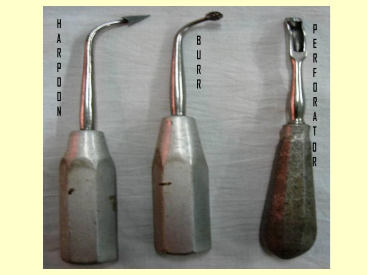
Intranasal antrostomy (INA) • Region of antral puncture in inferior meatus perforated with Tilley's antral harpoon. • Antrostomy enlarged with Tilley's antral burr or Myle’s nasoantral perforator.
Caldwell – Luc Surgery George Caldwell, 1893, New York Henri Luc, 1897, Paris
Indications Chronic refractory maxillary sinusitis Oro-antral fistula closure Foreign body removal from maxillary antrum Fungal maxillary sinusitis Elevation of orbital floor fractures Ethmoidectomy (trans-antral) Biopsy of suspicious neoplasm of maxillary antrum Orbital floor decompression Antrochoanal polyp (recurrent) Route to pterygo-palatine fossa (Vidian nerve, Max Artery) Dental / dentigerous cyst (maxillary antrum) removal
Exposure of incision site
Incision 4 cm long, sub-labial, horizontal incision made 3 mm above & parallel to the gingival margin, from lateral incisor to 2 nd molar tooth.
Incision deepened till periosteum
Anterior wall broken with osteotome
Hole made in anterior wall
Suction of maxillary sinus
Inferior meatal antrostomy
Packing of maxillary sinus
Packing of sinus & nose
Incision closed
Complications Facial: Cheek edema, ecchymosis, subcutaneous emphysema, infraorbital n. paresthesia Orbital: Hematoma, extraocular muscle trauma, diplopia, globe trauma, blindness Oral: Trauma to teeth roots, Superior alveolar nerve damage, Dental anesthesia, Oroantral fistula Vascular: Internal maxillary artery injury
Ethmoid Sinusitis Surgical Treatment Methods: 1. Intra-nasal microscopic ethmoidectomy 2. Extra-nasal Ethmoidectomy a. Lynch Howarth procedure b. Patterson trans-orbital procedure c. Trans-antral (Jansen Horgan procedure) 3. Functional Endoscopic Sinus Surgery
Lynch Howarth ethmoidectomy
Patterson ethmoidectomy
Trans-antral ethmoidectomy • Caldwell – Luc surgery done to reach maxillary antrum • Ethmoid cells approached via postero-superomedial angle of maxillary antrum
Frontal Sinusitis Surgical Treatment Methods: 1. Trephination of frontal sinus 2. Modified Lothrop procedure 3. Osteoplastic Flap surgery 4. Functional Endoscopic Sinus Surgery
Frontal sinus trephination
Frontal sinus trephination 2 -cm incision made 1 cm below medial end of eyebrow & deepened up to bone. Frontal sinus floor opened by drilling with burr. Opening enlarged with Citelli’s punch forceps to drain pus. Drainage tube inserted inside frontal sinus cavity & sutured in place. Regular lavage of the frontal sinus done through drainage tube for 48 -72 hours post-operatively.
Frontal sinus trephination
Osteoplastic flap procedure
Osteoplastic flap procedure
Lothrop Procedure Removal of frontal sinus (inferior septum + floor) + superior part of nasal septum
Lothrop Procedure
Sphenoid sinus Surgical Treatment Methods: 1. Trans-nasal trans-septal approach 2. Sublabial trans-septal approach 3. External ethmoidectomy approach 4. Endoscopic intra-nasal approach 5. Functional Endoscopic Sinus Surgery
Sublabial trans-septal approach
External ethmoidectomy approach
Endoscopic approach
Functional Endoscopic Sinus Surgery
F. E. S. S.
Anatomy of lateral wall
Steps of F. E. S. S. 1. Uncinectomy (Infundibulotomy) 2. Anterior ethmoidectomy 3. Middle meatal antrostomy 4. Perforation of basal lamella 5. Posterior ethmoidectomy 6. Sphenoid sinus exploration 7. Skull base disease clearance 8. Frontal recess exploration
Steps of F. E. S. S.
Left nasal cavity
Left middle meatus
Left middle meatus
Incision on uncinate process
Incision completed
Uncinate process removed
Opening of bulla ethmoidalis
Bulla ethmoidalis removed
Natural & accessory ostia exposed
Middle meatal antrostomy done
Opening made on basal lamella
Basal lamella removed
Posterior ethmoidectomy done
Anterior sphenoid sinus wall
Interior of sphenoid sinus
Skull base clearance done
Frontal recess opened
Final FESS cavity
Surgical Navigation
Complications Major (1%) Major epistaxis Orbital hematoma Diplopia Blindness or ed visual acuity Internal carotid injury Intracranial hemorrhage CSF leak / Meningitis Pneumocephalus Anosmia Nasolacrimal duct trauma Minor (7%) Minor epistaxis Hyposmia Adhesions (synechiae) Headache Periorbital echhymosis Periorbital hematoma Dental / facial pain
Thank You