SURGICAL REPAIR OF CLEFT LIP AND PALATE CAPT

 Affects 1")

 + Palatine Bones(Horizontal Lamina)")

 2. Pfeifer’s symbolic classification (1966) 3. Kernahan &")


 Lowest in AFRO-CARRIBEAN (0. 3/1000)")
 l Linkage analysis l TGF")


")


- Slides: 45
SURGICAL REPAIR OF CLEFT LIP AND PALATE CAPT MUBASHIR IQBAL
Introduction Facial clefting is the second most common congenital deformity (after clubfoot) Affects 1 in 750 births Problems are cosmetic, dental, speech, swallowing, hearing, facial growth, emotional Otolaryngologist holds key role in management
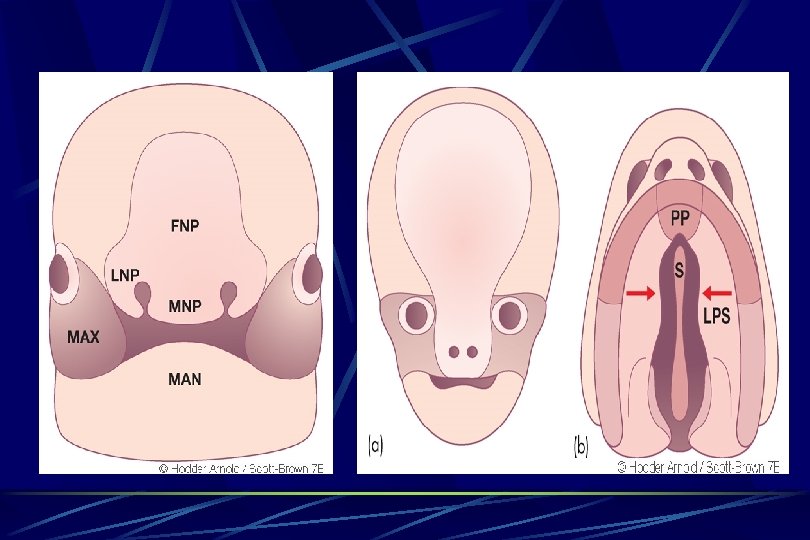
EMBROYOLOGY Ø 4 th week FACIAL PROMINENCES Ø Frontonasal process forms forehead , bridge of nose, medial & lateral nasal prominences Ø Maxillary process forms cheeks & lateral portion of upper lip Ø Lateral nasal process forms alae of nose Ø Medial nasal process forms nasal septum, philtrum, premaxilla & primary palate Ø Mandibular process forms lower lip
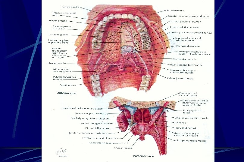
ANATOMY Hard Palate l Maxilla( Palatine Processes) + Palatine Bones(Horizontal Lamina)
ANATOMY Soft Palate l Fibromuscular shelf attached like a shelf to posterior portion of hard palate Tensor Veli Palatini Levator Veli Palatini(Primary Elevator) Musculus Uvulae, Palatoglossus Palatopharyngeus
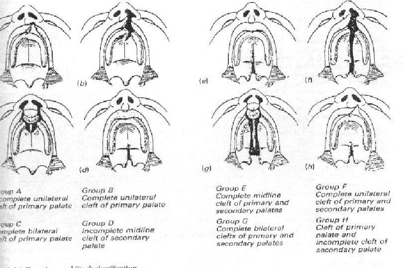
CLASSIFICATION 1. Veau’s simple classification (1931) 2. Pfeifer’s symbolic classification (1966) 3. Kernahan & Stark’s striped Y classification (1971) 4. Krien’s LAHSHAL classification (1987)
VEAU’s Veau Classification - 1931 Veau Class I: isolated soft palate cleft l Veau Class II: isolated hard and soft palate l Veau Class III: unilateral CLAP l Veau Class IV: bilateral CLAP l
PFEIFER’s SYMBOLIC
KERNAHAN’s STRIPED Y
KRIEN’s LAHSHAL
EPIDEMIOLOGY Highest incidence in NATIVE AMERICANS ( 3. 7/1000) Lowest in AFRO-CARRIBEAN (0. 3/1000) Cleft lip + palate more common in boys 2 : 1 Cleft lip common in boys 1. 5 : 1 Cleft palate alone more common in girls C/L left twice as common as right Unilateral/bilateral Complete/incomplete
GENETIC FACTORS Siblings ( 2. 2 to 10 %) l Linkage analysis l TGF alpha l TGF beta l MSX 1 l MTHFR l ENVIRONMENTAL FACTORS Smoking l Alcohol l Anticonvulsants l steroids l
ASSOCIATION with other SYNDROMES Increased Clefts with maternal diabetes mellitus and amniotic band syndrome Cleft Lip + Palate- 50% l Cleft Palate- 30% l Cleft Lip- 20% l Cleft Lip + Alveolus- 5% l SYNDROMES velocardiofacial syndrome, goldenhar syndrome, stickler & treacher collins syndromes
CLEFT FORMATION Cleft result in a deficiency of tissue Cleft lip occurs when an epithelial bridge fails Clefts of primary palate occur anterior to incisive foramen Clefts of secondary palate occur posterior to incisive foramen
CLEFT LIP Nasal floor communicates with oral cavity Maxilla on cleft side is hypoplastic Columella is displaced to normal side Nasal ala on cleft side is laterally, posteriorly, and inferiorly displaced Lip muscles insert into ala and columella
Palatal Clefts Soft palate muscles insert on posterior margin of remaining hard palate rather than midline raphe. Associated Dental Abnormalities Supernumery Teeth- 20% l Dystrophic Teeth- 30% l Missing Teeth- 50% l Malocclusion- 100% l
Unilateral Cleft Lip
Bilateral Cleft Lip
MANAGEMENT Team Approach Otolaryngologist has a pivotal role Initial Head and Neck Examination Speech Disorders Ear Disease Airway Problems Surgical Repair
Head and Neck Exam Head- facial symmetry Otologic- auricle and canal development and location, pneumatic otoscopy, forks Rhinoscopy- identifies clefting, septal anomalies, masses, choanal atresia Oral Exam- cleft, dental, tongue Upper airway- phonation, cough, swallow
Speech Disorders Errors in Articulation: Fricatives, Affricates Velopharyngeal Competence- Most important determinant of speech quality in cleft palate patients-75% achieve competence after initial palate surgery Incompetence- nasal emission or snort Evaluation- Direct exam , Fiberoptic Exam
Ear Disease Cleft Lip- Incidence similar to normal pop. Cleft Palate- Almost all with ETD, CHL ETD- Due to abnormal insertion of levator veli palatini and tensor veli palatini into posterior hard palate ETD- Returns to normal by mid-adolescent Cleft Palate- Increased Cholesteatoma(7%)
Ear Disease Otologic Goals For Cleft Palate Patients Adequate hearing l Ossicular chain continuity l Adequate middle ear space l Prevent TM deterioration l Indications for Myringotomy Tubes CHL, Persistent/Recurrent effusion, Retraction l Cleft palate: Multiple BMTs from 3 mo. - 12 yrs l
Airway Problems More common in Cleft Palate patients with concomitant structural or functional anomalies. e. g. Pierre-Robin Sequence Micrognathia, Cleft Palate, Glossoptosis l May develop airway distress from tongue becoming lodged in palatal defect l
Surgical Repair- Cleft Lip Adhesions 2 weeks of age l Converts complete cleft into incomplete cleft l Serves as temporizing measure for those with feeding problems l May interfere with definitive lip repair l Less often needed in recent years due to wider variety of specialty feeding nipples l
Surgical Repair- Cleft Lip Cleft lip repaired at 10 weeks Rotation-advancement method- Most common in the U. S. Nine Landmarks Rotation Flap cuts made first Advancement cuts made next Cleft side nasal ala cuts made last
Surgical Techniques Cleft Lip Repair l unilateral l rotation-advancement flap developed by Millard l complications l l dehiscence l infection thin white roll l excess tension
Milliard & Delaire repairs
Surgical Techniques Cleft Lip Repair l bilateral rotation advancement with attachment to premaxilla mucosa l complications l l dehiscence thin white roll
Alveolar Cleft Repair
Surgical Repair- Cleft Palate Several Techniques- Trend is towards less scarring and less tension on palate Scarring of palate may cause impaired midfacial growth(alveolar arch collapse, midface retrusion, malocclusion) Facial growth may be less affected if surgery is delayed until 18 -24 months, but feeding, speech, socialization may suffer.
Surgical Repair- Cleft Palate Bardach Method- Two Flap technique Medial incisions made, which separate oral and nasal mucosa l Lateral incisions made at junction of palate and alveolar ridge l Elevate flaps, preserve greater palatine artery. l Detach velar muscles from posterior palate l Close in 3 layers l
VEAU REPAIR(BARDACH)
LANGENBACH
VEAU WARDOL KILNER
Langenbeck, Veau, VWK
FURLOW Z-plasty
2 staged repair of cleft palate
Vomer flap repair of unilateral cleft palate
Non-Surgical Treatment Dental Obturator l For high-risk patients or those that refuse surgery Advantage- High rate of closure l Disadvantage- Need to wear a prosthesis, and need to modify prosthesis as child grows. l
Conclusions Cleft Lip and Palate are common congenital deformities that often affect speech, hearing, and cosmesis; and may at times lead to airway compromise. The otolaryngologist is a key member of the cleft palate team, and is in a unique position to identify and manage many of these problems.