
Surgical Radiology Hepatobiliarypancreatic Department of General Surgery Cairo
Surgical Radiology Hepatobiliary-pancreatic Department of General Surgery Cairo University
Contents • Normal appearance • Abnormalities Plain X-ray Oral cholecystography Ultrasound CT scan ERCP PTC MRCP Intraoperative cholangiography T-tube cholangiography HIDA scan
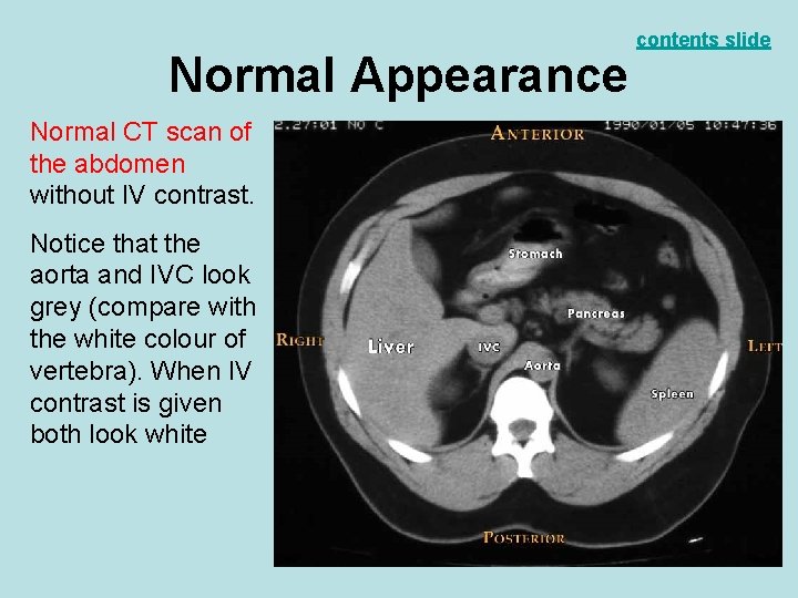
Normal Appearance Normal CT scan of the abdomen without IV contrast. Notice that the aorta and IVC look grey (compare with the white colour of vertebra). When IV contrast is given both look white contents slide
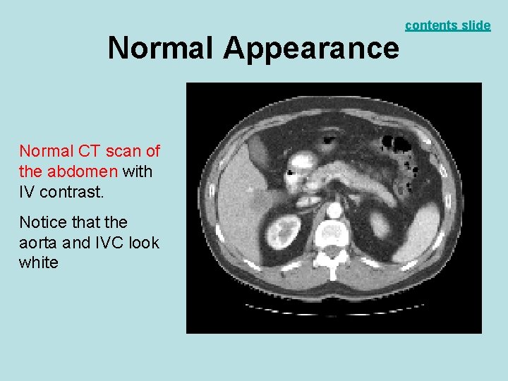
Normal Appearance Normal CT scan of the abdomen with IV contrast. Notice that the aorta and IVC look white contents slide
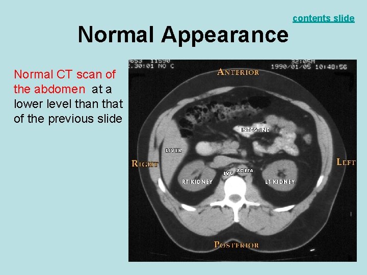
Normal Appearance Normal CT scan of the abdomen at a lower level than that of the previous slide contents slide
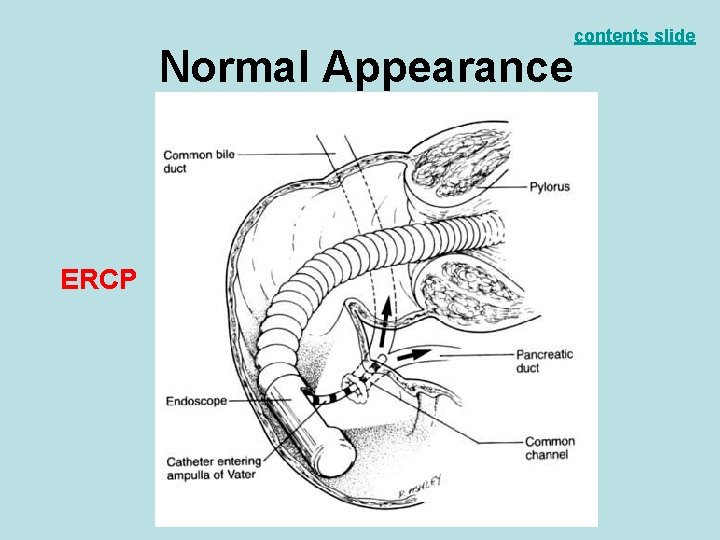
Normal Appearance ERCP contents slide
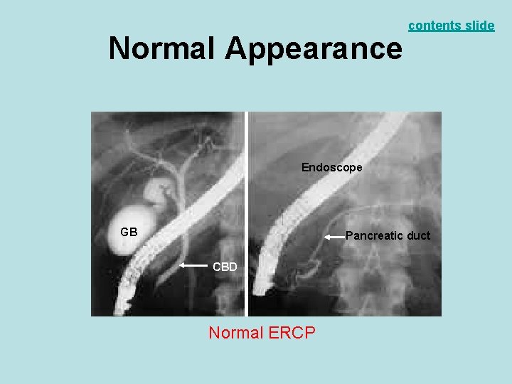
Normal Appearance contents slide Endoscope GB Pancreatic duct CBD Normal ERCP
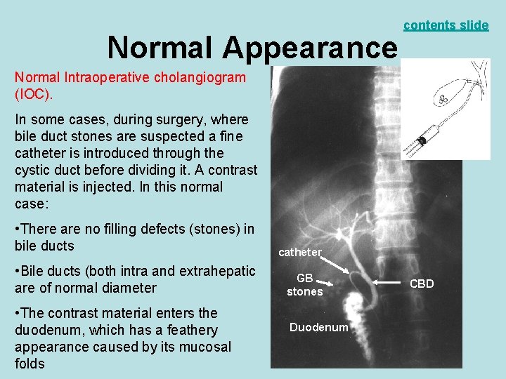
Normal Appearance contents slide Normal Intraoperative cholangiogram (IOC). In some cases, during surgery, where bile duct stones are suspected a fine catheter is introduced through the cystic duct before dividing it. A contrast material is injected. In this normal case: • There are no filling defects (stones) in bile ducts catheter • Bile ducts (both intra and extrahepatic are of normal diameter GB stones • The contrast material enters the duodenum, which has a feathery appearance caused by its mucosal folds Duodenum CBD
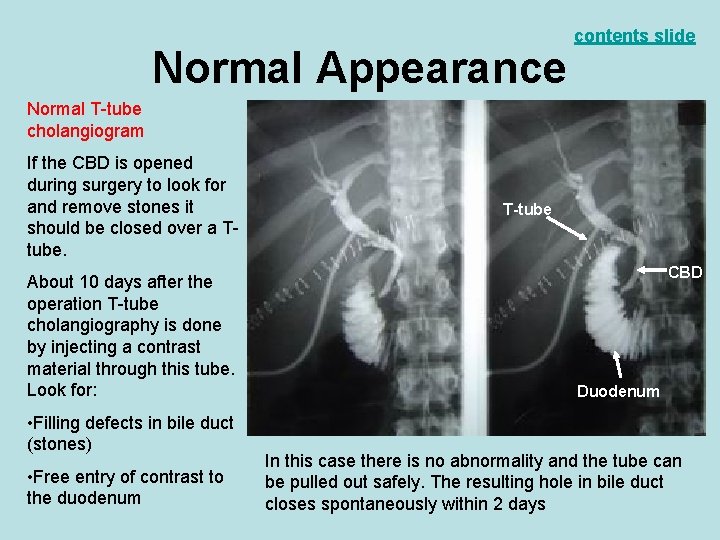
Normal Appearance contents slide Normal T-tube cholangiogram If the CBD is opened during surgery to look for and remove stones it should be closed over a Ttube. About 10 days after the operation T-tube cholangiography is done by injecting a contrast material through this tube. Look for: • Filling defects in bile duct (stones) • Free entry of contrast to the duodenum T-tube CBD Duodenum In this case there is no abnormality and the tube can be pulled out safely. The resulting hole in bile duct closes spontaneously within 2 days
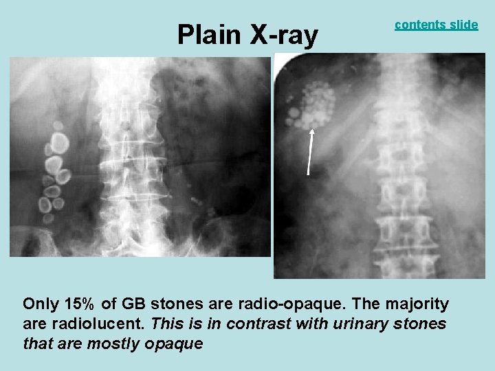
Plain X-ray contents slide Only 15% of GB stones are radio-opaque. The majority are radiolucent. This is in contrast with urinary stones that are mostly opaque
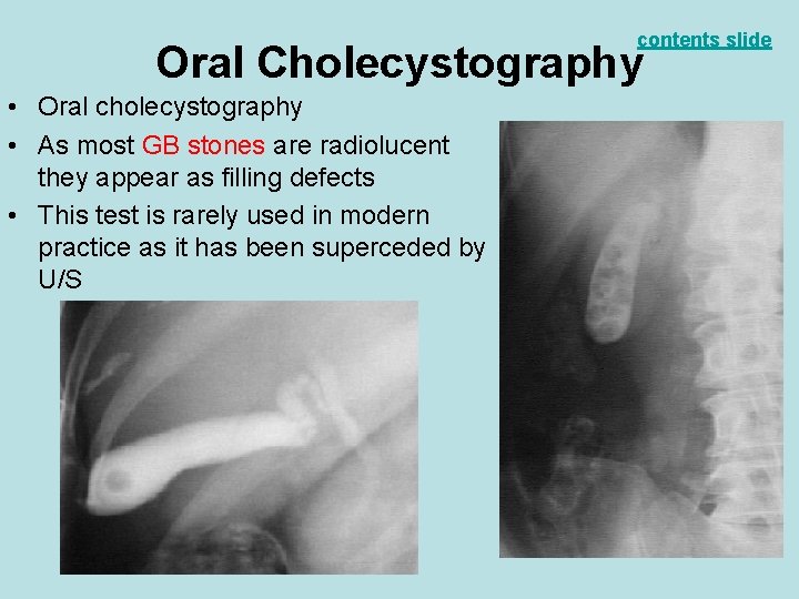
contents slide Oral Cholecystography • Oral cholecystography • As most GB stones are radiolucent they appear as filling defects • This test is rarely used in modern practice as it has been superceded by U/S
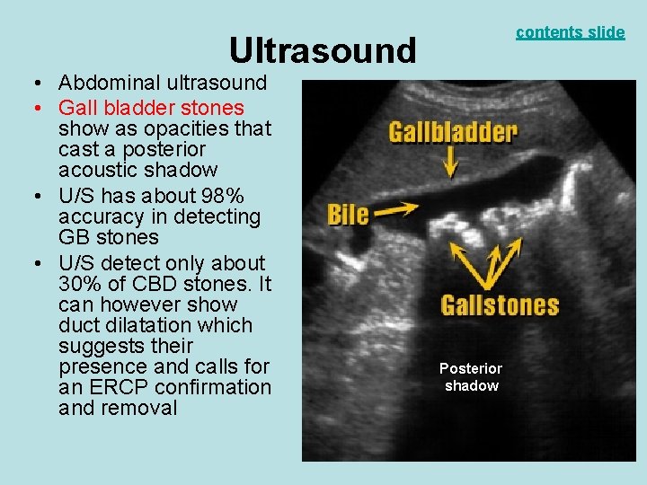
contents slide Ultrasound • Abdominal ultrasound • Gall bladder stones show as opacities that cast a posterior acoustic shadow • U/S has about 98% accuracy in detecting GB stones • U/S detect only about 30% of CBD stones. It can however show duct dilatation which suggests their presence and calls for an ERCP confirmation and removal Posterior shadow
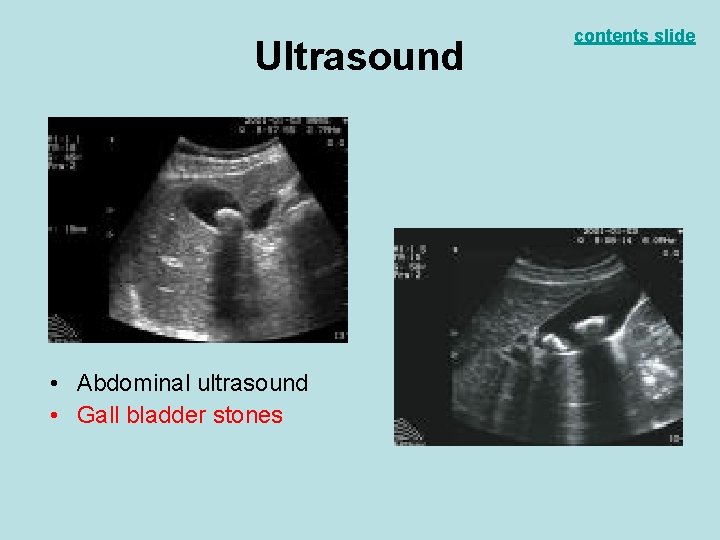
Ultrasound • Abdominal ultrasound • Gall bladder stones contents slide
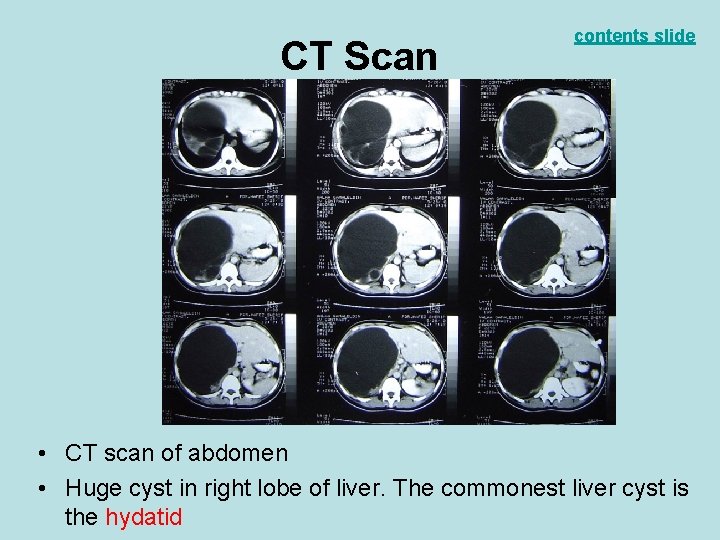
CT Scan contents slide • CT scan of abdomen • Huge cyst in right lobe of liver. The commonest liver cyst is the hydatid
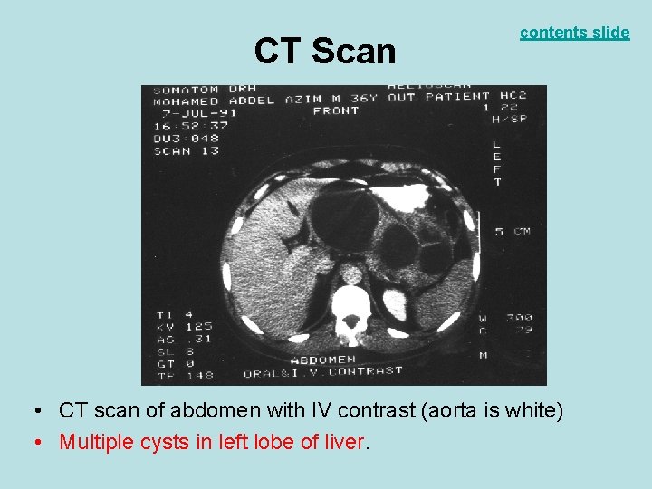
CT Scan contents slide • CT scan of abdomen with IV contrast (aorta is white) • Multiple cysts in left lobe of liver.
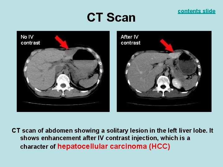
CT Scan No IV contrast contents slide After IV contrast CT scan of abdomen showing a solitary lesion in the left liver lobe. It shows enhancement after IV contrast injection, which is a character of hepatocellular carcinoma (HCC)
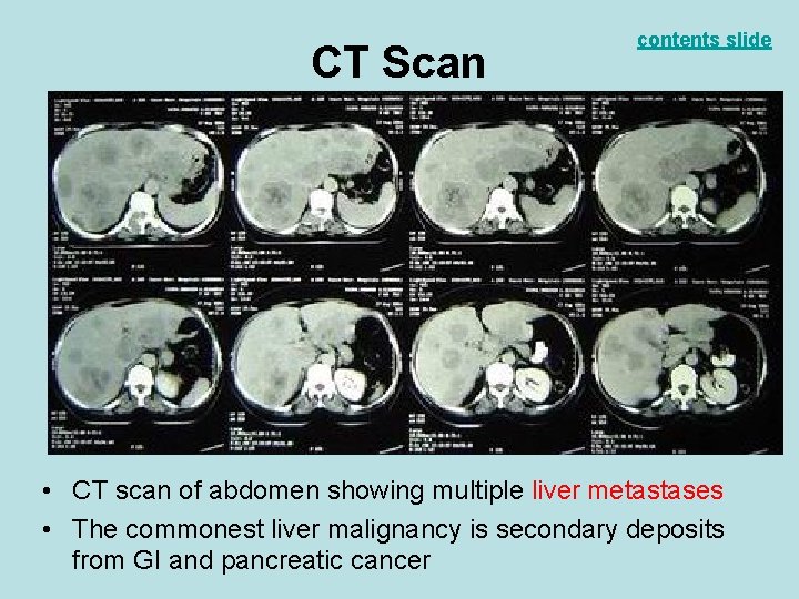
CT Scan contents slide • CT scan of abdomen showing multiple liver metastases • The commonest liver malignancy is secondary deposits from GI and pancreatic cancer
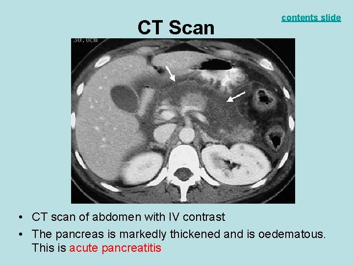
CT Scan contents slide • CT scan of abdomen with IV contrast • The pancreas is markedly thickened and is oedematous. This is acute pancreatitis
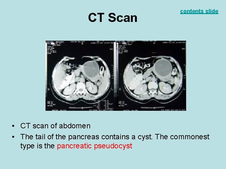
CT Scan contents slide • CT scan of abdomen • The tail of the pancreas contains a cyst. The commonest type is the pancreatic pseudocyst
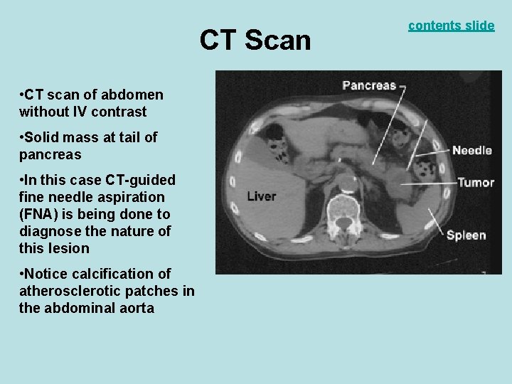
CT Scan • CT scan of abdomen without IV contrast • Solid mass at tail of pancreas • In this case CT-guided fine needle aspiration (FNA) is being done to diagnose the nature of this lesion • Notice calcification of atherosclerotic patches in the abdominal aorta contents slide
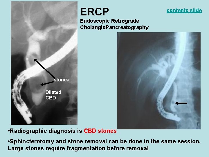
ERCP contents slide Endoscopic Retrograde Cholangio. Pancreatography stones Dilated CBD • Radiographic diagnosis is CBD stones • Sphincterotomy and stone removal can be done in the same session. Large stones require fragmentation before removal
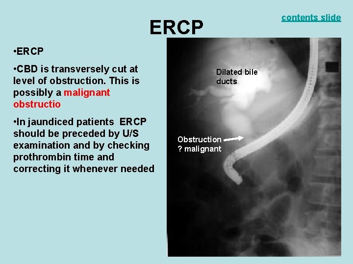
contents slide ERCP • CBD is transversely cut at level of obstruction. This is possibly a malignant obstructio • In jaundiced patients ERCP should be preceded by U/S examination and by checking prothrombin time and correcting it whenever needed Dilated bile ducts Obstruction ? malignant
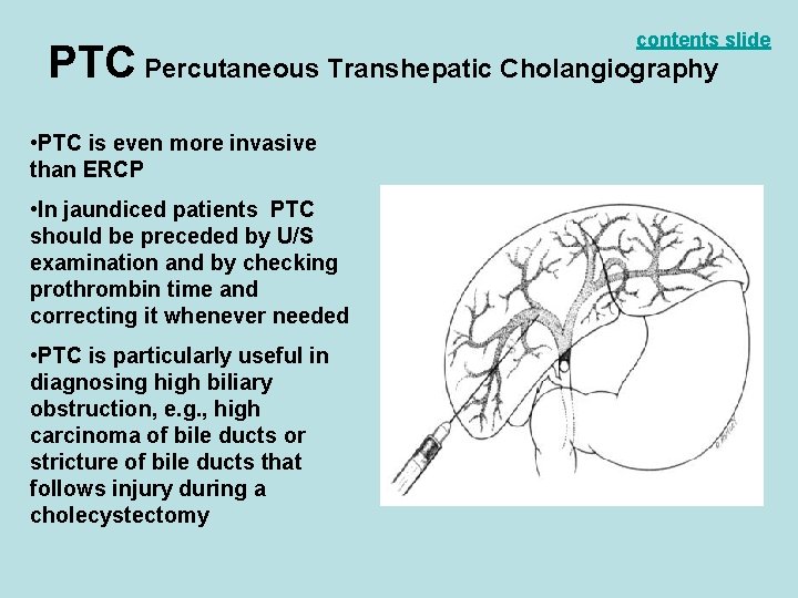
contents slide PTC Percutaneous Transhepatic Cholangiography • PTC is even more invasive than ERCP • In jaundiced patients PTC should be preceded by U/S examination and by checking prothrombin time and correcting it whenever needed • PTC is particularly useful in diagnosing high biliary obstruction, e. g. , high carcinoma of bile ducts or stricture of bile ducts that follows injury during a cholecystectomy
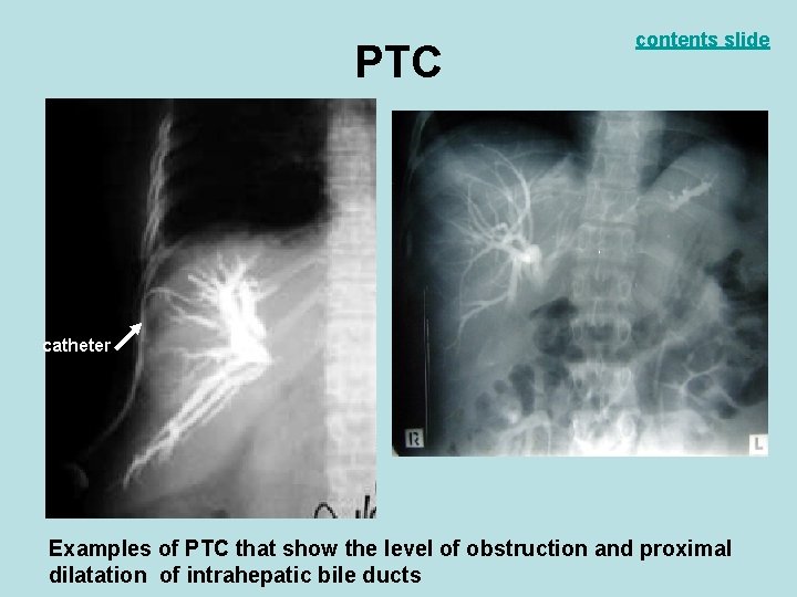
PTC contents slide catheter Examples of PTC that show the level of obstruction and proximal dilatation of intrahepatic bile ducts

contents slide MRCP Magnetic Resonance Cholangio. Pancreatography This is a form of magnetic resonance imaging Non-invasive, no contrast used, but computer generates the image as shown (CBD stone) Unlike ERCP this test is nontherapeutic Dilated CBD stone
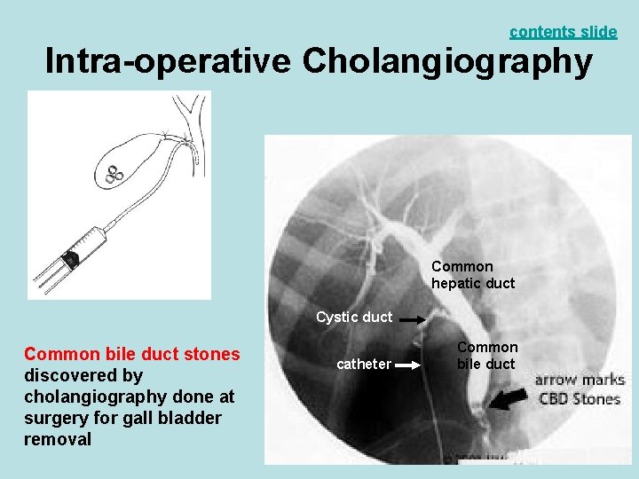
contents slide Intra-operative Cholangiography Common hepatic duct Cystic duct Common bile duct stones discovered by cholangiography done at surgery for gall bladder removal catheter Common bile duct
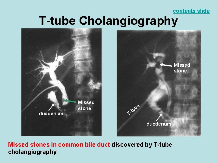
contents slide T-tube Cholangiography Missed stone duodenum Missed stone be u -t T duodenum Missed stones in common bile duct discovered by T-tube cholangiography
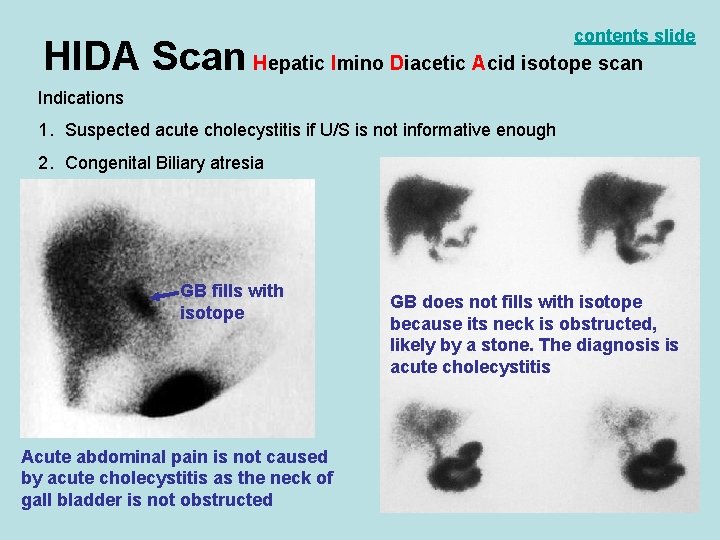
contents slide HIDA Scan Hepatic Imino Diacetic Acid isotope scan Indications 1. Suspected acute cholecystitis if U/S is not informative enough 2. Congenital Biliary atresia GB fills with isotope Acute abdominal pain is not caused by acute cholecystitis as the neck of gall bladder is not obstructed GB does not fills with isotope because its neck is obstructed, likely by a stone. The diagnosis is acute cholecystitis
- Slides: 28