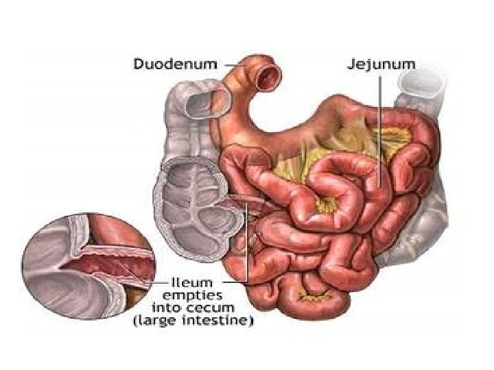
SURGICAL ANATOMY The duodenum has 4 parts superior


 2. Fistula formation: deep ulcer")


")

 *Is typically defined as a group of diseases characterized by")


- Slides: 56
SURGICAL ANATOMY *The duodenum has 4 parts: superior, descending, horizontal, and ascending * Except for its first part, the duodenum is largely retroperitoneal it has no mesentery and is covered by peritoneum only on its anterior surface. * The B. supply of small intestine by sup. Mesenteric artery From the second part of the duodenum to the distal third end of the transverse colon.
• The duodenojejunal flexure important point where the reflection of the peritonium from retro to intra make the preduodenal fossa >> one of the important site of internal hernia • The fixed part of the duodenum with the mobile part of the jejunum is the site of injury in blunt abdominal trauma. • The folding mucosa of the jejunum is giving the valvulae conniventes on XR. • The most absorptive area of the small intestine.
` Clinical anatomy clear demarcation is noted between the ileum and jejunum The jejunum has a thicker wall and a wider lumen than the ileum and mainly occupies the left upper and central abdomen.
The ileum has a thinner wall and a smaller lumen than the jejunum and mainly occupies the central and right lower abdomen and pelvis.
Investigation of GIT The small intestine is evaluated radiologically by: - Plain abdominal Xray - upper GI series using barium or Gastrografin follow-through. - enteroclysis ( in case of intestinal obstruction ), in which contrast is introduced directly into the proximal jejunum thorough a nasojejunal tube. - CT scanning can also be combined with enteroclysis. - Capsule endoscopy, video camera that is swallowed by the patient; as it passes through the gastrointestinal tract, it keeps transmitting digital images of the mucosa that are captured by a receiver device strapped to the patient.
DISEASES OF SMALL INTESTINE 1. CONGENITAL 2. INFLAMATORY 3. MECHANICAL CAUSES 4. VASCULAR 5. NEOPLASTIC
Congenital variants *A patent vitellointestinal duct: - Meckel diverticulum: . - Vitelline sinus at the umbilicus - Fibrous band between ileum and umbilicus, around which torsion of a small bowel loop may occur * Malrotation of the gut results in the location of the small intestine on the right side and the narrow base of the small bowel mesentery predisposes it to volvulus. * Atresia (duodenal, jejunal, and ileal), narrowing, or even complete obliteration of the lumen resulting in neonatal intestinal obstruction * Diverticulae and duplication can occur in any part of the small intestine.
CROHNS DISEASE American gastroenterologist t Burrill Bernard Crohn * Dr. Burrill B. Crohn, who first described the disease in 1932 - Crohn’s disease may affect as many as 780, 000 Americans. * Men and Women are equally likely to be affected. * the disease can occur at any age, is more prevalent among adolescents and young adults between the ages of 15 and 35 y. * The causes of Crohn’s disease are not well understood. Diet and stress may aggravate the dis. , It tends to run in families. - recently a gene called NOD 2 has been identified as being associated with Crohn's disease and the susceptibility to abnormal activation of the immune system is genetically inherited.
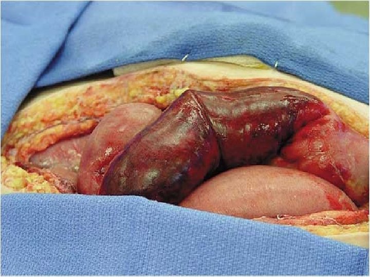
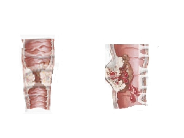
PATHOLOGY Affect any part of G. I. T. , comonly : Distal ileum only 33% Colon only 20% Ileum & colon 50% 1. Transmural pathology 2. Skipped areas are characteristic 3. dull purple red loops +areas of thick grey –white exudate 4. Deep mucosal ulcerations+ edema of the mucosa in between – cobble stone appearance 5. Enlarged mesenteric L. N.
6. extensive fat wrapping of intestine ( the fat existing between the two layers of the mesentery is going to crepe and cover part of the wall of the small intestine ). 7. dilated proximal segment Due to chronic fibrosis >> narrowing of the lumen >> dilation of the proximal part
CLINICAL FEATURES The symptoms of Crohn's disease are dependent on the location, the extent, and the severity of the inflammation 1. colicky intermittent pain is the most common symptom of Crohn's disease , becomes constant dull ache as the disease progresses. ( because of edema and fibrosis ) 2. Diarrhoe, the 2 nd most common symptom ( transudate and exudate from the inflamed mucosa ) 3. Hematochezia ( if the ulcer is deep ) 4. Low grade fever 5. malaise, wt loss 6. arthralgia 7. tenderness in Rt lower abdomen
• 8. Non classical anal fissures, recurrent perianal abscesses Transverse , , any where in the circumference , , below and above the dentate line The classical one : is posterior longitudinal and below the dentate line
Diagnosis of crohns disease There isn't any single test that can diagnose crohns dis. * Crohn disease is initially diagnosed on the basis of a laboratory, radiologic and histologic findings combination of clinical, * A diagnosis of Crohn's disease should be considered in any patient who presents with chronic diarrhea, abdominal pain, bowel obstruction, weight loss, fever, or night sweats
Laboratory study *Generally they are nonspecific but may be helpful in supporting the diagnosis ESR, CRP. Hb, *Serologic studies are sometimes used to facilitate differentiation of Crohn disease from ulcerative colitis - Antibodies to the yeast saccharomyces cerevisiae are found more commonly in Crohn disease than in ulcerative colitis, whereas perinuclear antineutrophil cytoplasmic antibody (p. ANCA), is found more commonly in ulcerative colitis than in Crohn disease
Radiology Various imaging modalities are available to aid in the diagnosis and management of Crohn disease 1. Plain abdominal X ray 2. Barium meal and follow through 3. C. T. scan 4. U/S *Endoscopic visualization and biopsy are essential in the diagnosis of Crohn disease A noncaseating granulomas in about 15 -30% of cases of biopsy samples and 40 -60% of surgical specimens.
Barium follow through , , the cecum and the terminal ilium ( narrow segment and dilated segment above it
COMPLICATIONS 1. Obstruction ( the most common complication ) 2. Fistula formation: deep ulcer might go through to the serosa the to : -peritoneum >> causing peritonitis -bowel >> enteroenteric fistula -Skin >> enterocutaneous fistula -Urinary bladder >> enterovesical fistula 3. Haemorrahge 4. localised abscesses 5. malignancy
TREATMENT There is NO CURE for crohns disease MEDICAL & SURGICAL therapy are palliative - to treat the acute flare-ups - to maintain remission 1. the use of medication, designed to suppress your immune system’s abnormal inflammatory response recommended 2. alterations in diet and nutrition , dietlow fibre 3. surgical procedures to repair or remove affected portions of your GI tract.
Mild to moderate Crohn's disease * Treated with a salicylate preparation which include mesalamine and sulfasalazine The dosage of oral mesalamine is 3 to 4 g per day The response to therapy should be evaluated after several weeks. *antibiotic therapy : Metronidazole in a dosage of 10 to 20 mg per kg per day has demonstrated benefit in the treatment of ileocolitis and colitis
Moderate to Severe Disease *Treated with steroids until symptoms resolve and weight loss is reversed. prednisone 40 mg daily over eight to 12 weeks have been shown to achieve a clinical response *The immunomodulators azathioprine (Imuran) and mercaptopurine (Purinethol) may be used, but full response may not be achieved for several months.
Recently, the U. S. Food and Drug Administration approved : infliximab, an antibody to human tumor necrosis factor alpha, to treat Crohn's disease In persons unresponsive to salicylates, antibiotics, corticosteroids, or immunosuppressants, infliximab has proved successful in closure of fistulas, steroid-refractory disease, and in the improvement of moderate to severe disease Other Considerations in treating crohns disease vitamins and mineral supplementation iron, calcium folic acid and vit. B 12
SURGICAL INDICATIONS 1. Recurrent intestinal obstruction 2. bleeding 3. Perforation 4. Abscess formation 5. intestinal fistula 6. malignant changes
The surgical procedure in crohn disease always conservative surgery *Resection of the affected bowel *Drainage of any septic foci *Strictureplasty : cutting the fibrotic area *Bypass *Endoscopic dilatation of symptomatic accessible strictures
Acute mesenteric ischemia (AMI)
The vascular problems • Ether in : 1 - superior mesenteric artery or vein 2 - inferior mesenteric artery 3 - celic artery Ether Occlusive non occlusive “ hypoperfusion” >> in patients with heart failure , acute pancreatitis , any type of shock
Acute mesenteric ischemia (AMI) *Is typically defined as a group of diseases characterized by an interruption of the blood supply to varying portions of the small intestine, leading to ischemia and secondary inflammatory changes. If untreated, this process will eventuate in life threatening intestinal necrosis. *The incidence is low, estimated at 0. 09– 0. 2% of all acute surgical admissions. AMI may be classified as either arterial or venous. AMI as arterial disease may be subdivided into: nonocclusive mesenteric ischemia and occlusive mesenteric arterial ischemia OMAI may be further subdivided into: acute mesenteric arterial embolism and acute mesenteric arterial thrombosis
THE RISK FACTORS FOR ACUTE VISCERAL ISCHEMIA 1. Age greater than 50 years 2. Congestive heart failure, Digitalis therapy 3. Recent transmural myocardial infarction 4. Cardiac dys arrythmias especially atrial fibrillation 5. Hypercoagulable state 6. Hypovolaemia with hypoperfusion.
PATHOLOGY 1. The early changes , alteration in capilary permeability. 2. sloughing of epith. Cells at the tip of villi. 3. A membrane of necrotic epith. +fibrin + infla. Cells+bacteria. 4. Edema of sub mucosa with bleeding. 5. Release of endogenous subs. i. e. bact. endotoxin, platelet activating factor. 6. Transmural necrosis
CLINICAL FEATURES Symptoms are nonspecific initially, before evidence of peritonitis presents. Thus, diagnosis and treatment are often delayed until the disease is advanced 1. Medical history may be significant for presence of stroke, MI, or peripheral artery disease 2. Patients may present with a long history of weight loss, postprandial pain, and phagophobia. 3. abdominal pain : severe, acute, unrelenting abdominal pain 4. vomiting 5. frank blood in the stools.
Signs : On abdominal examination , the findings may be minimal. soft abdomen , , no tenderness , , true or sluggish bowel sound , , normal vital signs > if signs of peritonitis present, consider bowel perforation Its difficult to be diagnosed >> because of non- synchronization between the symptoms and the sign
DIAGNOSIS 1. Clinical features: the high index of suspicion 2. Leucocytosis : >15000 3. ECG 4. Moderatly Elevated serum amylase 5. Metabolic acidaemia 6. A doppler ultrasound, 7. CT scan, CT angiogram. 8. Angiography, selective S. M. A.
On plain films Late findings include > intramural air > air in the portal venous system. If bowel perforation occurs: > free air in the abdomen may be observed
CT scan findings with a specificity greater than 95% include : > SMA or SMV thrombosis, > intestinal pneumatosis, > portal venous gas,
Treatment in acute cases 1. Analgesic to alleviate severe pain. 2. treatment is usually an emergency procedure since severe intestinal damage can occur rapidly. 3. vascular surgeon may recommend thrombolytic therapy by injecting clot-dissolving medication into a blood vessel, and is often given at the same time as a diagnostic angiogram. Catheterization with injection of papaverine ( smooth muscles dilator ) 4. If there is evidence of intestinal damage or too little time is available for the thrombolytic agent to work, surgery may be needed to remove the clot and restore blood flow to the intestinal arteries.
Postoperative 1. should include close monitoring of blood pressure and hemoglobin level to evaluate for sepsis or hemorrhage. 2. Patients should continue to have postoperative , anticoagulation therapy in order to reduce thrombotic events heparin Its not enough when the surgeon cut or remove the gangrenous part and do anastomosis , because sometimes the process of coagulation is going on and on causing extension of the thrombosis and a new segment get infarcted >> so we need what's called the second look operation
SMALL INTESTINAL TUMOUR Despite comprising 75% of the length and 90% of the surface area of the GI tract, the small bowel harbors relatively few primary neoplasms account 5% of all G. I. NEOPLASM and fewer than 2% of GI malignancies. .
Factors suggested to explain the scarcity of small bowel lesions and the infrequency of their malignant transformatio: * Rapid Transit of contents fluidity * High turn over rate of epith. Cells * Alkalinity of small intestinal Media. * High level of IGA. * High level of benzyl peroxidase”detoxify potential carcinogene”
BENIGN TUMOURS - adenoma, the most common - liomyoma - lipoma - fibromas - fibromyxomas - neurofibromas - ganglioneuromas - hemangiomas 90% of the carcinoma of the small intestine arise from adenoma “ adenoma- carcinoma syndrome “
CLINICAL FEATURES: 1. they invariably present difficult problems in diagnosis 2. Symptoms are often absent until the tumor has progressed to produce a complication. “ delayed complications” 3. Even then, the presentation is often vague and nonspecific, intermittent pain, chronic anemia 4. May present as one of the possible complications: - intestinal obstruction - intussusception - bleeding
MALIGNANT NEOPLASM of small intestine 1. Accounts for about 2% of all G. I. cancer 2. 64% of all small bowel tumours are malignant 3. 40% of the malignant tumours 0 f small intestine are adenocarcinoma
MALIGNANT NEOPLASM Four types of cancer are found in the small intestine 1. Adeno carcinoma : 30 – 50% 2. Carcinoid tumour : 13 - 34 % 3. Sarcoma 11% The most common is gist “ gasto intestinal stromal tumor “ Which is the leiomyosarcoma 4. Lymphoma
ADENO CARCINOMA 1. similar to adenocarcinomas in the colon, arise from premalignant adenomas. This occurs both sporadically and in the context of familial adenomatous polyposis 2. occurs frequently in the proximal small bowel Approximately 50% arise in the duodenum, 30% in the jejunum, 20% in the ileum. 3. either ulcerating or annular
Clinical features symptoms 1. Abdominal pain , Nausea, vomiting symptoms of intestinal obstruction are common presenting symptoms 2. weight loss. 3. Bleeding is less common, manefested by anaemia physical signs : 1. distended abdomen may be found due to obstruction. 2. Peritoneal signs indicate perforation. 3. Mass in the abdomen
Investigations Lab Studies: 1. CBC count may show mild anemia related to 2. Liver function tests may reveal hyperbilirubinemia, obstruction from peri ampulary tumour 3. Elevated transaminase levels also may be found in 4. Carcinoembryonic antigen levels may be elevated. . chronic blood loss. which may be related to biliary the presence of liver metastases.
Imaging Studies 1. Plain abdominal x ray films may reveal partial or complete smallbowel obstruction. 2. Upper GI series with small-bowel followthrough show abnormalities in 53 -83% of patients with small-bowel cancer. 3. Abdominal CT scan may elucidate the site and extent of local disease and the presence of liver metastases
Treatment for adenocarcinomas Treatment depends on the location, size and stage of the tumor, and whether it has spread. . . * surgical resection, with removal of lymph nodes considered acceptable. - In patients with involvement of the proximal pancreatoduodenectomy may be necessary. Chemotherapy and radiation may be helpful Margins of 5 cm are duodenum,
INTESTINAL LYMPHOMA 1. Lymphomas of the small bowel may be a. primary: arising from the small bowel lymphoid tissue b. secondary: due to a systemic lymphoma 2. Most commonly found in the ileum & ileocaecal region 3. Intestinal lymphoma of the small intestine is a recognized complication of celiac sprue, and can occur in immunodeficiency syndromes. 4. Symptoms include crampy-like l abdominal pain, weight loss, features of malabsorption, bleeding, or even bowel obstruction.
CARCINOID TUMOUR 1. A well-differentiated neuroendocrine tumor 2. Arise from Argentaffin cells at the base of intestinal crypt, its ability to secrete humoral agents i. e. serotonin 3. The most common distal small bowel malignancy 4. The most common tumor of the appendix, 5. The incidence of metastasis is quite low. 6. Common sites of metastatic spread include the regional mesenteric and para-aortic lymph nodes and the liver.
Cont. carcinoid tumour Sex: The incidence of carcinoid is similar between males and females Age: The occurrence rate of carcinoid tumors peaks at approximately age 62 years * In contradistinction to adenocarcinoma, the carcinoid tends to arise in the distal small intestine rather than in more proximal sites. surgically confirmed : 3% were in the duodenum 5% in the jejunum 32% in the proximal ileum 60% in the distal ileum
Clinical features Due to their slow rate of growth and indolent course, carcinoids tend to remain asymptomatic for long periods Nonhormonal symptoms abdominal pain intermittent obstruction, palpable abdominal mass. Carcinoid syndrome * greatest for midgut primary tumors and occurs in <10% of patients are the hormonal manifestations of carcinoid tumors : flushing, diarrhea, bronchoconstriction, and cardiac disease
TREATMENT 1. Surgery for the complications. 2. Chemotherapy is the treatment of choice
Tumors of the Appendix 1. Tumors of the appendix are infrequent, and when they are found, it is almost always during a routine appendectomy 2. Benign tumors of the appendix consist of leiomyomas, neuromas, and lipomas. 3. Malignant tumors of the appendix include adenocarcinomas and carcinoids and the appendix is the most common site of carcinoid tumor. The most common tumor is adenocarcinoma