Surgical anatomy physiology Injury to abdominal organs especially

Surgical anatomy & physiology * Injury to abdominal organs, especially those in the retroperitoneal space, when bleed the space can hold a great deal of blood, up to four liters. *Solid organs, such as the liver and spleen bleed profusely as do the major abdominal blood vessels, the aorta and vena cava. * Injury to hollow presents a serious risk of infection, especially if there is a delay in diagnosis

Introduction accidents is the main cause of abdominal trauma in the civilian population al injuries rank third as a cause of traumatic death after head and chest injuries 3. The primary cause of death in abdominal trauma is hemorrhage and sepsis after 48 hours

Abdominal trauma : 1 - intraperitoneal organ injury 2 - retroperitoneal organ injury ( isolated retroperitoneal organ injury is very difficult to be discovered “ no abdominal signs “ ) Penetrating trauma : 1 - intra thorax 2 - pelvic abdomen 3 - abdominal proper

Types of abdominal trauma 1. Blunt abdominal trauma is the most common injury pattern with RTA accounting for approximately 75%. a. vehicular trauma a. auto to auto b. auto to pedestrain b. Direct blow to the abdomen c. Fall from a height 2. Penetrating abdominal trauma a. low & high velocity missiles b. stabs: knives, ice picks, industrial implement

Mechanism of injury In blunt abdominal trauma : 1. Decelaration : The differential movement among adjacent structures especially at relatively fixed points of attachment such as the ligament of Treitz, the ileocecal valve, and the phrenocolic ligament. 2. Compresion with crush : when intra-abdominal contents are crushed between the anterior abdominal wall and the vertebral column or posterior thoracic cage. 3 - compression with rupture : Common in the second part of the duodenum and the ileocecal valve

The mechanism in pentrating abdominal trauma 1. Mechanical distruption of tissue along the path of stab or bullet pasage. “ according to the width and the size of the knife 2. In high missile injuries : ( velocity > 1000 m / sec ) 1. KE =1/2 M (v 1 - v 2 2. Cavitation within solid organs result in shattering 3. The colon less tolerable to hight velocity missile than small bowel because of its fecal content.

• High missile injuries : • Mechanical disruption according to the mass of bullet >> but because of high velocity it is going to liberate energy >> ( compression with crush >> cavitation. • As the solidity of the organs increase as the effect of liberation increase. >> so the left side is more prone to injury than the right side of the colon.

Assessment in blunt trauma Motor vehicle accident History: type of collision, extent of vehicular damage, b. status of other passengers, dead person c. patient position in vehicle, belted d. A record of hypotension reading by prehosp. team *Fall - heigh - the site

The assessment in pentrating injury: History -Time - Type of weapon, knife , hand gun, shot gun. -length of knife -no. of stabs, no. of shot fired

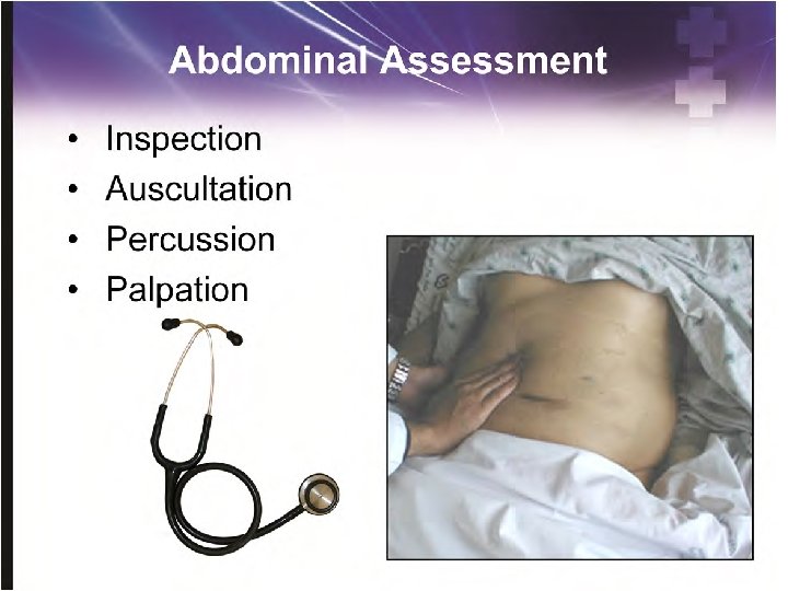
Clinical evaluation in blunt trauma INSPECTION - fully exposed patient - Echymotic area , abrasion - steering wheel shaped contusion, - seat belt sign : indicates intra-abdominal injury in about one third of patients. - skin discoularation - abdominal distension

INSPECTION

IN PENTRATING ABD. INJURY Any wound in The boundries of the abdomen considered as apotential abdominal injury From the 5 th intercostal space down to the inferior gluteal fold posteriorly and to the inguinal ligament anteriorly

PALPATION 1. Haemodynamic instability. 2. Signs of peritoneal irritation: gaurdining, rigidity tenderness, rebound. 3. Crepitus at lower thoracic cage 4. Pelvic instability 5. Abdominal distension 6. Evisceration 7. Per digital rectal exam.

Diagnostic aids for evaluation 1. repeated “serial”physical exam. 2. local wound exploration 3. Ultra sound ‘FAST” to check if there is any fluid in the abdomen or not 4. CT. abdomen CT of the abdomen is the preferred diagnostic examination for the evaluation of blunt abdominal trauma in the hemodynamically stable patient. ( the most diagnostic in retroperitoneal organ injury ) 5. Diagnostic peritoneal lavage

Diagnostic peritoneal lavage To find out if there is blood in the peritoneal cavity to suspect a bleeding and to find if there is any injury to the intestine The procedure is still performed when alternative diagnostic methods such as computerized tomography (CT) or ultrasound imaging are unavailable

INDICATIONS FOR DPL 1. Equivocal clinical examination 2. Difficulty in assessing patient. 3. Persistent hypotension despite adequate ressuscitation 4. Multiple injuries

DPL Technique

Positive signs in DPL 1. > 5 mls of blood aspirated before fluid is infused. 2. Bloody irrigated fluid 3. the presence of bile, 4. enteric contents. 5. Hematological & biochemical tests for the aspirated fluid: a. RBC > 100, 000/cmm b. WBC > 500 /cmm c. Amylase > 175 units

Organs injury The Spleen

CLINICAL ASPECTS OF SPLENIC RUPTURE Symptoms: - May be painless, or LUQ/diffuse abd pain. - referred L shoulder pain in splenic laceration: Kehr’s sign - Syncopy due tohypotension. Signs - Physical examination is insensitive and non specific. - Pt may have signs of lt upper quadrant tenderness or signs of generalized peritoneal irritation. - May present with tachycardia , tachypnea, hypotension or shock

Plain radiographic findings in splenic injury: 1. left lower rib fracture 2. left hemidiaphragm elevation 3. left lower lobe atelectasis, 4. Left pleural effusion 5. medial displacement of the gastric bubble 6. inferior displacement of the splenic flexure gas pattern.

SPLENIC ORGAN INJURY SCALING I – subcapsular hematoma <10% of surface. Laceration < 1 cm deep. II – subcapsular hematoma 10 -50% of surface. Laceration 1 -3 cm deep. III – subcapsular hematoma >50% of surface or expanding. > 3 cm parenchymal depth. IV –Laceration > 25 % of spleen or laceration involving the hilum. . V – completely shattered spleen or hilar vessel injury with devascularization.

Splenic injury Grade II

Splenic injury Grade IV

Management options in splenic injury 1. Conservative –Observation - may followed by a selective splenic artrey embolization 2. Surgery a. Splenic preservation - splenorraphy - partial splenectomy b. splenectomy

Liver injury

Symptoms of a liver injury right upper quadrant pain, increase with deep breathing. nausea or vomiting, tachycardia and fainting, Physical examination : tenderness to palpation in the right upper quadrant of the abdomen. Abnormalities of blood pressure and pulse will be noted (low blood pressure and pulse over 100).

Liver injury scale Grade I : Sub capsular hematoma < 10%of surface area, non expanding. Laceration < 1 cm parenchymal depth, non bleeding. Grade II : Sub capsular hematoma 10 -50% parenchymal Laceration 1 -3 in depth , <10 cm in length. Grade III : Sub capsular hematoma >50 % 3 cm parenchymal depth. Grade IV : Ruptured intra parechymal hematoma with active bleeding. Parechymal distruption involving 25 -50%of hepatic lobe. Grade V : Parenchymal distruption >50%of hepatic lobe Vascular injuries : hepatic veins, inf. Vena cava.

Liver injury Grade II Grade IV

Treatment of liver injury Nonoperative management: is safe, effective, and clearly the treatment modality of choice in hemodynamically stable patients. The weakness in this treatment is the possibility of missing an associated intra abdominal injury Operative management : - simple suture techniques - resectional debridement to control hemorrhage. - Anatomic resection, -hepatic artery ligation - Mesh wrapping or perihepatic packing, -fibrin glue application

Retroperitoneal organs injury

Retroperitoneal injuries & retro peritoneal hematoma 1. Frequently over looked and carry significant morbidity. 2. Diagnosis require a high index of suspicion and an organized diagnostic approach any patient who has sustained a direct high-energy blow to the epigastrium , ie from a crushed steering wheel in an adult. 3. the findings of retro peritoneal hematoma on CT or at operation.

Zones of retroperitoneal hematoma Zone I : . Occupy the centro medial portion of the retro peritoneum, . Include : dodenum &pancrease and major bloosd vessels. Zone II : Is lateral to zone I, . Include : kidney & retro peritoneal portion of colon. Zone III : Include entire pelvis

Retroperitoneal hematoma , CT

Retroperitoneal hematoma location Blunt Pentrating Zone I Explore Zone II Observe Explore Z 0 ne III Observe Explore

Abreviated laprotomy Multiple trauma patients are more likely to die from their intraoperative metabolic failure that from a failure to complete operative repairs. The patients die from , a triad of : a. coagulopathy, b. hypothermia and c. metabolic acidosis. The principles of the first 'damage control' procedure are control of haemorrhage, prevention of contamination and protection from further injury.
- Slides: 38