Surgical aids to Orthodontic The extraction of fully


- Slides: 51
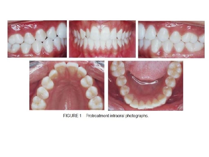
Surgical aids to Orthodontic The extraction of fully erupted teeth or the surgical removal of impacted, partially erupted, displaced, supernumerary, dilacerated, submerged teeth or surgical exposure of impacted tooth, frenectomy and corticotomy are conditions which require surgical aids prior to orthodontic treatment. The dental history, medical history, clinical examination, radiographs and consultation with the orthodontist. are taken for preoperative assessment and surgical treatment plan. The most conditions which require surgical aids prior to orthodontic treatment are:
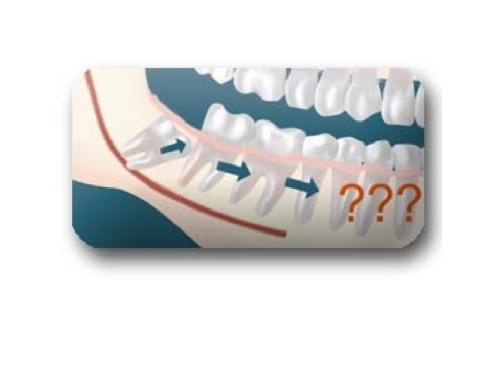
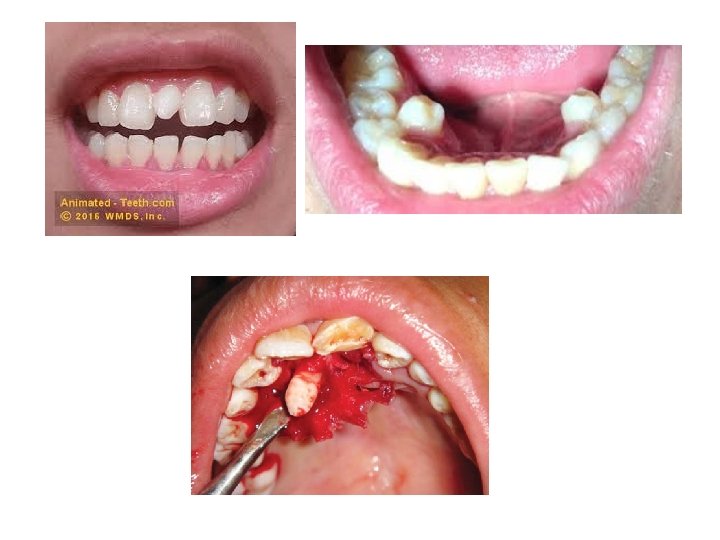
I- Impacted teeth Localization of unerupted teeth: The exact position of the impacted tooth and its precise relationship to other adjacent structures must be determined. It is of particular importance to determine whether a buried tooth is lying labiobuccally or palatally to the standing teeth, so that the surgeon know whether to use a labiobuccal or a palatal approach for surgical removal. 1 - Clinical examination is often of some value for localization of impacted tooth, Inspection and palpation of the alveolar bone may reveal the presence of bulges of the buried tooth. Tilting and displacement of adjacent teeth may provide a clue to the position of the unerupted canine; that's, if the crown of the upper lateral incisor is displaced distally and labially, the impacted canine is usually found to be related to labial surface of the apex of the lateral incisor and vice versa.
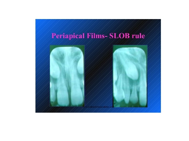
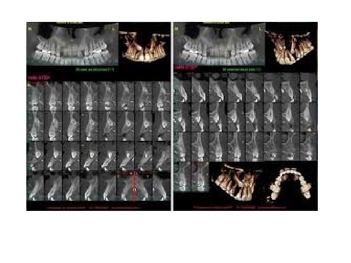
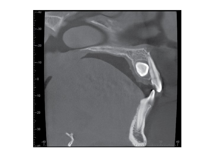
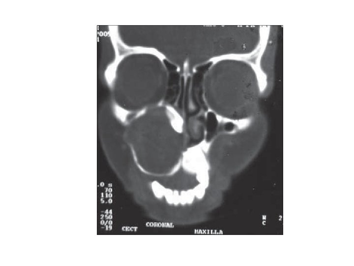
2 - Careful radiographic assessment is essential for localization of the impacted tooth and at least two views taken at right angle to each other are required, radiographic examination should reveal the direction of the canine as well as the position of its crown and root apex in relation to the adjacent teeth, in the vertical (depth), mesiodistal, and labiopalatal dimensions. The most helpful radiographic views for localization impacted teeth are: A Periapical films These reveal the size, shape, direction and root pattern of the unerupted tooth, also the presence of cysts, odontomes, or supernumerary teeth can be detected in a periapical film, and which gives the relationship between the buried tooth and the adjacent erupted teeth in the vertical plane. The relative radioopacity of the impacted tooth may assist in the determination of the tooth position which indicate it is closer to the x ray film, but a more reliable method for localization of impacted tooth can be made by the use of the so called "parallax method" or "cone shift technique". In this technique a periapical radiograph of the area is taken and the X ray tube is then moved in either a mesial or a distal direction before a second periapical film is taken. The two radiographs are then compared and if the buried tooth is seen to move in the' same direction as the X ray tube, it is lying palatally to the standing teeth, while if it moves in the opposite direction it is lying on the labial side to them.
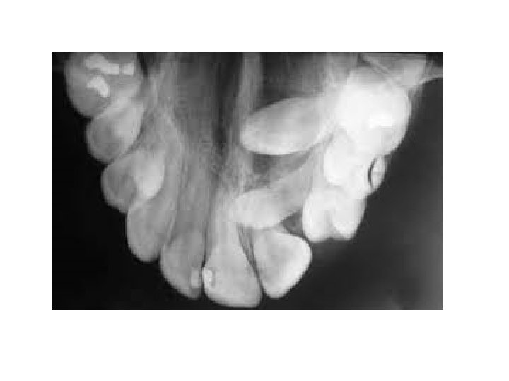
B-Occlusal films A number of occlusal views are available for use and the value of each should be clearly appreciated. There are three types of occlusal views: Anterior Occlusal, True Occlusal and Vertex Occlusal views. The best view for localization of the impacted tooth is Vertex Occlusal. When taking the "vertex occlusal" view, the x ray tube is arranged so that the central ray passes along or parallel to the long axis of the central incisors. If the central incisors are shown in cross section, the film is an accurate one and may be used with confidence to determine the true position of a buried tooth whether buccally or palatally to the standing teeth. As the rays have to pass through the skull and facial bones to reach the film, this type of radiograph lacks both contrast and detail and is useful only for determining the relative position of the impacted teeth. C Cone beam CT It is very useful for precise localization of the impacted tooth and it's relation to the adjacent vital structures. D- Other x-ray views Cephalometric and posteroanterior and orthopantamogram (OPG)views have been taken for orthodontic purposes that may provide much useful information about the condition of the teeth and if there are supernumerary teeth or presence of any bone lesion.
Modalities of managements of impacted teeth are: ØLeave it in situ ØSurgical extraction ØSurgical exposure ØTransplantation Indications of leave it in situ: - If the impacted tooth is asymptomatic and its extraction might cause damage to the adjacent teeth or vital structure No presence of any pathological lesion associated with it, however; any impacted tooth left in situ should be radiographed at regular intervals, for checking. Indications of surgical extraction of impacted teeth: Before the construction of complete, partial dentures or bridges. Prophylactic extraction to limit the orthodontic alignment of crowded teeth, . . for e. g. prophylactic extraction of impacted lower third molar if there is suspicion of anterior teeth crowding in future. If the impacted tooth cause resorption of the roots of an adjacent tooth. If the impacted tooth associated with dentigerous cyst or any pathological lesion. If the first premolar and lateral incisor are suitably positioned and the malposed impacted canine is difficult to be aligned by orthodontic treatment. The surgical removal of an impacted canine may produce an acceptable aesthetic result.
Indications of surgical exposure: 1 Adequate space in the arch or create it for the impacted tooth either by orthodontic means or by the extraction of a tooth, for example extraction of the first premolar to accommodate the impacted canine. 2 The potential path of eruption is unobstructed. 3 The patient's age is less than 17 years.
Indications of transplantation: Presence of sufficient space or create it to accommodate the transplanted tooth. The potential path of eruption is obstructed (the impacted tooth is malposed and it is difficult to reposition it by orthodontic appliance). The patient's age over 17 years.
Management of the impacted canines for orthodontic reasons If the canine not erupted in a patient who is 13 years of age, the case should be investigated. A careful history should be taken, the eruption dates of other teeth determined, whether the deciduous canine is retained or missing and, if the permanent tooth has been removed or if there is a family history of either malposition or absence of teeth. In the majority of cases the canine should be either surgically exposed in an attempt to aid its eruption or surgically removed, or transplanted and the choice of treatment must be based on both surgical and orthodontic considerations.
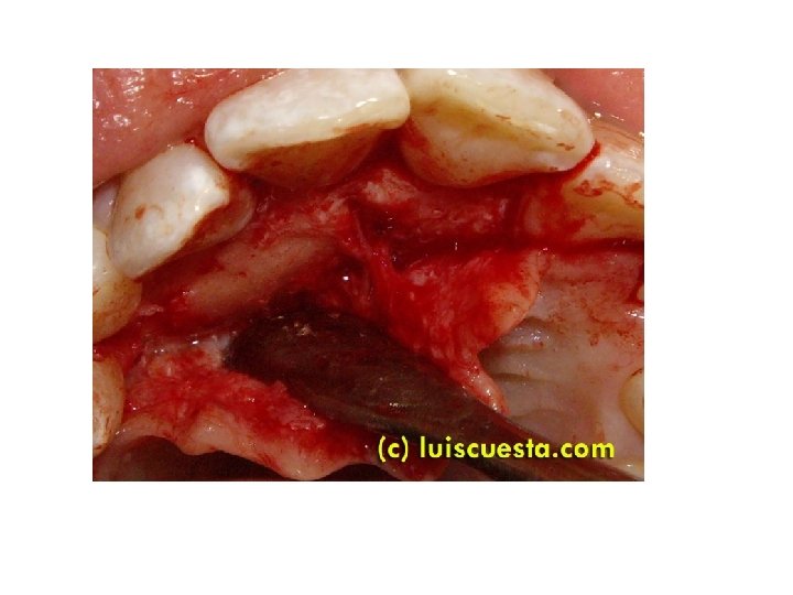
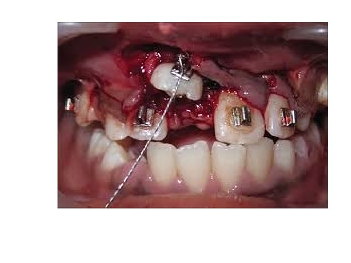
Surgical procedure for the removal of impacted canine: The dental surgeon should base his decision whether to employ a labial or a palatal approach when removing an unerupted canine after careful preoperative clinical and radiographic evaluation of the tooth. The labial approach done mostly by three sided flap, reflect the mucoperiosteal flap, sufficient amount of bone around the crown is removed by surgical round bur, then elevate the tooth by elevators or sometimes it may need sectioning of the tooth and removed in two pieces, and finally suture the flap.
When a palatal approach is employed the palatal gingival margins are incised from the second premolar to second premolar. Then reflect the mucoperiosteal flap, the flap will be found to be tethered in the midline by the nerves and vessels passing through the incisive foramen. In most cases the flap should be freed by cutting the neurovascular bundle close to the bone with a sharp, . scalpel. The hemorrhage which results is easily controlled by applying pressure with a gauze pack to the bone for a few minutes. Then sufficient bone which covering the crown is removed with a round surgical bur to expose the entire crown of the tooth, without risking damage to the standing teeth, then as mentioned elevate the tooth or section it and removed. Sutures are inserted between the teeth and the knots tied on the labiobuccal side to avoid tongue worry. The patient is seen one week postoperatively and at this visit the sutures are removed and the vitality of the teeth related to the operative site tested.
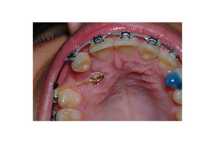
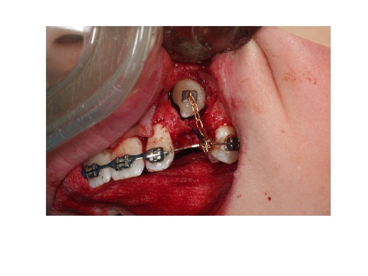
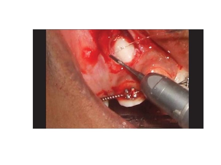
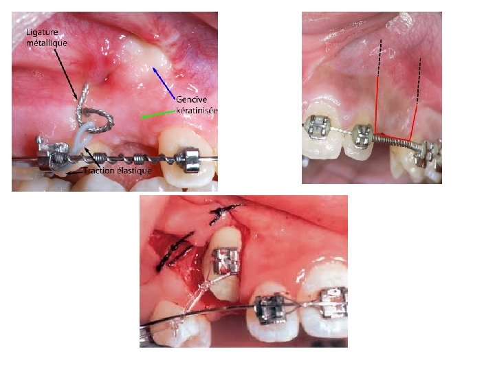
2 - Surgical procedure for exposure of impacted canine: After the clinical examination and radiographs have been carefully examined, the surgical approach either labiobuccally or palatally. After flap reflection, bone is removed carefully by round bur so as to expose the tip of the cusp, the cingulum (palatal approach), the labial surface (buccal approach) and the greatest mesial and distal convexities of the crown. At the end of the operation the crown of the tooth will be left lying in a saucer shaped bony cavity, the eruption of an exposed canine can be aided by cutting away the bone which obstructs the path of movement, to form an "eruption channel". Then after suturing we must make a window in the mucoperiosteal flap apposing the surgically exposed labial or palatal surface, then cover the exposed crown by iodoform pack or periodontal pack to prevent the soft tissues growing over it and fix the pack in position by suture. Pack and suture are removed after one week. Most correctly assessed and adequately exposed canines erupt without orthodontic traction aid, but if traction is to be employed it is best applied by the use of a bracket cemented to the crown of the canine at the time of operation or after removal of the pack. Surgical exposure of unerupted teeth followed by orthodontic treatment is a very successful method of bringing teeth into occlusion.
Surgical exposure of other teeth: Incisors: The surgical exposure of an unerupted incisor tooth is performed in a similar fashion to that of impacted canine, care being taken to expose the entire incisal edge, labial surface, and/or cingulum, mesial and distal convexities of the buried tooth. When incisors are palpable above the reflection of the labial sulcus they may be exposed with ease by incising the mucosa exposing the crown and then excise the mucosa to make a surgical window. This however, may give a shallow labial sulcus with a deficiency of attached mucoperiosteum. So the gum should be incised slightly on the palatal side of the buried tooth and the mucoperiosteal flap is widely undermined and rotated upwards to line a labial sulcus, a better result is obtained, this flap named "apically repositioned flap" in this manner the amount of attached labial mucoperiosteum is increased and gingival health is preserved. Premolar & molar teeth: when exposing a cheek tooth; the bone and soft tissues should be cleared from the cusps, the occlusal surface, and the greatest convexity of the crown. The exposed crown packed with iodoform pack and the pack is removed one week after operation.
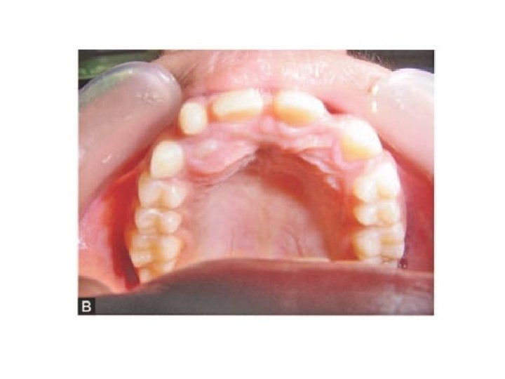
3 - Transplantation of impacted tooth Transplantation of misplaced canines and third molars are widely accepted technique and may be a superior alternative to a partial denture or a fixed bridge in a young patient (over 17 years of age) in whom surgical exposure and prolonged orthodontic treatment to reposition a canine is unacceptable to the patient on aesthetic and social grounds, however, recently the dental implant is superior alternative to the transplantation. Transplantation is sometimes preceded by simple orthodontic treatment designed to achieve either a small increase of space in the maxillary canine region or to re align an over erupted opposing mandibular canine, or both, usually by utilizing removable orthodontic appliances.
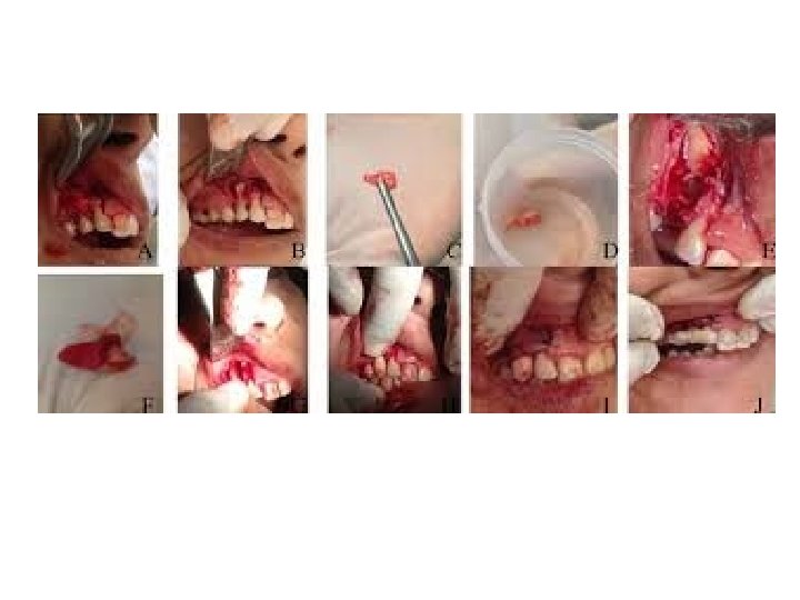
Transplantation of a canine should be based upon a preoperative assessment including clinical examination, radiographs and study models. Periapical and vertex occlusal views should be utilized for the localization of the impacted tooth. The surgical approach may be buccally or palatally according to location of the impacted tooth. Transplantation is usually successful if care is taken to preserve the • periodontal membrane of the transplanted tooth and to minimize the time between its removal and re implantation in a new socket.
The crown of the canine is exposed by careful removal of bone using round surgical burs. Sufficient bone must be removed to enable the tooth to be delivered from its socket in such a way as to avoid damage to its cementoenamel junction, periodontal membrane and cementum. Once delivered, the tooth is stored under the base of the flap, or stored in milk. Care must be taken to ensure that the tooth does not become dry.
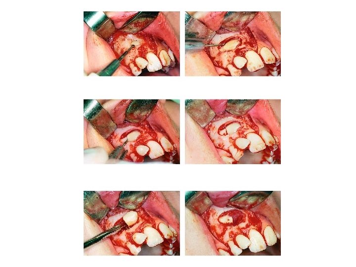
During the creation of the new socket efforts should be made to preserve the labiobuccal bone and mucoperiosteum. It may be necessary to create socket to place the transplanted tooth in a labiopalatal position which is acceptable from a functional and esthetic points. The socket is created by using assorted burs from dental implant kit, taking care not to damage the roots of the adjacent teeth. The new socket should not be excessively large but should be made slightly larger than the root of the canine "friction fit" in order to minimize the risk of damage to the periodontal membrane during the insertion and positioning of the tooth. The tooth is gently inserted into the new socket. The position of the tooth is adjusted using finger pressure alone, in order to ensure a good appearance and that the canine is free from traumatic occlusal contact with its apposing canine during all excursions of the mandible. When the canine is positioned correctly the bone fragments removed earlier or use bone graft which packed around the tooth as necessary and into any residual bony defect. The mucoperiosteal flap is trimmed to fit the neck of the canine and sutured into place.
A decision must then be made whether or not to splint the transplanted tooth as in some cases this is not necessary provided that the tooth is firm, free of the bite and the patient's care in the postoperative period. If required, interdental wire splint is placed and fixed to the adjacent teeth. If further stability is required composite filling can be used to fix the transplanted tooth to the adjacent teeth. Healing is usually uneventful and gingival reattachment and bone regeneration occurs rapidly. The splint should be removed after 2 to 4 weeks. It is important to repeatedly check the occlusion in the immediate postoperative period and at each subsequent visit. The patient is usually seen one week, one month, two months and three months after surgery and then reviewed at six monthly intervals for 2 or 3 years. Regeneration of the bone around the root of the tooth is usually complete in 6 — 12 months. Transplanted tooth should not be root filled at the same time because it leads to damage of the periodontal ligament, which are both affect the success rate of transplantation. Only if periapical pathological changes occur should endodontic treatment is undertaken.
Ankylosis or mainly resorption may occur in the transplanted tooth and may be either external, internal or periapical, and its extent and progression may determine the prognosis of the transplanted tooth, it usually occurs within 3 years of transplantation. The resorption is sometimes arrested by root filling the tooth, but may progress until the tooth is lost. ,
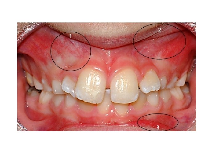
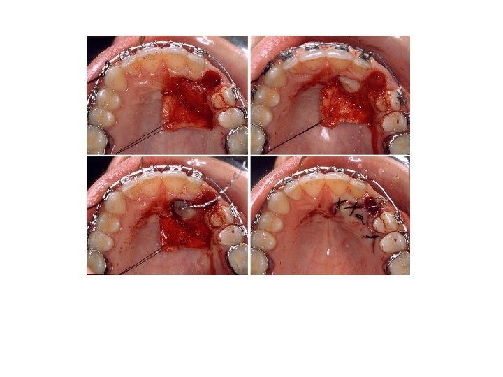
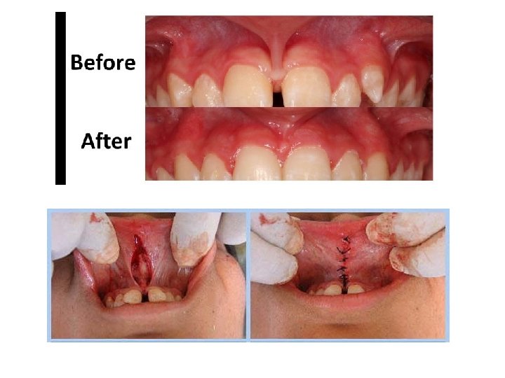
II- Abnormal labial frenum In some cases the upper central incisors are separated by a diastema, through which the labial frenum passes to gain attachment to the incisive papilla, which is seen to blanch when the lip and frenum are tensed. A periapical radiograph often reveals a marked midline suture in these cases. Usually delay the surgical excision until the eruption of the lateral incisors and canines is complete and the diastema has failed to close naturally. The operation of fi -enectomy is designed to eliminate the fibrous tissue which is present in the midline suture between the roots of the maxillary central incisors and removal of fibrous band that raises the mucosa which is an entirely different procedure to frenoplasty or frenotong, which are operations, designed to eliminate only the fibrous band which raises the mucosa to form a frenum.
The technique of frenectomy can often be performed under local anesthesia. The upper lip is held out at right angles to the anterior surface of the maxilla by an assistant throughout the operation. This not only tenses the frenum and facilitates dissection but aids haemostasis. An incision down to bone is made on either side of the frenum, when the palatal ends of these incisions are joined together the attachment of the underlying fibrous tissue to the bone is severed and the frenum springs upwards into the lip. The detached frenum and the fibrous tissue underlying it are then dissected out of the undersurface of lip, care being taken to preserve as much mucous membrane as is practicable. The median suture is then completely cleared of fibrous tissue by the use of a small round bur. The mucosa on the undersurface of the lip is then undermined and approximated with interrupted sutures. The exposed bone covered by iodoform gauze and suture over the resultant gingival defect and left in situ for one week.
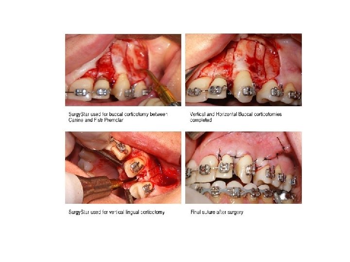
III Corticotomy is a surgical aid to orthodontics which can be utilized to shorten the duration of orthodontic appliance therapy. It is of especial value in the treatment of young adults and can be used to facilitate the movement of one or more teeth in the anterior maxilla. An incision is made through both the labial and the palatal gingival margins of the tooth or teeth to be moved on either side. A broad based labial mucoperiosteal flap is elevated from the underlying bone and a large palatal flap is then reflected. A series of vertical cuts of predetermined width in the labial and palatal compact bone (cortical plates) on each side of individual malaligned teeth are made with a bur. The apical ends of these vertical cuts are joined by horizontal cuts through the compact bone _alone thus leaving the teeth to be supported by cancellous bone only. The mucoperiosteal flaps are then replaced and repaired with interrupted interdental sutures. Orthodontic pressure, usually utilizing strong elastic traction, is applied to the teeth and after a few days their alignment is rapidly effected. At the end of the active phase of orthodontic. treatment a retainer is fitted and is worn at all times for the first month after surgery. After this it is usually necessary to wear the retainer at nights for the next 6 months.
IV- Other conditions which require surgical aids to ortho. : 1 - Supernumeraries The failure of one or more of the maxillary incisor teeth to erupt may be due to the presence of a supernumerary tooth (extra tooth). Supernumerary occur most frequently in the maxillary incisor region and in this area the vast majority of supernumeraries are found to lie on the palatal side of both the erupted and the unerupted normal teeth. More than one supernumerary may be found in the maxillary incisor region, and usually have abnormal forms, being either tuberculate or conical in shape. Tuberculate supernumeraries are almost invariably found to have incompletely formed roots and to lie on the palatal side of adjacent normal teeth, while conical supernumeraries are often found with completely formed roots and when it lies in the midline of the maxilla between the upper central incisors named 'mesiodens'. Extra teeth are found less frequently in the premolar and third molar regions and in these sites there is a higher incidence of supernumeraries which closely resemble adjacent teeth and are often called 'supplemental teeth'. In almost every case supernumeraries should be removed as soon as possible without damaging the adjacent teeth. The decision as to whether a palatal or labial approach should be employed is taken after a careful clinical and radiographic assessment of the case has been undertaken.
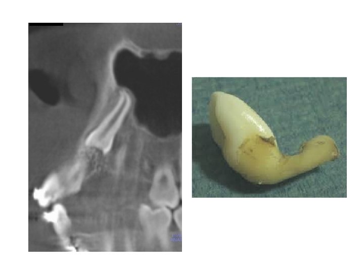
2 -Dilacerated teeth Dilacerations is any tooth with a hooked or bent root (the whole or part of the crown set at an angle to the root). The condition is believed by many to be the result of trauma applied to the deciduous predecessor and transmitted to the forming permanent tooth, or may be of developmental origin and is probably related to ectopic development of the tooth germ. Dilacerated teeth are usually best removed via labial and palatal flaps. Usually the crown is divided from the root and each section can be: delivered along its optimum line of withdrawal.
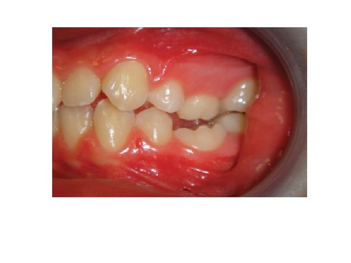
3 - Submerged teeth Children aged between 3 and 12 years may have one or more of their primary molars in infra occlusion, i. e. with the occlusal surface at a level lower than that of the neighboring teeth. Such teeth are usually described a being submerged teeth. The second deciduous molar and the second premolar are most frequently found to have become submerged and the condition is much more common in the mandible than in the maxilla. Submerged deciduous teeth are often associated with absence of the permanent successor. They are frequently the site of food stagnation, and the removal of such a tooth may be indicated either for orthodontic reasons or for the relief of discomfort. These teeth may become ankylosed, which should be removed surgically.
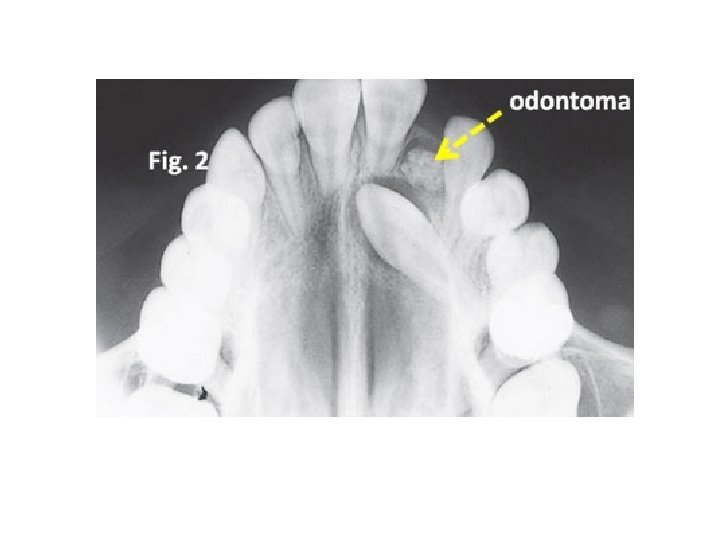
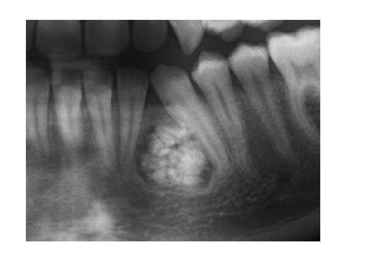
4 - Odontomes arise as a result of an aberration in the tissues responsible for the formation of teeth. They divided into complex and compound odontomes. The complex odontome is a malformation in which all the dental tissues are represented in a disorderly pattern. They tend to occur in the posterior parts of the mandible. The compound odontome is a malformation in which all the dental tissues are represented in an orderly pattern so that the lesion consists of many small tooth like structures, and occurs most frequently in the anterior maxilla. Odontomes are often diagnosed in the second decade of life and are commonly associated with delayed eruption, displacement of related permanent teeth or presence of swelling. The increased use of routine radiographic examination discovered the presence of these lesions earlier before symptoms appearance. Once detected, an odontome is usually best removed as soon as possible to perform surgery without damaging adjacent teeth or tooth germs.