
Surgery of Cerebrovascular Diseases Subarachnoid Hemorrhages Etiologies intracranial
Surgery of Cerebrovascular Diseases
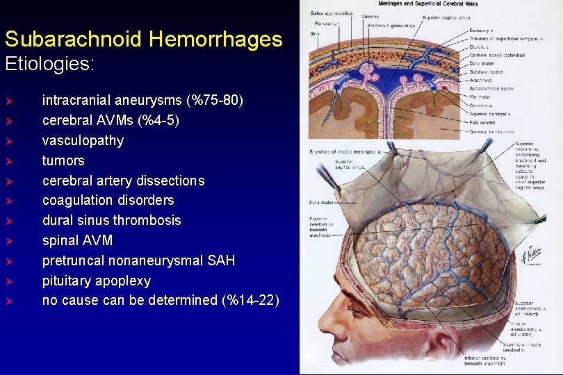
Subarachnoid Hemorrhages Etiologies: Ø Ø Ø intracranial aneurysms (%75 -80) cerebral AVMs (%4 -5) vasculopathy tumors cerebral artery dissections coagulation disorders dural sinus thrombosis spinal AVM pretruncal nonaneurysmal SAH pituitary apoplexy no cause can be determined (%14 -22)
Subarachnoid Hemorrhage Ø Ø Ø Ø Incidence: 6 -8/100 000 10 -15% of patients die before reaching medical care Overall mortality is 45% peak age for aneurysmal SAH is 55 -60 years. 20% of cases occur between ages 15 -45 yrs 30% of aneurysmal SAHs occurs during sleep SAH is complicated by intracerebral hemorrhage in 20 -40%, by intraventricular hemorrhage 13 -28%, and by subdural blood in 2 -5% rupture incidence is higher in spring and autumn
SAH Risc Factors: Ø Ø Ø Hypertension Oral contraceptives Cigarette smoking Cocain Alcohol? Pregnancy
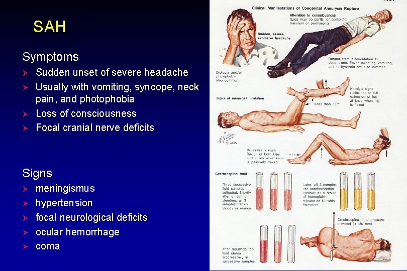
SAH Symptoms Ø Ø Sudden unset of severe headache Usually with vomiting, syncope, neck pain, and photophobia Loss of consciousness Focal cranial nerve deficits Signs Ø Ø Ø meningismus hypertension focal neurological deficits ocular hemorrhage coma
SAH Diagnosis Ø Ø Ø Non-contrast high resolution CT will detect SAH in 95% of cases if scanned within 48 hours of SAH If CT is negative: Lumbar punction in questionable cases CT angiography MR angiography Cerebral angiography
Grading SAH (Hunt and Hess) Grade 1: asymptomatic, or mild headache and slight nuchal rigiditity Grade 2: moderate to severe headache, nuchal regidity, cranial nerve palsy Grade 3: mild focal deficit, lethargy, or confusion Grade 4: stupor, moderate to severe hemiparesis, early decerebrate rigidity Grade 5: deep coma, decerebrate rigidity, moribound appearance *add one grade for serious systemic disease or severe vasospasm on angiography
Grading SAH (Yaşargil) Grade 0: Grade 1: Grade 2: Grade 3: Grade 4: Grade 5: a, unruptured aneurysm b, unruptured aneurysm, neurological deficit (+) a, asymptomatic b, focal neurological deficit (+) a, headache, nuchal rigidity b, focal neurological deficit (+) a, lethargy, confusion, disorientation, agitation b, focal neurological deficit (+) semi coma deep coma

Grading system of Fisher Grade 1: no subarachnoid blood detected (5. 8% mortality) Grade 2: diffuse or <1 mm blood (10. 3% mortality) Grade 3: localized clot and/or >1 mm blood (32. 8% mortality) Grade 4: intracerebral or intraventricular clot (45% mortality)

SAH Initial Management Concerns rebleeding hydrocephalus Ø Ø § § Ø Ø acute (obstructive) hydrocephalus (20 -27%) chronic (communicating) hydrocephalus (14 -23%) delayed ischemic neurological deficit (DIND) attributed to vasospasm Hyponatremia with hypovolemia (10 -34%) DVT and pulmonary embolism seizures (10. 5%)
SAH Admitting Order ØAdmit to ICU ØBed rest with head of bed at 30º ØLow level of external stimulation, restricted visitation, no loud noises ØIV fluids: 2 ml/kg/h or 150 ml/h (normal saline + 20 m. Eq KCl/L) ØMedications ØProphylactic anticonvulsants ØSedation, Analgesics, Dexamethasone ØAntiemetics, H 2 blockers, stool softener ØOxygenation ØCardiac rhythm monitor ØSystolic blood pressure 120 -150 mm Hg by cuff

SAH (Grade 1 -2) Ø Ø Cerebral angiography If there is cerebral aneurysm, early surgery
SAH (Grade 3 -4) Ø Ø Ø Arterial line Central venous catheter Urinary catheter Nasogastric tube (if necessary) External ventricular catheter (if necessary) Endotracheal intubation (if necessary)
SAH Rebleeding (70% mortality) ØFirst 24 hours (4%) Ø 1. 5% daily for 13 d. Ø 15 -20% rebleed within 14 d Ø 50% will rebleed within 6 months ØThereafter the risk is 3%/yr Ø 50% of deaths occur in the 1 st month ØThe rebleeding risk increases in patients with higher grades ØVentriculostomy and possibly lumbar spinal drainage increase the risk of rebleeding
Cerebral Vasospasm Ø Ø Ø A delayed focal ischemic neurologic deficit following SAH. Clinically characterized by confusion or decreased level of consciousness with focal neurological deficit Findings usually develop gradually, and my progress or fluctuate Radiographic cerebral vasospasm is identified in 30 -70% of arteriograms Symptomatic cerebral vasospasm occurs in only 20 -30% of patients Pathogenesis of cerebral vasospasm is poorly understood

Cerebral Vasospasm Ø Ø Almost never before day 3 post-SAH Maximal frequency of onset during days 6 -8 post SAH Rarely can occur as late as day 17 Usually resolves in 2 -4 weeks
Vasospasm Diagnosis: ØDelayed onset or persisting neuro deficit ØOnset 4 -20 days pos-SAH ØDeficit appropriate to involved arteris ØRule-out other causes of deterioration Ørebleeding Øhydrocephalus Øcerebral edema Øseizure Ømetabolic disturbances (hyponatremia…) Øhypoxia Øsepsis ØAncillary tests Øtranscranial doppler ØCBF studies ØSPECT Øcerebral angiography
Cerebral Aneurysms Etiology: Ø § § Ø Ø congenital predisposition (defect in the muscular layer) Atherosclerotic or hypertensive Unrupture aneurysm: 0. 5 -1% Risk of bleeding 1 -2%/per year
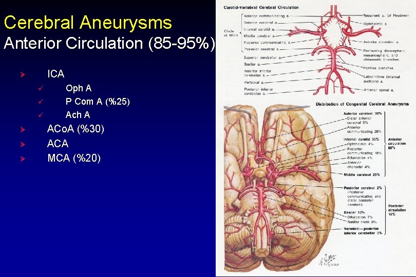
Cerebral Aneurysms Anterior Circulation (85 -95%) ICA Ø ü ü ü Ø Ø Ø Oph A P Com A (%25) Ach A ACo. A (%30) ACA MCA (%20)
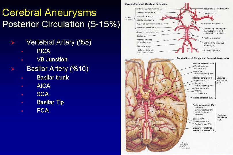
Cerebral Aneurysms Posterior Circulation (5 -15%) Vertebral Artery (%5) Ø § § PICA VB Junction Basilar Artery (%10) Ø § § § Basilar trunk AICA SCA Basilar Tip PCA
Vascular Malformations ØArteriovenous malformations ØCavernous malformation ØVenous angioma ØCapillary telangiectasia
Arteriovenous Malformations ØDilated arteries and veins with dysplastic vessels, no capillary bed and no intervening neural parenchyma ØUsually prents with hemorrhage, less often with seizures ØCongenital lesions ØLifelong risk of bleeding of 2 -4%/per year ØDemonstrable on angiography, MRI, or CT ØPrevalence 0. 14%
Arteriovenous Malformations Presentation 1. 2. 3. 4. 5. 6. 7. Hemorrhage (50%) (10% mortality, 30 -50% morbidity). Seizures Mass effect Ischemia Headache Bruit Increased ICP
Arteriovenous Malformations Treatment 1. Microsurgery 2. Embolisation 3. Stereotactic Radiosurgery
Cavernous Malformations (Cavernomas) Ø Ø Ø Usually not demonstrable on angiography Usually present with seizures, rarely with hemorrhage No intervening neural parenchyma, no arteries Low-flow Surgery best for symptomatic accessible lesions
Venous Angiomas Ø Abnormally medullary vein Ø Usually demonstrable on angiography as a starburst pattern Ø Represents the venous drainage of the area, and intervening brain is present Ø Seizures rare, hemorrhage even more rare Ø Low flow, low pressure Ø Should not be treated
- Slides: 26