Surfactant deficiency decreased production and secretion is the
 is the primary cause of RDS. �")


 §")


 � Infection � Pulmonary air leak � ETTcomplication(vocal cord injury,")
- Slides: 44
� Surfactant deficiency (decreased production and secretion) is the primary cause of RDS. � The failure to attain an adequate FRC and the tendency of affected lungs to become atelectatic correlate with high surface tension and the absence of pulmonary surfactant.
� The timing of surfactant production in quantities sufficient to prevent atelectasis depends on an increase in fetal cortisol levels that begins between 32 and 34 weeks of gestation. � By 34 to 36 weeks, sufficient surface-active material is produced by the type II cells in the lung, is secreted into the alveolar lumen, and is excreted into the amniotic fluid.
� An L/S ratio of 2: 1 usually indicate pulmonary maturity. � The presence of minor phospholipids, such as phosphatidylglycerol, also is indicative of fetal lung maturity and may be useful in situations in which the L/S ratio is borderline or possibly affected by maternal diabetes, which reduces lung maturity.
� Prematurity � Maternal diabetes � Multiple births � Cesarean delivery � Precipitous delivery � Asphyxia � Cold stress � Maternal history of previously affected infants � male gender
� Chronic or pregnancy-associated hypertension � Maternal heroin use � Prolonged rupture of membranes � Antenatal corticosteroid prophylaxis
� Alveolar atelectasis, � Ventilation to Perfusion mismatch � Hypercapnia, hypoxia, and acidosis � Pulmonary arterial vasoconstriction � Increased right-to-left shunting through the foramen ovale and ductus arteriosus and within the lung itself. � Reduced Pulmonary blood flow � Hyaline membrane formation edema and atelectasis
� RDS may develop immediately in the delivery room in extremely immature infants at 26 to 30 weeks of gestation. � Some more mature infants (34 weeks’ gestation) may not show signs of RDS until 3 to 4 hours after birth, � Manifestations of RDS include cyanosis, tachypnea, nasal flaring, intercostal and sternal retractions, and grunting.
� During the first 72 hours, infants with untreated RDS have increasing distress and hypoxemia. � In infants with severe RDS, the development of edema, oliguria, apnea, pallor , and respiratory failure necessitates assisted ventilation. � Thereafter, uncomplicated cases show spontaneous improvement that often is heralded by diuresis and a marked resolution of edema.
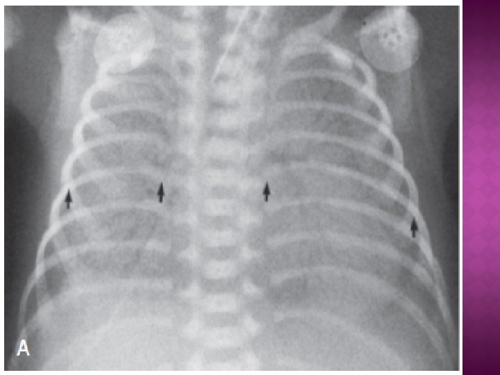
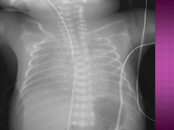
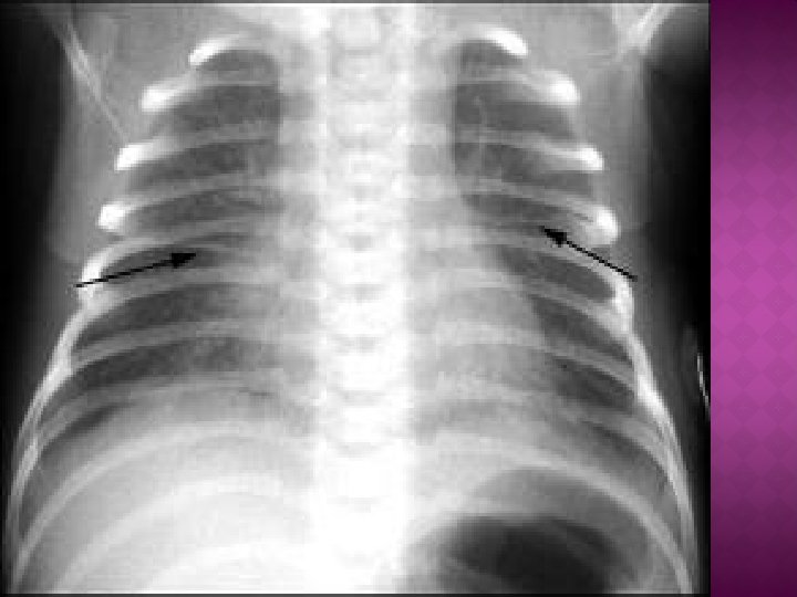
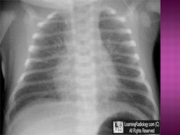
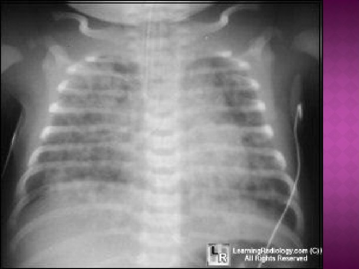
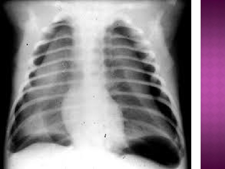
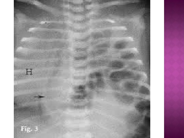
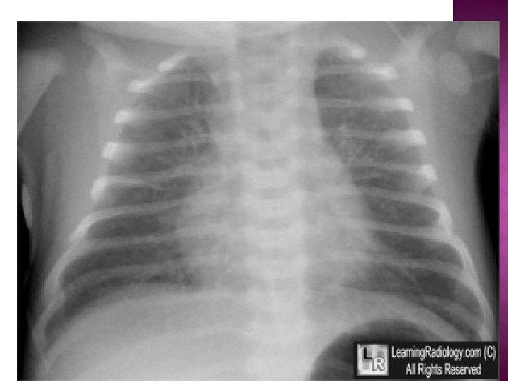
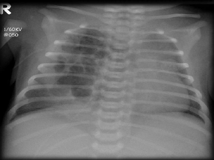
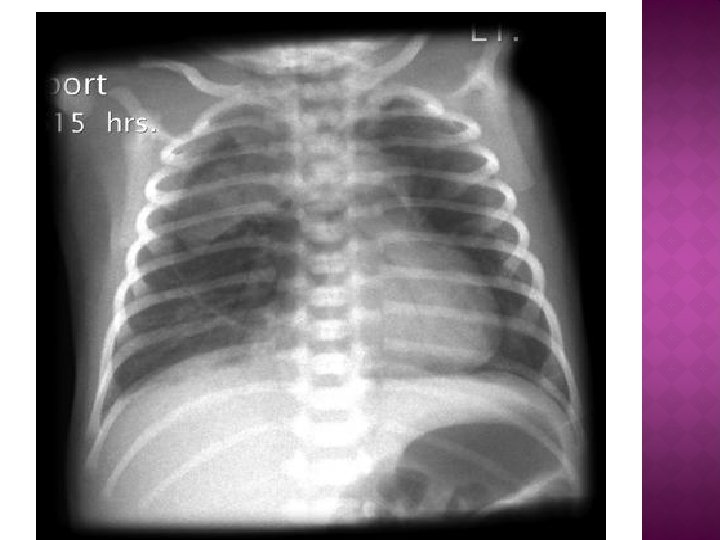
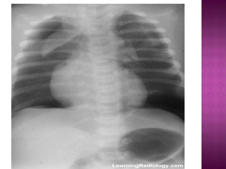
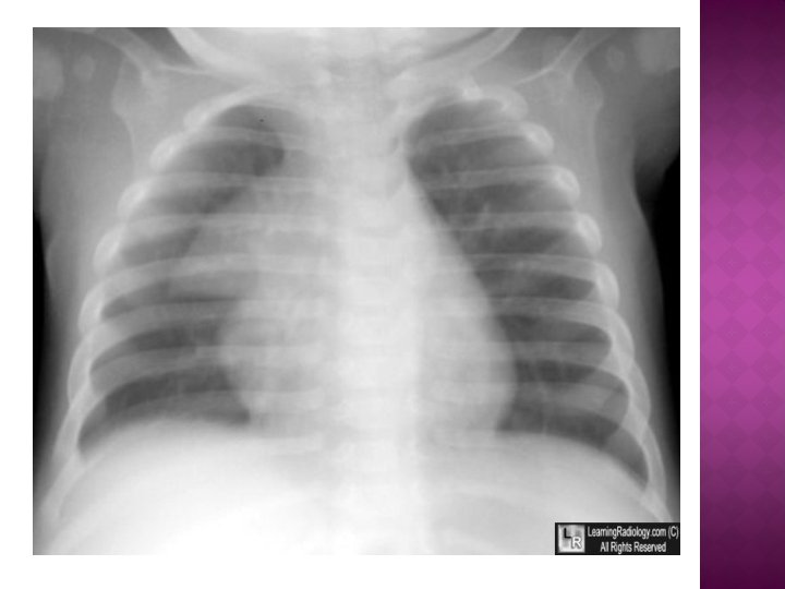
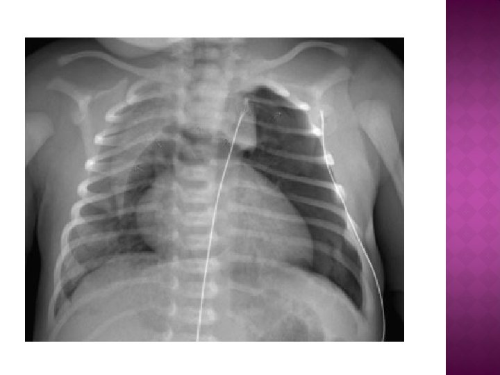
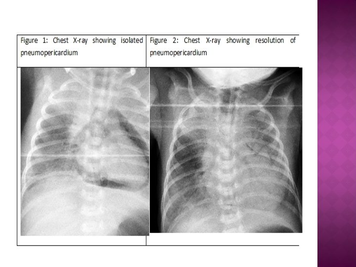
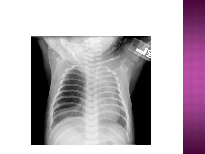
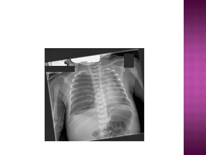
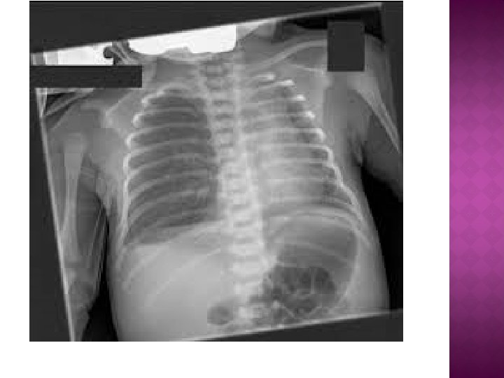
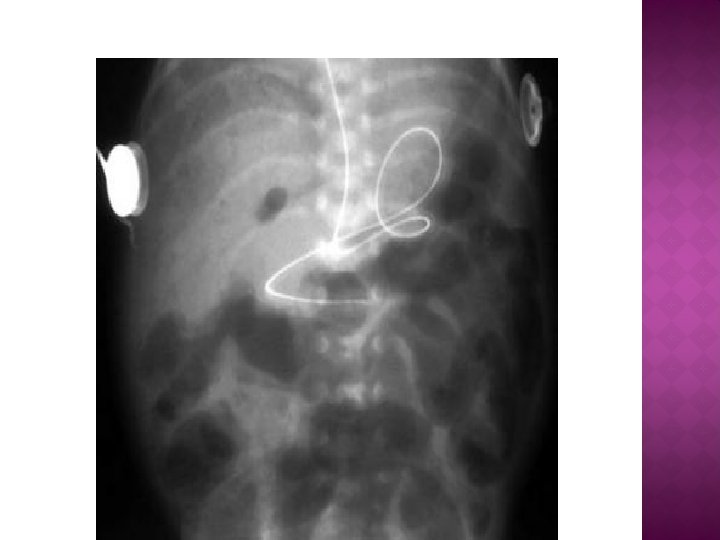
� On radiographs, the lungs may have a characteristic but not pathognomonic appearance that includes � A fine reticular granularity of the parenchyma(occasionaly whiteout lung) and air bronchograms, which are often more prominent early in the left lower lobe because of superimposition of the cardiac shadow.
§ Severe impairment of gas exchange § Alveolar air leaks (interstitial emphysema, Pneumothorax) § Pulmonary hemorrhage § Intraventricular hemorrhage (IVH) § Bronchopulmonary dysplasia (BPD)
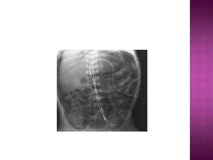
� Careful and frequent monitoring of heart and respiratory rates, oxygen saturation, Pao 2, Paco 2, p. H, serum bicarbonate, electrolytes, glucose, and hematocrit, blood pressure, and temperature. � Intravenous � Antibiotics fluids, electrolytes and calories
� Administration of antenatal corticosteroids to women between 23 and 37 wk of gestation significantly reduces the incidence and mortality of RDS as well as overall neonatal mortality. � Antenatal steroids also reduce (1) the need for and duration of ventilatory support and admission to a neonatal intensive care unit (NICU) and (2) the incidence of severe IVH, necrotizing enterocolitis � and developmental delay.
� After birth, RDS may be prevented or its severity reduced by intratracheal administration of exogenous surfactant immediately after birth in the delivery room within a few hours of birth or use of EARLY CPAP in delivery room
� Warm humidified oxygen with nasal canula or hood should be provided at a concentration initially sufficient to keep arterial oxygen pressure between 50 and 70 mm Hg (90 -95% saturation) � If oxygen saturation cannot be kept > 90% at inspired oxygen concentrations of 40% or greater or respiratory distress increased, applying CPAP at a pressure of 8 -10 cm H 2 O � Surfactant therapy if fio 2>40% on CPAP 810 cm. H 2 o. INSURE(INtubate, SURfactant, Extuba te)
� Arterial blood p. H <7. 25 and arterial blood Pco 2 of 60 mm. Hg or higher � Oxygen saturation <90%oxygenconcentrations of 50 -70% and CPAP of 8 -10 cm H 2 o � persistent apnea
� The Pao 2 level should be maintained between 60 and 70 mm Hg (oxygen saturation 90%), and the p. H should be maintained above 7. 25.
� Retinopathy of prematurity(ROP) � Infection � Pulmonary air leak � ETTcomplication(vocal cord injury, subglottic stenosis, asphyxia, … � UAC complication(thromboemboli, hemorrhage ischemic necrosis of abdominalviscera, hypertention � PDA left to right shunting(ventricular volume overload, pulmonary edema) � Bronchopulmonary dysplasia(oxygen dependency at day 28 or 36 wk postmenestral age