SUPPORT AND MOVEMENT THE SKELETAL SYSTEM REVIEW Clinical

 • Provide a framework")


 • Form skull • Protects brain/structures inside skull")

 • Scapula • Sternum (breastbone)")

 -> ulna and radius (lower arm)")


- Slides: 22
SUPPORT AND MOVEMENT: THE SKELETAL SYSTEM REVIEW Clinical Health J. Sugahara
Bare Bones • Skeletal System = joints and bones (206) • Provide a framework for the body • Protect vital organs like the brains and spinal cord • Serve as levers, when muscles are attached to help us lift and move • Store calcium, which may be reabsorbed into the blood if there isn’t enough in the diet • Produce blood cells in the red bone marrow.
Two kinds of Bones • Compact • Homogeneous • Spongy • Cancellous bones • Spaces or honeycombed
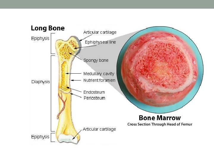
Bone Structure
Categories of Bones • Long • Short • Flat • Irregular • Sesamoid
Long Bones
Short Bones • Small, cube-shaped bones of wrists, ankles and toes • Consist of an outer layer of compact bone w/ inner layer of cancellous bone (latticework structure) • Provide support/stability w/ little to no movement
Flat Bones • Large, somewhat flat surface that cover organs or provide a surface for large areas of muscle
Irregular Bones • Specialized bones with specific shapes • Ears, vertebrae, face
Sesamoid Bones • Bones embedded in tendon (tough, connective tissue that connects muscle to bone) • Knee, hands and feet
Interior – Red and Yellow Marrow • Marrow – tissue comprising center of large bones • Red Marrow • at birth all marrow red • Produces red blood cells, platelets and most white blood cells • Yellow Marrow • Produces some white blood cells • Color due to fat • More and more marrow becomes yellow as you age
Bones of the Head (cranial bones) • Form skull • Protects brain/structures inside skull • Join at points called sutures • Frontal – forehead, roof of eye sockets • Ethmoid – nasal cavitiy and orbits of eyes • Parietal – top and upper parts of the sides of the skull • Temporal – lower part of the skull and the lower sides, incl. openings for ears • Occipital – back and base of the skull • Sphenoid – base of the cranium, holds frontal, occipital and ethmoid bones
Spinal Column • 5 sets of vertebrae • Cervical – 7 vertebrae of neck • Thoracic – 12 vertebrae that connect to ribs • Lumbar – 5 bones of middle back • Sacrum – curved bone of the lower back, 5 separate bones at birth = fuse in early childhood • Coccyx – tailbone, 4 fused bones • Vertebra separated by a thick cartilaginous disc, helps in movement and flexibility
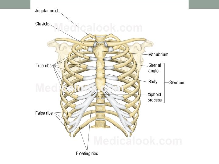
Bones of the Chest • Top • Clavicle (collarbone) • Scapula • Sternum (breastbone) • 12 pairs of ribs • 7 joined to both vertebral column and sternum – “True ribs” • 3 joined to vertebral column and 7 th rib – “False ribs” • Last two – “false/floating ribs” – do not attach to sternum or other ribs
Bones of the Pelvis • Pelvic girdle – large bone that forms hips and supports the trunk of the body • Ilium, ischium and pubes • Point of attachment for legs • Area where two pubic bones join = “pubic symphysis”
Bones of the Extremities • Humerous (upper arm) -> ulna and radius (lower arm) -> eight carpals (wrist) -> metacarpals (palm) -> phalanges (fingers) • Attaches at scapula, clavicle • Femur (thigh) ->patella (kneecap -> tibia (shin) and fibula -> tarsals (ankle) -> phalanges (toes)
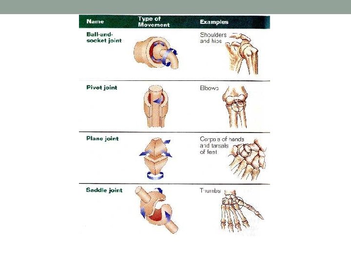
Joints • Points where bones connect • Connected to other bones with ligaments (bands of fibrous tissue) • Movement • Diarthroses – joints that move freely – knee • Amphiarthroses – cartilaginous joins that move slightly – between vertebrae • Synarthroses – do not move – between skulls bones • Symphyses cartilaginous joints that unit two bones firmly – pubic symphysis • Synovial – covered in a membrane that secretes a fluid lubricant and helps joint move easily - hip
Take C. Notes on Skeletal System 1. Pp 192 – 196 2. Don’t forget to write your summary (on the bottom of the first page if possible) 3. Include colored drawings of the following structures: 1. Bone Structure (figure 6 -23) 2. Different types of fractures *You will need to look up pictures of the fractures that are not listed as well.
Sources • http: //www. emsjunkie. com/anatomy-physiology/skeletal- system/