SUPPORT AND MOVEMENT IN VERTEBRATES SKELETAL SYSTEMS Vertebrates

SUPPORT AND MOVEMENT IN VERTEBRATES

SKELETAL SYSTEMS • Vertebrates are supported, move and locomote using endoskeletons. • Endoskeletons which are made of bone, consist of: 1. A main axis (skull and vertebral column) 2. The appendages attached to this main axis by the pectoral and pelvic girdles.
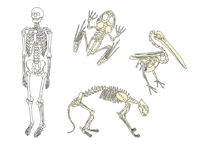

• BONES OF THE HUMAN SKELETON ARE QUITE SIMILAR TO THOSE OF OTHER ANIMALS IN THE PHYLLUM CHORDATA.

FUNCTIONS OF THE SKELETON* • 1. 2. 3. 4. 5. 6. The skeleton serves six main functions: Support Protection Movement and locomotion Muscle attachment Production of red and white blood cells Stores calcium and phosphorus.

SUPPORT 1. Holds the body upright, off ground 2. Prevents organs crushing each other. 3. Keeps body’s shape.

PROTECTION • Bones protect delicate organs from injury e. g. 1. Skull brain 2. Vertebral column spinal cord. 3. Ribs heart and lungs.

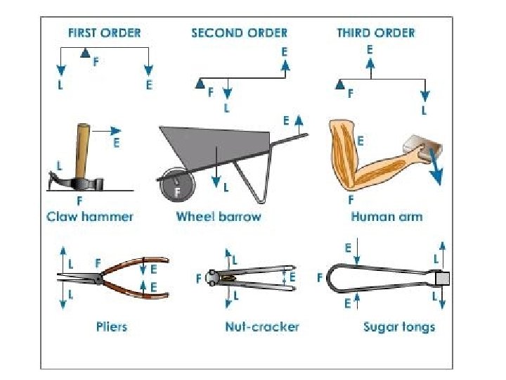
MOVEMENT AND LOCOMOTION • Bones along with muscles act as levers for movement and locomotion.

MUSCLE ATTACHMENT • One end of the muscle is attached to a bone that is fixed or does not move (origin). • The other end of the muscle must be attached securely at the bone to be moved (insertion). • • Muscles work in antagonistic muscle pairs.

PRODUCTION OF RED AND WHITE BLOOD CELLS • The bone marrow in the long bones of the legs and the short bones that make up the ribcage make new red cells (3 months) and white blood cells.

CALCIUM AND PHOSPHORUS STORAGE • Calcium and phosphorus salts are stored in bones and teeth strengthening them. • Acids in juices and sodas can wear the minerals away over time.

WHAT MAKES THE SKELETON MOVE

• *The following components of the skeleton help it to move: 1. 2. 3. 4. Bones and cartilage Joints Muscles Ligaments and tendons

BONE • Bones are living, hard, connective tissue made up from a matrix of tough collagen protein fibres strengthened by calcium phosphate and calcium carbonate in which are found many bone cells. • Bone secreting cells are arranged in rings around a central canal. • These canals carry nerve and blood capillaries so that bone is a living tissue.

BONE CELLS

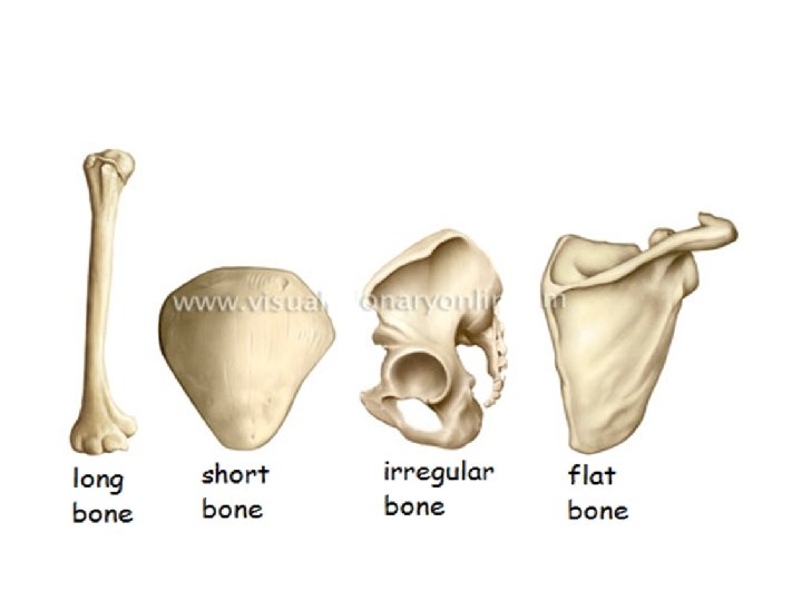
GROUPS OF BONES • Four categories of bones: 1. Long bones- femur and humerus 2. Short bones- ribs, wrist, hand, foot bones 3. Flat bones- cranial bones 4. Irregular bones- vertebral bones
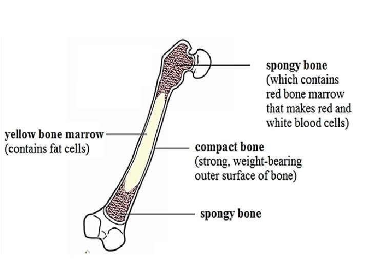

• SECTION THROUGH A BONE

CARTILAGE • Is flexible connective tissue with a smooth, glossy appearance. • In adult skeletons, cartilage remains at the ends of bones, between joints; at the tips of the nose and tops of the ears, making these areas very flexible.

FUNCTIONS OF CARTILAGE • The functions of cartilage are: 1. To spread load at joints 2. To provide cushioning at joints as well 3. To prevent friction from bones rubbing together at these areas.

JOINTS • A joint is an area where two bones meet. The flow chart below shows the different types of joints.
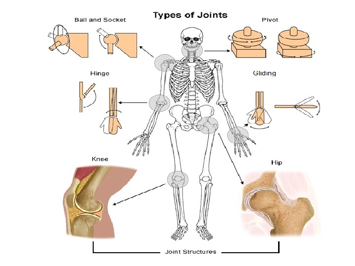
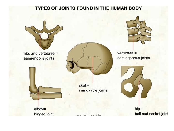

IMMOVEABLE JOINTS • Two bones are joined firmly by fibres and have very limited or no movement at all. • The bones in the skull are joined in this way and the joints are called sutures. • Also found between vertebrae where the bones are joined by cartilage with fibres in called intervertebral discs.

MOVEABLE JOINTS • The diagrams below show two synovial joints

TYPES OF MOVEABLE JOINTS 1. Hinge- allows movement in one plane such as when one opens and closes a door. 2. Ball and socket- a ball at the end of one bone fits into the socket of another; this joint allows circular movement. 3. Pivot- allows rotation 4. Gliding- where one bone slides over the surface of the other.

MUSCLES • There are three types of muscles in the body
 and getting longer (relaxing).")
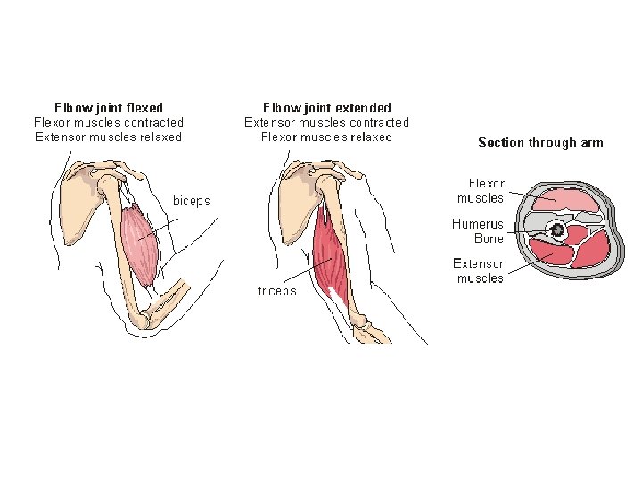
ANTAGONISTIC MUSCLES • Muscles cause movement by getting shorter (contracting) and getting longer (relaxing). They need a lot of energy from respiration to do this so they have many mitochondria. • One end of the muscle is attached to a bone that does not move (anchor) and the other end is attached to the bone to be moved (insertion). Muscles move bones like levers. • Muscles work in pairs to move bones. If one muscle is in charge of bending at a joint, another muscle must be in charge of straightening. • Since these muscles work against one another, they are called antagonistic muscles.

MOVEMENT OF THE FOREARM • When the biceps in the arm contracts, it gets short and fat while the triceps behind it relaxes and the arm will bend. • When the biceps relaxes, it gets long and thin, while the triceps contracts, getting short and fat causing the arms to straighten out. • The biceps bends the arm so is called the flexor muscle. • The triceps straightens it so is called the extensor. • These pairs of muscles are found all over the body e. g. the thigh the bend and straighten the leg.
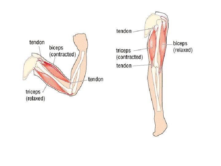

LIGAMENTS AND TENDONS • Ligaments join bones to bones at joints. They are very elastic and flexible to allow for this. • Tendons attach muscles to bone. They are made of tough, non-elastic fibres since they must not stretch when the muscles contract.

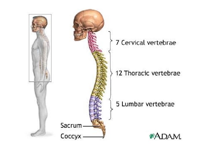
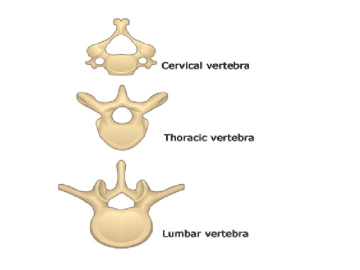
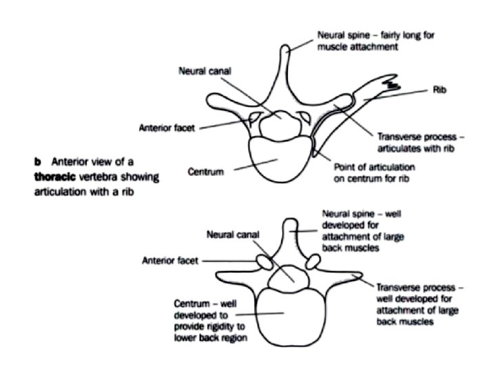
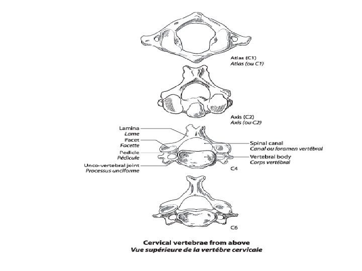
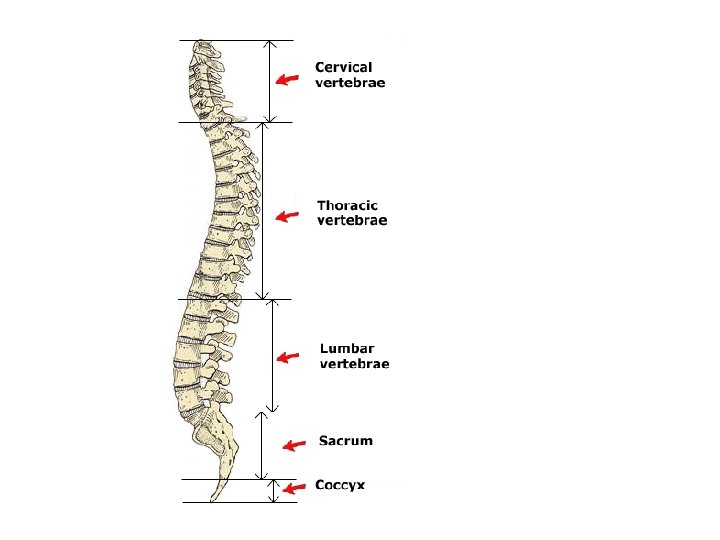
THE VERTEBRAL COLUMN • Protects the spinal cord and keeps the body upright and anchors the rib bones (thoracic vertebrae). • There are 34 vertebrae in all. • There are categories of vertebra: cervical (neck), thoracic (chest), lumbar (lower back) and caudal (tailbone). • Each of these categories have different appearances which make them unique. (LAB 16).

IMPORTANCE OF LOCOMOTION So animals can: Find mates Escape danger Get away from competition (food shortage, space shortage, etc. ) 4. Populate new areas 5. Respond to stimuli such as climatic changes, loud noses, bright lights. • 1. 2. 3.

LAB # • Draw representations if the three vertebral bones comprising the vertebral column of a mammal. Neat drawings, proper labels and annotations required. • Cervical • Thoracic • Lumbar
- Slides: 42