Superior Mesenteric Artery Syndrome By Ali Azizi Alborz

Superior Mesenteric Artery Syndrome By: Ali Azizi Alborz Hedayati
; left gastric artery (lga); splenic")
Superior Mesenteric Artery Syndrome Male adult. Celiac trunk (ct); left gastric artery (lga); splenic artery (sa); common hepatic artery (cha); superior mesenteric artery (sma)

Superior Mesenteric Artery Syndrome

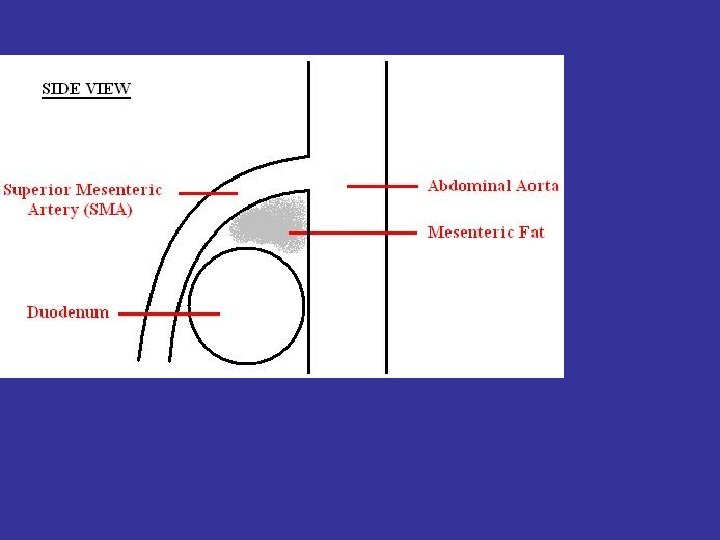
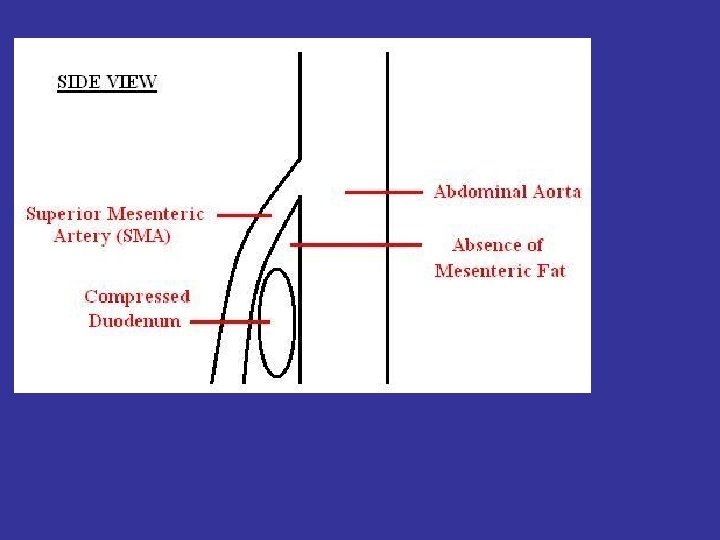
Superior Mesenteric Artery Syndrome Compression of 3 rd portion of duodenum between the aorta and superior mesenteric artery (SMA) Females >males Pathophysiology Narrowing of angle between SMA and aorta SMA usually forms an angle of 45 degrees with the aorta

Superior Mesenteric Artery Syndrome Etiologies 1. 2. 3. 4. 5. 6. 7. Prolonged bed rest in supine position (body cast, whole-body burns, surgery) Substantial and, frequently, rapid weight loss Anorexia nervosa or malnutrition Loss of abdominal muscle tone (as in pregnancy) May be congenital Seen in those with asthenic build Exaggerated lumbar lordosis

Superior Mesenteric Artery Syndrome Clinical findings 1. 2. 3. 4. 5. Epigastric pain Nausea Repetitive vomiting Abdominal cramping Typically findings are worst in supine position and may be relived by changing to the prone or left lateral decubitus positions 6. Associated with a higher than normal incidence of peptic ulcer disease and hyperchlorhydria

Superior Mesenteric Artery Syndrome Imaging findings Usually requires upper GI or CT of abdomen for diagnosis Megaduodenum dilation of 1 st and 2 nd portion of duodenum and frequently stomach Best seen in supine position Compression of duodenum between aorta and SMA

Upper gastrointestinal tract series shows dilatation of second portion of duodenum.

. (Upper gastrointestinal series showing duodenal dilation (white arrow

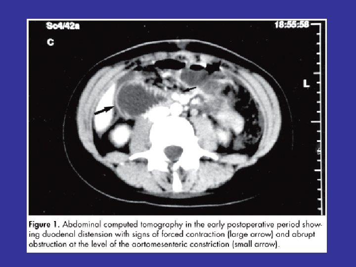
Axial CT scans of the upper abdomen show a dilated 2 nd portion of the duodenum (D) just proximal to a narrowed segment of the 3 rd portion of the duodenum (green arrow) compressed between the superior mesenteric artery (red arrow)and the aorta (black arrow ) SMA>red

Computed tomography shows narrowing between superior mesenteric artery and aorta, with dilatation of second portion of duodenum.
 by the abdominal aorta")
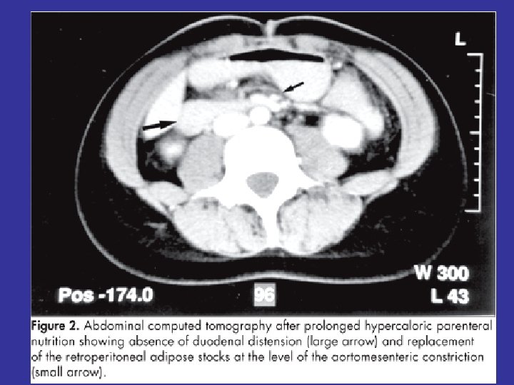
Abdominal and pelvic CT scan showing duodenal compression (black arrow) by the abdominal aorta and the superior mesenteric artery

Thank you
- Slides: 17