Superficial Fascia of the Thigh It is differentiated

 T. S of middle of right thigh")
 ØIt is an oval opening in the")


. ØIt")


")
 Canal : n n v v It is an intermuscular cleft lies")


 emerging on the")


- Slides: 69
Superficial Fascia of the Thigh ØIt is differentiated into a superficial fatty layer & deep membranous layer. ØFatty layer of superficial fascia : it is downward continuation of that on anterior abdominal wall ØDeep membranous layer becomes adherent to the deep fascia of the thigh (Fascia Lata) along a horizontal line ½ inch below the inguinal ligament.
Deep Fascia of the Thigh (Fascia Lata) T. S of middle of right thigh ØSuperiorly : it is attached to the pelvis + inguinal ligament. ØLaterally : it is thickened to form the iliotibial tract which is attached above to iliac tubercle /and below to lateral condyle of tibia. -Iliotibial tract receives insertion of : tensor fascia latae + gluteus maximus muscle. ØInferioly : it is attached to capsule of knee joint + patella + upper end of tibia & fibula. ØPosteriorly ( in Gluteal region) : it forms sheaths, which enclose the tensor fasciae latae & gluteus maximus muscles. ØIts deep surface is attached to back of femur by 3 intermuscular septa…. Lateral/ medial /& posterior.
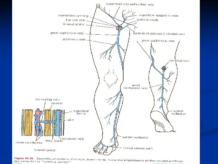
Deep Fascia of the Thigh (Saphenous Opening) ØIt is an oval opening in the deep fascia of the thigh. opening – ØIt lies in front of thigh just below inguinal ligament. ØIt transmits : great saphenous vein + branches of femoral artery (superficial epigastric, superficial circumflex iliac & superficiial external pudendal) + lymph vessels. ØIt is located 1, 5 in. below and lateral to pubic tubercle. It has margin called Falciform margin which is sharp superiorly, laterally and inferiorly. It is filled with loose C. T called cribriform fascia.
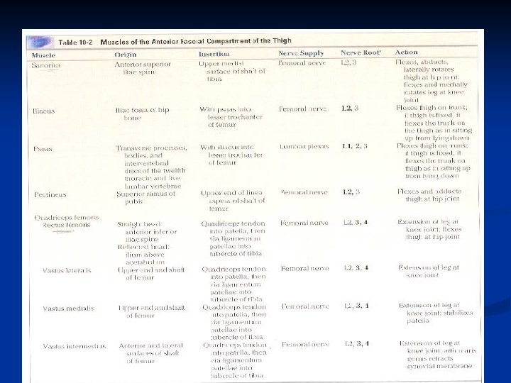
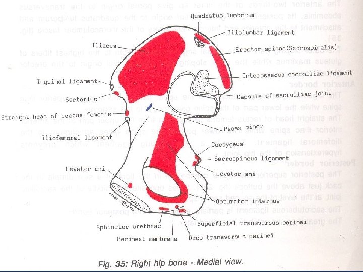
Fascial Septae and Compartments of Thigh Ø 3 fascial septa pass from inner aspect of deep fascial sheath of thigh to linea aspera of femur, dividing the thigh into 3 compartments : anterior, medial and posterior. ØContents of Anterior compartment : 1 -Muscles : sartorius, iliacus, psoas, pectineus, and Quadriceps femoris Ms. 2 -Blood supply : femoral vessels. 3 -Femoral nerve.
Muscles of Anterior Compartment of Thigh
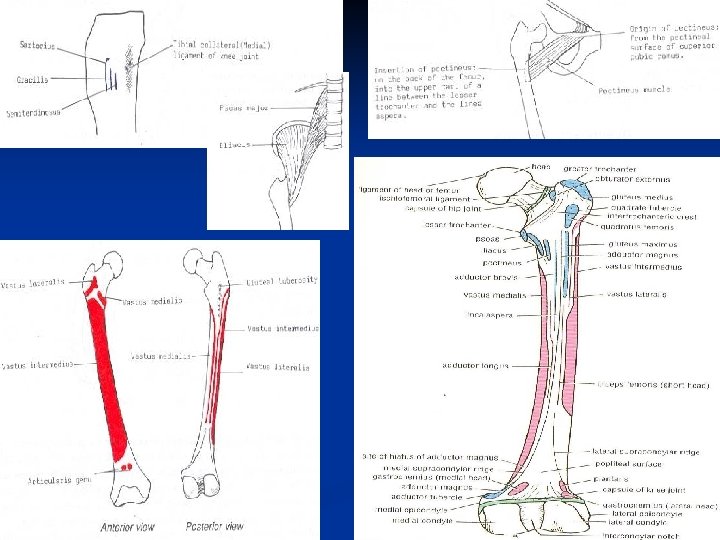
Quadriceps Femoris Insertion
Femoral Sheath ØIt is funnel-shaped tube of fascia that surround the upper 1 in. of femoral vessels & lymphatics, below inguinal ligament. ØThe upper end of sheath opens into abdomen, while lower end fuses with the walls of femoral vessels (tunica adventitia). ØIts anterior wall is formed of downward continuation of fascia transversalis of anterior abdominal wall. ØIts posterior wall is formed of downward continuation of the fascia iliaca of posterior abdominal wall. Ø it is divided by 2 fibrous septa into 3 compartments : 1 -lateral compartment : contains femoral artery. 2 -intermediate comp. : contains femoral vein. 3 -medial comp. : femoral canal transmitting lymph vessels.
Femoral Canal ØIt is the medial compartment of femoral sheath. It is about ½ in. long. ØIt contains lymph vessels + fatty C. T ØIts upper end opens into abdomen and is called femoral ring, which is closed by femoral septum (condensation of extraperitoneal tissue of fatty tissue & lymph). ØBoundaries of Femoral Ring : -Anteriorly : inguinal ligament. -Posteriorly : superior ramus of pubis. -laterally : femoral vein. -Medially : lacunar ligament. ØIts lower end is closed by fusion of its walls. ØApplied anatomy : femoral hernia.
Femoral hernia ØFemoral hernia is more common in female because of wider pelvis and femoral canal, it should always be treated surgically. ØThe neck of the herniated sac lies below and lateral to pubic tubercle, while In Inguinal hernia, the neck of swelling lies above and medial to pubic tubercle. ØIt may be difficult to push it up to return to abdomin. –irreducible hernia. . ØAfter coughing or straining, a piece of bowel may be forced through the neck, and its blood vessels may be compressed by femoral ring performing strangulated hernia.
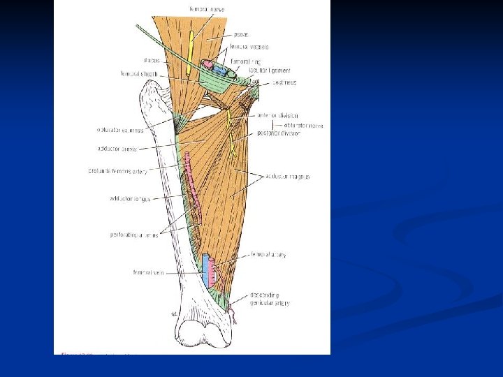
Femoral Triangle ØIt lies in the upper part of medial side of the thigh. ØBoundaries : -superiorly (base) : inguinal ligament. -laterally : sartorius. -medially : medial border of adductor longus. ØFloor : from lateral to medial : -iliopsoas, pectineus & adductor longus. ØRoof : skin & fascia of the thigh. ØContents : femoral nerve & its branches, femoral sheath, femoral artery & its branches, femoral vein & its tributaries, and deep inguinal lymph nodes.
Femoral Artery ØIt begins behind the midinguinal point, (between ant. sup. iliac spine & pubic tubercle of symphysis pubis), as a continuation of external iliac artery. ØIt descends through the femoral triangle, then vertically toward the adductor tubercle of femur. Øends at opening in adductor magnus muscle to enter the popliteal fossa as popliteal artery.
Relation of Femoral Artery ØAnteriorly : its upper part is covered by skin & fascia. Its lower part is covered by sartorius muscle. ØPosteriorly : iliopsoas, pectineus, and adductor longus. Femoral vein is behind femoral artery in the lower part of femoral triangle. Ø Medially : femoral vein (in upper part of femoral triangle) ØLaterally : femoral nerve & its branches.
Branches of Femoral Artery ØSuperficial circumflex iliac artery : to the region of anterior superior iliac spine. ØSuperficial epigastric artery : to region of umbilicus. ØSuperficial external pudendal artery : to supply skin of scrotum (or labium majus). ØDeep external pudendal artery : to supply skin of scrotum (or labium majus). ØProfunda femoris artery : is a large and important branch arising from lateral side of femoral artery, about 1, 5 in. below inguinal ligament. It passes medially behind femoral vessels to enter medial side
Branches of Femoral Artery ØProfunda femoris artery : -it ends by becoming 4 th perforating artery. -at its origin it gives off medial and lateral femoral circumflex arteries. -during its coarse it gives off three perforating arteries. ØDescending genicular artery : near its termination to supply knee joint.
Femoral vein ØIt enters thigh by passing through opening in adductor magnus as a continuation of popliteal vein. ØIt ascends in the thigh, lying at first laterally to femoral artery, then posterior and finally medially. ØIt leaves thigh by passing behind inguinal ligament to become the external iliac vein. ØTributaries : great saphenous vein + branches correspond to that of artery. ØSuperficial circumflex iliac, superficial epigastric, and external pudendal veins drain into great saphenous vein.
Femoral Nerve ØIt is the largest branch of lumbar plexus (L 2, 3&4). ØIt arises from lateral side of psoas major ms. within abdomen to descend in interval between posas & iliacus. ØIt enters thigh behind inguinal ligament, lying outside the femoral sheath, but within the femoral triangle lateral to femoral artery. ØIt terminates about 1, 5 in. below inguinal ligament, by dividing into anterior & posterior divisions. ØIt supplies all Ms. of anterior compartment of thigh.
Branches of Femoral Nerve ØAnterior division : - 2 cutaneous branches : medial cutaneous N. of thigh + intermediate cutaneous N. -2 muscular branches : to sartorius + pectineus. ØPosterior division : -1 cutaneous branch : Saphenous N. -Muscular branches : to quadriceps femoris Ms. -Articular branches to knee joint.
Saphenous Nerve ØIt descends medially crossing the femoral artery. ØIt emerges on the medial side of knee between the tendons of sartorius & gracilis. ØIt then descends on the medial side of leg in company with great saphenous vein. ØIt passes in front of medial malleolus and along medial border of foot to end in big toe.
Superficial inguinal Lymph Nodes ØThey are arranged into Horizontal & Vertical groups. ØHorizontal group : -lies below and parallel to inguinal ligament. -The medial members of this group receive afferent vessels from : 1 -superficial lymph vessels from anterior abdominal wall below umbilicus. 2 -lymph vessels from perineum, + urethra + lower ½ of anal canal + external genitalia (except lymph drainage of testes ends in lumbar (para-aortic) L. Ns. at level of L 1 vertebra. -The lateral members of this group receive afferent vessels from back below level of iliac crest (skin of gluteal region)
Superficial inguinal Lymph Nodes ØVertical group : -Lies along terminal part of great saphenous vein. -They receive most of afferent superficial lymph vessels of the lower limb (except lateral sides of foot & leg drained into popliteal L. Ns. + gluteal region drained by lateral membres of horizontal group. ØEfferent lymph vessels from vertical & horizontal groups of superficial inguinal L. Ns. : pass through saphenous opening to end in deep inguinal L. Ns. (lying along medial side of femoral vein).
Deep Inguinal Lymph Nodes ØThey are located beneath the deep fascia along medial side of femoral vein. ØThey receive afferent lymph vessels from : -superficial inguinal L. Ns. -popliteal L. Ns. -deep structures of thigh. ØTheir efferent lymph vessels pass through femoral canal to end into external iliac lymph nodes.
Contents of Medial compartment of thigh n Muscles: Gracilis , adductor longus , adductor brevis , adductor magnus , obturator externus. q Blood supply: Profunda femoris Ar. +Obturator Ar. q Nerve supply : Obturator nerve.
Muscles of medial fascial compartment of thigh
Muscles of medial fascial compartment of thigh - Adductor part ( pubic part ) - Hamstring part ( ischial part) + + Trochanteric fossa ( lateral area )
Adductor (Subsartorial) Canal : n n v v It is an intermuscular cleft lies in the middle third of medial side of the thigh beneath sartorius muscle. Its upper end lies at the apex of femoral triangle, while its lower end lies at the opening of Adductor magnus. What are the walls of Adductor canal ? What are the contents of Adductor canal ?
Boundaries and Contents of Adductor Canal Cross section of the middle of thigh ØAnteromedial wall (roof) : sartorius & its fascia. ØPosterior wall ( floor) : addctor longus + adductor magnus. ØLateral wall : vastus medialis. ØContents : 1 - Terminal part of femoral artery. 2 - Femoral vein. 3 - Saphenous nerve. 4 - N. to vastus medialis. 5 - Terminal part of obturator N. 6 - Deep lymph vessels.
Profunda Femoris Artery ØIt arises from the lateral side of femoral artery, about 4 cm. Below inguinal ligament. ØIt descends with anterior division of femoral N. between adductor longus & brevis, then lying on adductor magnus, where it ends as 4 th perforating artery. ØBranches : 1 - Medial femoral circumflex artery : passes backward between Ms. that form floor of femoral triangle. It takes part in cruciate anastomosis. It gives off : ascending, transverse, muscular and acetabular branches. 2 - Lateral femoral circumflex artery : passes laterally and gives off : ascending, transverse and descending branches.
Profunda Femoris Artery 3 - Four perforating arteries : 3 of these are branches of profunda, the 4 th perforating is the terminal part of profunda. -They terminate by anastomosing with one another at the back of femur. -The 1 st perforating artery takes part in cruciate anastomosis by anastomosing with the inferior gluteal artery. - The 4 th perforating artery ( terminal part of profunda ) anastomosing with the muscular branches of popliteal artery. Profunda Femoris Vein ØIt receives tributaries corresponding to the branches of the artery. ØIt drains into femoral artery.
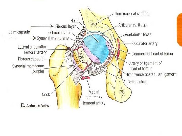
Obturator Artery ØIt arises in the pelvis from internal iliac artery. ØIt enters thigh by passing through the obturator canal (upper part of obturator foramen ) with company the obturator nerve. Ø It divides into medial & lateral branches, which form a circle on the outer surface of obturator membrane ØIt gives off : muscular branches + articular branch to hip joint and head of femur by passing through the acetabular notch and along the ligament of head of femur into the fovea.
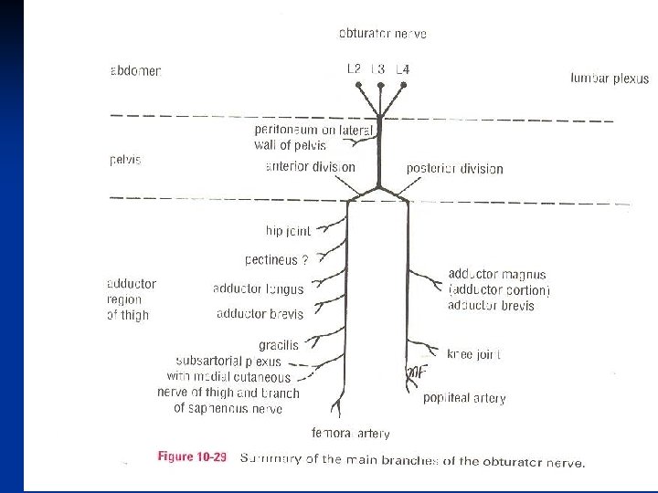
Obturator Nerve ØIt arises from lumbar plexus (L 2, 3, 4) emerging on the medial border of psoas muscle within the abdomen. ØIt runs on lateral wall of pelvis to reach upper part of obturator foramen, where it divides into anterior & posterior divisions. ØAnterior division : - it descends in front of adductor brevis/ behind pectineus + adductor longus. -it gives muscular branches : to gracilis, adductor brevis, adductor longus / occasionally to pectineus. -it gives articular branches : to hip joint. -it supplies skin of medial side of thigh.
Obturator Nerve ØPosterior division : -it pierces obturator externus to descends behind the adductor brevis and in front adductor magnus. -it terminates by passing through opening in adductor magnus ( adductor hiatus) to supply knee joint. - it gives muscular branches : to obturator externus, adductor part of adductor magnus /occasionally adductor brevis.
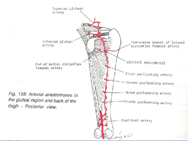
Cruciate Anastomosis ØIt lies in the back of thigh at the level of lesser trochanter of femur. ØIt provides a connection between internal iliac & femoral arteries. ØThe following arteries take part in the anastomosis : 1 - inferior gluteal artery ( from intenal iliac artery). 2 - medial femoral circumflex artery ( transverse branch). 3 - lateral femoral circumflex artery : (transverse branch). 4 - 1 st perforating artery ( branch of profunda artery).
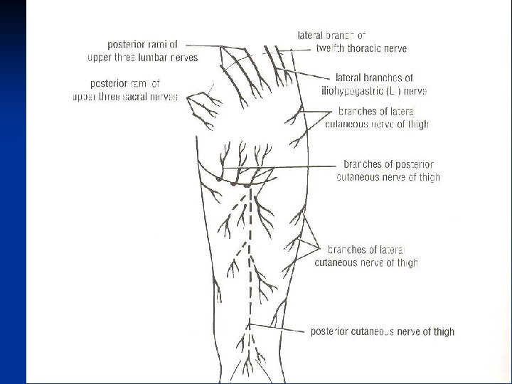
Cutaneous Nerves of Front of Thigh ØFrom the lumbar plexus : 1 - Ilio-inguinal N. (L 1 ). 2 - Femoral branch of genitofemoral N. ( L 1, 2 ). 3 - Lateral cutaneous N. of thigh. / L 2, 3 ØFrom Femoral N. : 1 - Intermediate cutaneous N. of thigh. 2 - Medial cutaneous N. of thigh. ØObturator N. / on medial side of thigh. ØPatellar plexus : it is a network of nerves in superficial fascia in front of patella and ligamentum patellae. -It is formed of : -infrapatellar branch of saphenous N. -Lateral, intermediate and medial cutaneous nerves of thigh.
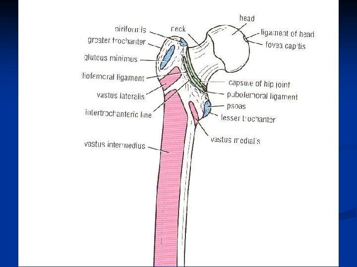
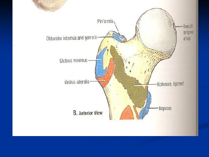
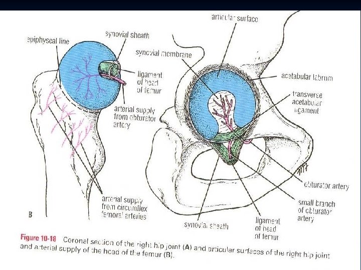
Blood supply of femoral head and Neck Fracture ØIn the young, epiphysis of of head is supplied by small branch of obturator artery, which passes to head along ligament of head of femur. ØThe neck of femur receives a profuse blood supply from medial femoral circumflex artery. These branches pierce capsule and ascend the neck. ØAs long as epiphyseal cartilage remains, no communication occurs. ØIn adult, after disappearance of epiphyseal cartilage, anastomosis occurs. ØFracture of the neck interfere with blood supply from neck to head, so
The neck of femur /coxa valga/coxa vara n n n The angle btween neck and shaft of femur (angle of femoral inclination) is about 160 in child /and 125 in adult. Increase in the angle is referred to as coxa valga/as in congenital dislocation of hip/ adduction of hip joint is limited. A decrease in the angle is referred to coxa vara/ as in fracture of neck of femur / abduction of hip is limited.
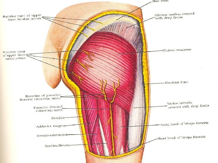
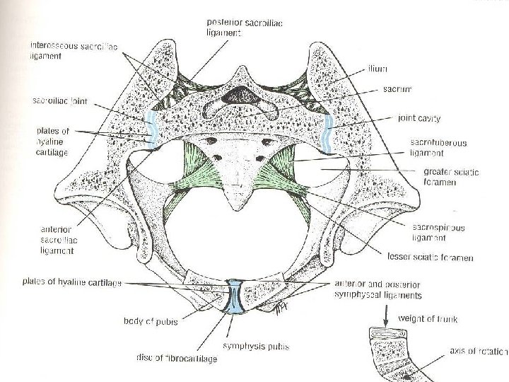
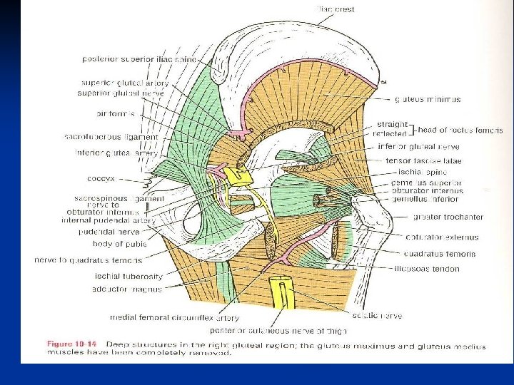
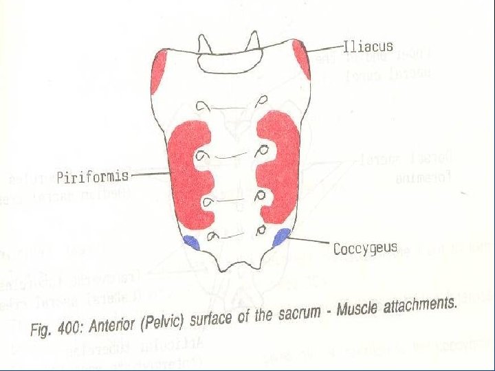
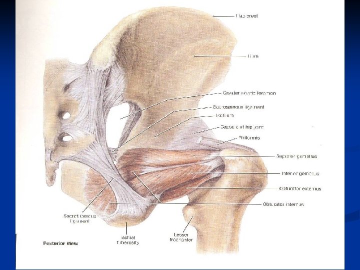
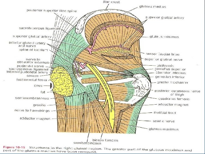
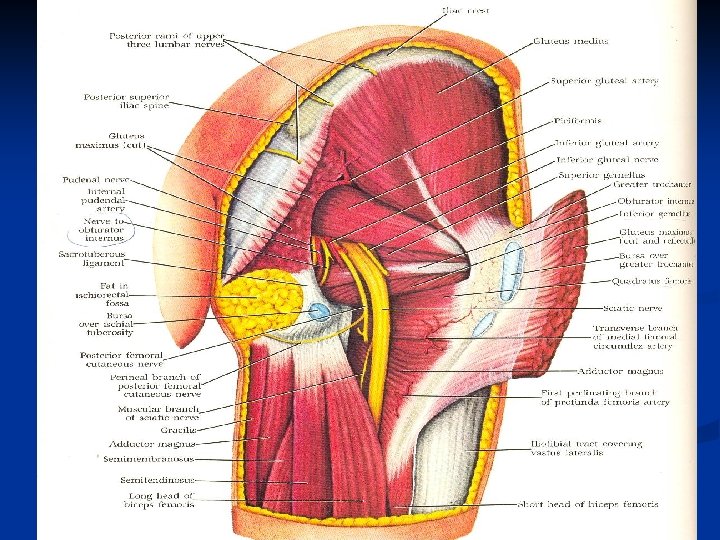
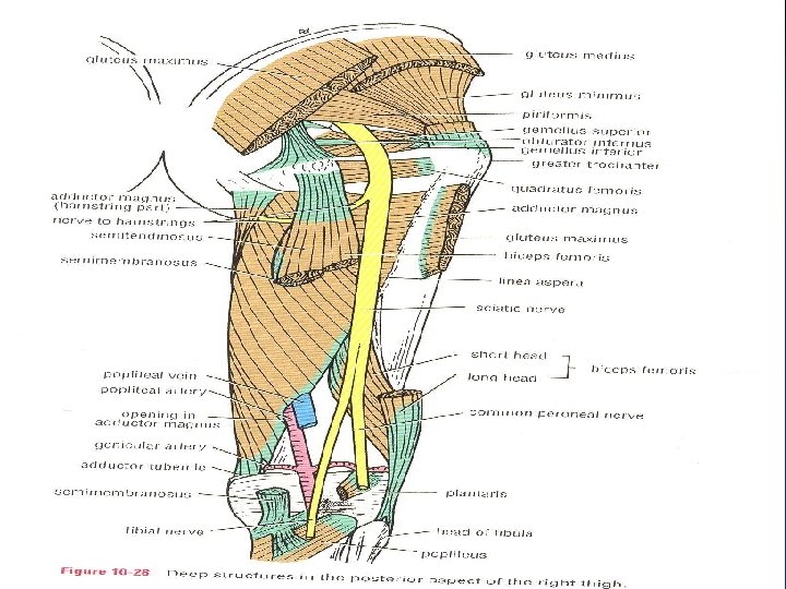
The gluteal region The ligaments : Sacrotuberous, Sacrospinous. n The foramina : 1 -Greater Sciatic Foramen. 2 -Lesser Sciatic Foramen. v The muscles : Gluteus Max. , Med. , Minimus. Tensor fascia latae , Piriformis, Obturator int. , sup. &inf. gemelli, Quadratus femoris. v The nerves: Sciatic N. , Post. cut. N. of the thigh, Sup. &inf. gluteal N. , N. to Q. F. , Pudendal N. , N. To Ob. int. v The arteries: Sup. , &inf. gluteal arteries. n
What are structures passing through the greater sciatic foramen? Piriformis. n Structures above piriformis: Sup. gluteal N. &vessels. n Structures below piriformis: Ø Inf. gluteal N. &vessels. Sciatic N. Ø Post. cut. N. of the thigh. N. to Q. femoris. Ø N. to obt. internus. ---Pudendal N. ---Int. pudendal vessels n
What are structures passing through the lesser sciatic foramen? Tendon of obturator internus muscle. n N. to obturator internus. n Pudendal N. n Internal pudendal vessels. n
The Sciatic Nerve in gluteal region. Origin : sacral plexus (L 4, 5, S 1, 2, 3) in pelvis. n Course and relation: it leaves pelvis through grater sciatic foramen below piriformis. It crosses from above downwards: ischium, obturator int. &and 2 gemilli, quadratus femoris. It leaves buttok by passing deep to long head of biceps femoris to enter the back of thigh. v Branches: No branches in the gluteal region. n
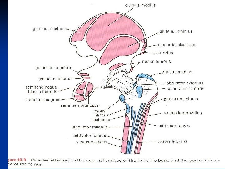
Structures deep to Gluteus Maximus v Muscles: 1 -gluteus medius. 2 -lateral rotators of the thigh. v Ligaments: 1 - sacrotuberous. 2 -sacrospinous. v Bones: Lat. : greater trochanter. Med. : ischial tuberosity. v Three synovial bursae. v Foramina : 1 -greater sciatic F. 2 -lesser sciatic F.
Anastomosis in the gluteal region The cruciate anastomosis: provide connection between the internal iliac &femoral arteries. n It lies at the level of Lesser trochanter. n The following arteries take part in the anastomosis : 1 -Inf. gluteal , 2 -1 st perforating , 3 -Med. femoral circumflex. 4 -Lat. femoral circumflex. n
Anastomosis in the gluteal region The Trochanteric anastomosis : provides the main blood supply of the head of femur. the arteries pass along femoral neck beneath the capsule of hip joint. n The following arteries take part in the anastomosis: 1 -Sup. gluteal. Ar. 2 -Inf gluteal. Ar 3 -Med. Femoral circumflex Ar. 4 -Lat. Femoral circumflex Ar. n