Summary n n n RMP ionic basis factors

Summary n n n RMP, ionic basis, factors affecting RMP AP, ionic basis, characteristics Change in excitability during an action potential Characteristics of local response Functional states of voltage-gated ion channel

Chapter 2 Basic Functions of cells 谢俊霞 教授

Striated muscle n Striated muscle § Skeletal muscle § Cardiac muscle n Smooth muscle

Neuromuscular transmission Anatomy of neuromuscular junction n Sequence of events during transmission n Characteristics of end-plate potential n Factors affecting neuromuscular transmission n

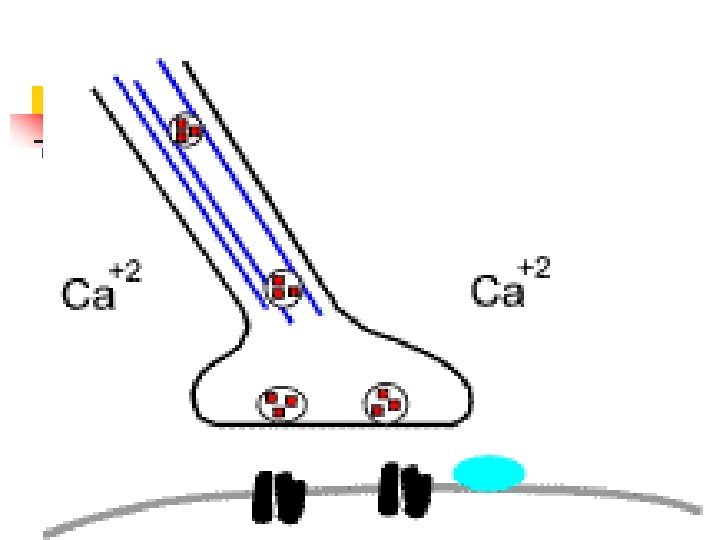
Anatomy of neuromuscular junction

Anatomy of neuromuscular junction n Prejunctional membrane n Synaptic vesicle n Active zone n Junctional cleft n Endplate membrane Junctional fold n N 2 -Ach receptor cation channel n Acetylcholinesterase

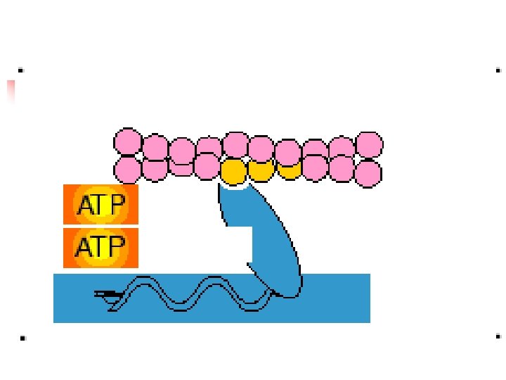
Sequence of events during transmission

Sequence of events during transmission Action potential arrives at Prejunctional membrane Action potential causes calcium channels to open (Ca 2+ enters ) Ca 2+ cause synaptic vesicle to move and release Ach diffuses across junctional cleft Ach binds to N 2 Ach receptor on endplate membrane Na+, K+ channels open (Na+>K+ ) Causes depolarisation of the endplate membrane (EPP) Action potential is produced in the muscle membrane
: depolarization of motor end plate")
Sequence of events during transmission n End-plate potential (EPP): depolarization of motor end plate of skeletal-muscle fiber in response to acetylcholine; initiates action potential in muscle plasma membrane.

Characteristics of endplate potential

Sequence of events during transmission

Sequence of events during transmission A 1 Transmitter-gated channels A 2 Voltage-gated channels Ach binding Channel opening Na+ channel opening Na+ inflow K+ outflow Na+ inflow Depolarization Result: end-plate potential Result: action potential

B 1 Transmitter-gated and voltage B 2 Effect of transmitter-gated channels on voltage-gated channels are in parallel channels

Miniature end-plate potential

Quantal release

Summary - Neurotransmission

Nicotinic acetylcholine receptor cation channel

Nicotinic acetylcholine receptor cation channel

Factors affecting neuromuscular transmission n Ca 2+ concentration at presynaptic terminal: Ca 2+ chelate n Activity of ACh receptor: tubocurarine; α -bungarotoxin n Acetylcholinesterase (ACh. E) inhibitor: pyridostigmine
 n Wendy Chu: a 23 -yr-old photographer for a local newspaper. Over")
Myasthenia Gravis(MG) n Wendy Chu: a 23 -yr-old photographer for a local newspaper. Over the last 8 months, she experienced ‘strange’ symptoms: severe eyestrain reading for longer than 15 min, tired when she chewed, brushed, extreme fatigue on the job. Physician initiated a trial of pyridostigmine, an acetylcholinesterase inhibitor, immediately felt better, antibody test was positive, confirming the diagnosis of MG.

Ultrastructure of striated muscle n Myofibril and sarcomere
 (Terminal cisterna) Longitudinal SR,")
Ultrastructure of striated muscle n Sarcotubular system (Junctional SR, JSR) (Terminal cisterna) Longitudinal SR, LSR (SR) (T tubule) (Terminal cisterna) Triad (T tubule) (Terminal cisterna) JSR: Ca 2+ release channel (ryanodine receptor, RYR) LSR: Ca 2+ pump T tubule: L-type Ca 2+ channel

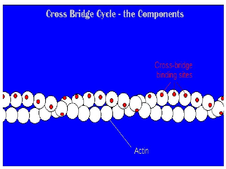
Molecular mechanisms of contraction n Myofilament sliding theory: process of muscle contraction in which shortening occurs by thick and thin filaments sliding past each other.

Molecular components of myofilament

Process of muscle contraction Cross-bridge cycling

The action potential triggers contraction How does the AP trigger contraction? n n This question has the beginning (AP) and the end (contraction) but it misses lots of things in the middle! We should ask: n n how does the AP cause release of Ca from the SR, so leading to an increase in [Ca]i? how does an increase in [Ca]i cause contraction?

Excitation-contraction coupling n Excitation-contraction coupling: mechanism in muscle fibers linking plasmamembrane depolarization with cross-bridge force generation.

Excitation-contraction coupling n Intracellular Ca 2+ is the key of excitation-contraction coupling

Excitation-contraction coupling
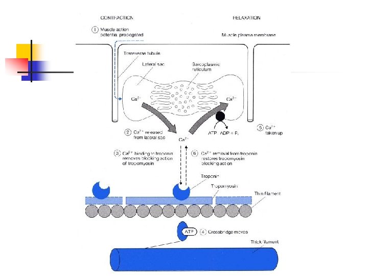

Difference of Ca 2+ release between skeletal and cardiac muscle Calcium-induced Ca 2+ release, CICR

Time relationships between action potential and the resulting shortening and relaxation of the muscle fiber
![Time relationships between action potential, intracellular [Ca 2+] and twitch tension Calcium transient](http://slidetodoc.com/presentation_image_h2/892670084b95cff6b2a3470e60398832/image-38.jpg "Time relationships between action potential, intracellular [Ca 2+] and twitch tension Calcium transient")
Time relationships between action potential, intracellular [Ca 2+] and twitch tension Calcium transient

Calcium transports n n Calcium pump Na+-Ca 2+ exchanger Low intracellular Ca 2+: 0. 1~0. 2μM 1 Ca 2+/ATP 2 Ca 2+/ATP 1 Ca 2+ 3 Na+ Endoplasmic reticulum Cell n Calcium pump

Factors affecting the performance of contraction n Performance of contraction: force; shortening; velocity Isometric contraction: contraction of muscle under conditions in which it develops tension but does not change length. Isotonic contraction: contraction of muscle under conditions in which load on the muscle remains constant but muscle shortens.

Isometric contraction and isotonic contraction Distance shortened Tension Latent period Isometric twitch Latent period Isotonic twitch

Preload Optimal initial length: the length at which the fiber develops the greatest tension.

Afterload

Contractility n Intracellular Ca 2+ level n ATPase activity of myosin

Summation n n Summation of number of motor unit Summation of frequency

Motor unit n Motor unit: one motor neuron plus the muscle fibers it innervates.
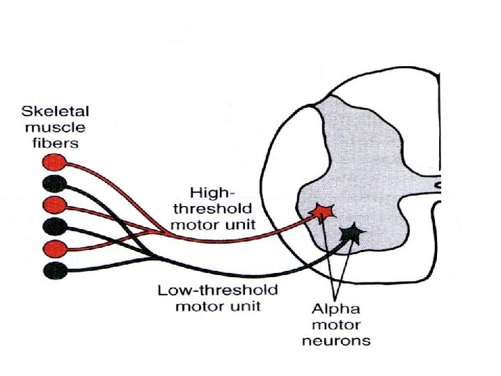

Size Principle n When a contraction occurs, small motor units fire first, as the strength of contraction increases, larger units are recruited, the orderly recruitment of motoneurons is referred to as size principle

Twitch and tetanus Twitch: mechanical response of muscle to single action potential. n Tetanus: maintained mechanical response of muscle to high-frequency stimulation. n § Incomplete tetanus § Complete tetanus

Twitch and tetanus

Summary n Neuromuscular transmission n Excitation-contraction coupling n Cross-bridge cycling

Summary n n n n n Physiology Steps in the scientific method Homeostasis Regulation Nervous regulation: reflex Humoral regulation Autoregulation Control system Feedback control system: negative feedback; positive feedback Feed-forward control system

Summary n n n n n Liquid mosaic model Simple diffusion Facilitated diffusion via carrier Facilitated diffusion through ion channel Voltage-gated ion channel Ligand-gated ion channel Mechanically-gated ion channel Primary active transport Secondary active transport Exocytosis and endocytosis

Summary Signal transduction mediated by Ø Chemically-gated ion channel Ø G-protein coupled receptor • c. AMP-PKA pathway • IP 3 -Ca 2+ pathway • DG-PKC pathway • G protein-ion channel pathway Ø Enzyme coupled receptor

The end

Smooth muscle Ultrastructure of smooth muscle

Ultrastructure of smooth muscle Spindle-shaped cell with a diameter ranging from 2 to 10 μm n Thin filament: thick filament=15: 1 n Dense body, dense area n Intermediate filament n

Molecular mechanisms of contraction n Two sources of Ca 2+ contribute to the rise in cytosolic Ca 2+ that initiates smooth muscle contraction Sarcoplasmic reticulum Extracellular Ca 2+
: smooth muscle")
Types of smooth muscle n n Single-unit smooth muscle (visceral smooth muscle): smooth muscle that responds to stimulation as single unit because gap junctions join fibers, allowing electrical activity to pass from cell to cell. Autorhythmicity Multi-unit smooth muscle: smooth muscle that exhibits little, if any, propagation of electrical activity from fiber to fiber and whose contractile activity is closely coupled to its neural input.

Modes of contraction n Phasic contraction: rapid cyclic contraction and relaxation. Phasic smooth muscle n Tonic contraction: smooth muscle can maintain a low level of active tension for long periods without cyclic contraction and relaxation. Tonic smooth muscle

Innervation of smooth muscle n n n Autorhythmicity Varicosity Non-synaptic chemical transmission End
- Slides: 61