Sudden Painless Loss of Vision Maj Ahsan Mukhtar

 Classified Eye Specialist")


 • – corneal edema – hyphema – vitreous")
 • – corneal edema – hyphema – vitreous")
 • – corneal edema – hyphema – vitreous")
 • – corneal edema – hyphema – vitreous")

 • – corneal edema – hyphema – vitreous")
 • – corneal edema – hyphema – vitreous")


 – BRVO with")
 •")


 • Patients usually>50 yrs • Strong association with hypertension")
 • extensive retinal haemorrhages in all quadrants • retinal")
 • Sudden and severe loss of vision in")


 • Recovery over 6 weeks,")


- Slides: 55
Sudden Painless Loss of Vision Maj Ahsan Mukhtar FCPS, FRCS (Ophth) Classified Eye Specialist Registrar VR Surgery AFIO
Objectives • Know Imp points in History and Exam • Enumerate common Causes • Know the clinical appearance of various diseases
History • True sudden vision loss OR sudden realisation of visual loss? • One eye or both eyes? • Onset and progression • Associated visual symptoms – flashes suggest retinal traction (but can be cortical e. g. CVA, migraine) – floaters suggest vitreous debris
History • Past ocular history – trauma and myopia are risk factors for retinal detachment • Systems review – in elderly patients, ask about headache and polmyalgia (temporal arteritis) – history of diabetes – cardiovascular disease, TIA symptoms suggest emboli
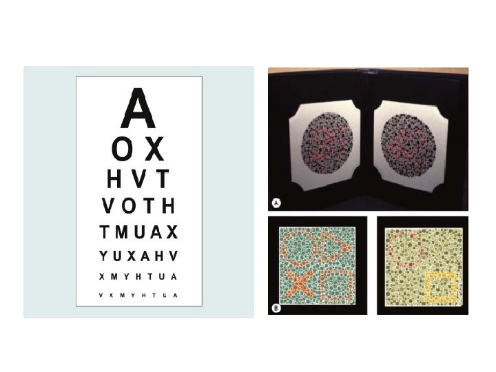
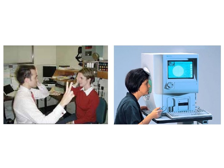
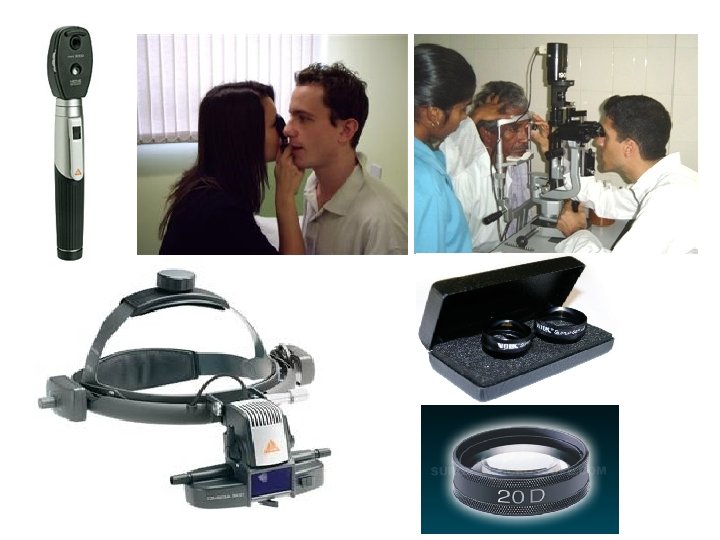
Examination • • • External ocular appearance Visual acuity Colour vision assessment Pupil Examination Visual field assessment Fundoscopy Palpation of temporal arteries Cardiovascular examination Neurological examination
RAPD • Darken the room • Have the patient fix on a distant target (e. g. the top letter on a Snellen chart) • Alternate a bright light rapidly (<1 second) between the two eyes, spending 2 seconds on each eye
CAUSES
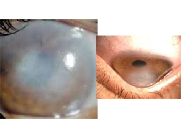
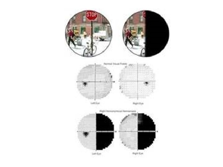
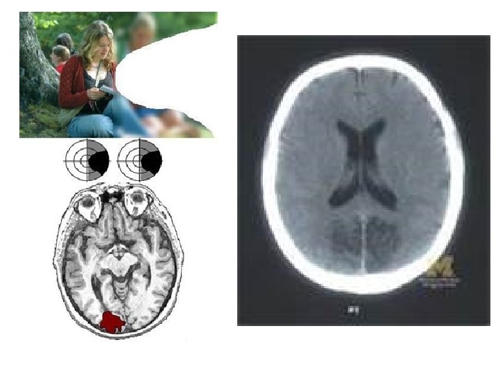
• Media opacity (No RAPD) • – corneal edema – hyphema – vitreous hemorrhage • Retinal disease (RAPD) – retinal detachment – macular disease (e. g. , macular degeneration); • – retinal vascular occlusions • Optic nerve disease (RAPD) – optic neuritis, retrobulbar • neuritis, and papillitis • – papilledema – glaucoma – ischemic optic neuropathy – giant cell arteritis Hypoxia – shock – g-LOC (an aviation related problem) – simply standing up suddenly, especially if sick or otherwise infirm Visual pathway disorder – homonymous hemianopia – cortical blindness Trauma Functional disorder
• Media opacity (No RAPD) • – corneal edema – hyphema – vitreous hemorrhage • Retinal disease (RAPD) – retinal detachment – macular disease (e. g. , macular degeneration); • – retinal vascular occlusions • Optic nerve disease (RAPD) – optic neuritis, retrobulbar • neuritis, and papillitis • – papilledema – glaucoma – ischemic optic neuropathy – giant cell arteritis Hypoxia – shock – g-LOC (an aviation related problem) – simply standing up suddenly, especially if sick or otherwise infirm Visual pathway disorder – homonymous hemianopia – cortical blindness Trauma Functional disorder
• Media opacity (No RAPD) • – corneal edema – hyphema – vitreous hemorrhage • Retinal disease (RAPD) – retinal detachment – macular disease (e. g. , macular degeneration); • – retinal vascular occlusions • Optic nerve disease (RAPD) – optic neuritis, retrobulbar • neuritis, and papillitis • – papilledema – glaucoma – ischemic optic neuropathy – giant cell arteritis Hypoxia – shock – g-LOC (an aviation related problem) – simply standing up suddenly, especially if sick or otherwise infirm Visual pathway disorder – homonymous hemianopia – cortical blindness Trauma Functional disorder
• Media opacity (No RAPD) • – corneal edema – hyphema – vitreous hemorrhage • Retinal disease (RAPD) – retinal detachment – macular disease (e. g. , macular degeneration); • – retinal vascular occlusions • Optic nerve disease (RAPD) – optic neuritis, retrobulbar • neuritis, and papillitis • – papilledema – glaucoma – ischemic optic neuropathy – giant cell arteritis Hypoxia – shock – g-LOC (an aviation related problem) – simply standing up suddenly, especially if sick or otherwise infirm Visual pathway disorder – homonymous hemianopia – cortical blindness Trauma Functional disorder
Methyl alcohol metabolized very slowly, stay longer period Oxidised in to formic acid & formaldehyde oedema Degenaration of ganglion cell of retina Complete blindness
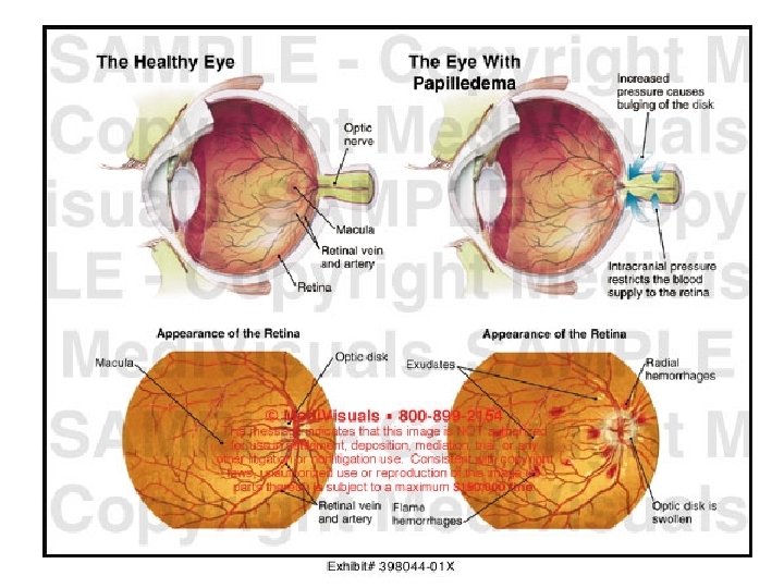
• Media opacity (No RAPD) • – corneal edema – hyphema – vitreous hemorrhage • Retinal disease (RAPD) – retinal detachment – macular disease (e. g. , macular degeneration); • – retinal vascular occlusions • Optic nerve disease (RAPD) – optic neuritis, retrobulbar neuritis, and papillitis • – papilledema • – glaucoma – ischemic optic neuropathy – giant cell arteritis Hypoxia – shock – g-LOC (an aviation related problem) – simply standing up suddenly, especially if sick or otherwise infirm Visual pathway disorder – Visual field defects – homonymous hemianopia – cortical blindness Trauma Functional disorder
• Media opacity (No RAPD) • – corneal edema – hyphema – vitreous hemorrhage • Retinal disease (RAPD) – retinal detachment – macular disease (e. g. , macular degeneration); • – retinal vascular occlusions • Optic nerve disease (RAPD) – optic neuritis, retrobulbar • neuritis, and papillitis • – papilledema – glaucoma – ischemic optic neuropathy – giant cell arteritis Hypoxia – shock – g-LOC (an aviation related problem) – simply standing up suddenly, especially if sick or otherwise infirm Visual pathway disorder – homonymous hemianopia – cortical blindness Trauma Functional disorder
THANK YOU
Sudden Painless Visual Loss • Alarming to both the patient and clinician alike • Requires careful history and examination to determine underlying cause • Visual Obscuration may range from – a symptom of dry eye – or it may herald the onset of irreversible visual loss or stroke
Aims • Focused history to identify the anatomic site of the pathology • Focused examination • Know the causes • Understand the importance of Simple examination techniques such as – visual acuity measurement – confrontational visual field testing – pupil assessment – fundoscopy
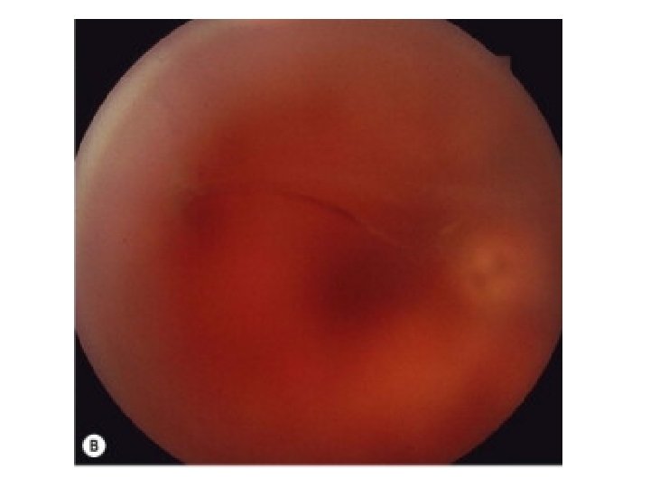
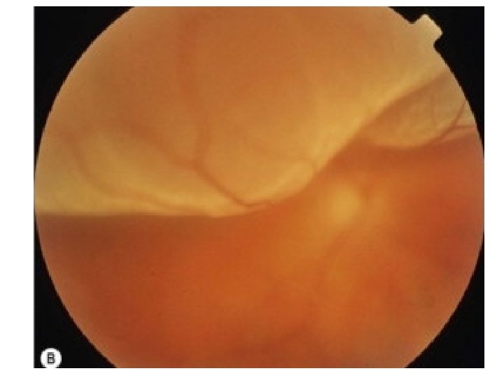
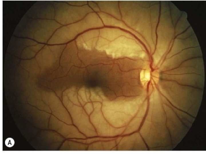
Retinal Detachment • Patients may notice an enlarging shadow in peripheral vision(not just a floater) • Sudden loss of central vision occurs when the macula detaches • Flashes and floaters are common associated symptoms • Ocular history of trauma, surgery and myopia.
Retinal Detachment Acuity normal = macula "on" Acuity poor = macula "off' RAPD Visual field defect corresponding to area of detached retina • Fundus examination is diagnostic (but may be difficult to pick with direct ophthalmoscope) • •
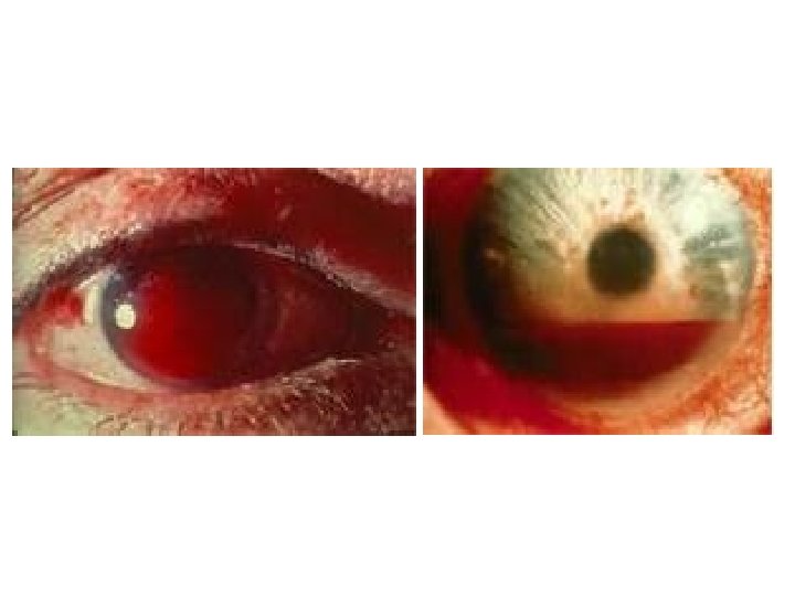
Vitreous Haemorrhage • Causes – Proliferative diabetic retinopathy (new vessels present) – BRVO with new vessels – Retinal tears (tear through a retinal vessel)
Vitreous Haemorrhage • History – Blurred vision with floaters – Diabetes(may be undiagnosed) • Vision: varies with severity of haemorrhage (6/6 to PL) • Pupils: NO RAPD (unless retina detached as well) • Fundus: reduced reflex and difficult to see retinal detail
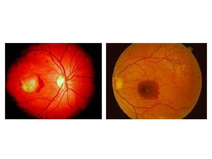
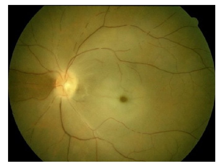
Central Retinal Artery Occlusion • • • Sudden total loss of vision Previous episodes of amaurosis fugax Cardiovascular disease Vision may be NPL Afferent pupil defect Total field loss
Central Retinal Artery Occlusion • Cloudy swelling of infarcted posterior retina • Cherry red spot at fovea (where retina thinnest) • Segmentation of blood columm in retinal veins (slow flow) • Look for emboli in the retinal arteries
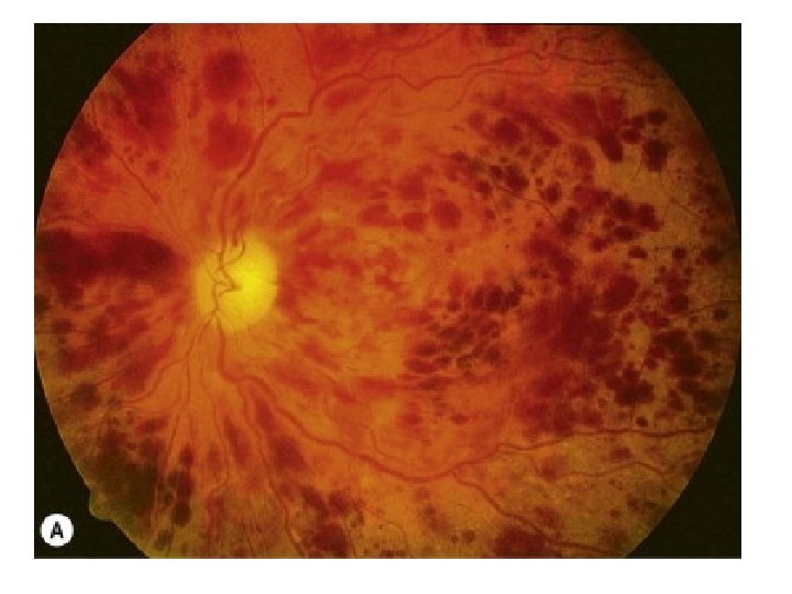
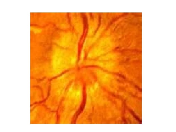
Central retinal vein occlusion (CRVO) • Patients usually>50 yrs • Strong association with hypertension and cardiovascular disease • Sudden painless b. Iur of vision • Vision varies with severity (from 6/6 to hand movements) • Afferent pupil defect if severe CRVO (HM vision)
Central retinal vein occlusion (CRVO) • extensive retinal haemorrhages in all quadrants • retinal venous distension • optic disc swelling
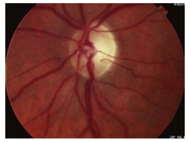
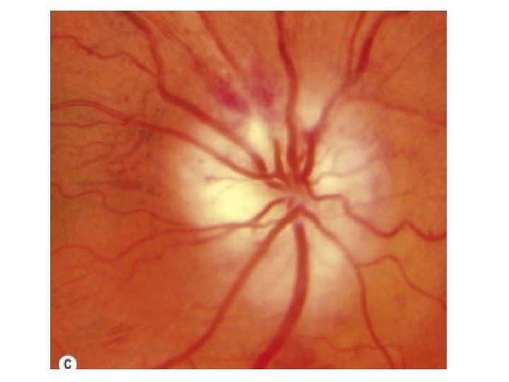
AION • Elderly patients (age >65) • Sudden and severe loss of vision in one eye initially • Systemic symptoms are headaches, scalp tendemess, malaise, jaw claudication • Vision 6/60 or worse RAPD • Extensive visual field loss • Pale swollen optic disc (anterior ischaemic optic neuropathy), rarely CRAO.
AION • • • Aim to prevent loss of the other eye! Urgent ESR (expect >60) Prednisolone l 00 mg stat Urgent referral Temporal artery biopsy will confirm the diagnosis
Optic Neuritis • Typically affects one eye of young women • Vision progressively dims over 48 hours (not truly "sudden") • Ache around eye at onset (worse with eye movement) • Reduced acuity and colour vision • A relative afferent pupil defect (RAPD) is present
Optic Neuritis • Fundus may be normal (retrobulbar neuritis) • Recovery over 6 weeks, more rapid if IV methylprednisolone. • Strong association with MS (MRI Brain will help predict risk)
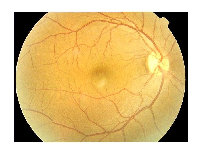
Alcohol Amblyopia Acute onset Resulting in optic atrophy & permanent blindness Etiology • Intake of wood alcohol spirit in cheap adulterated beverages • Inhalation of fumes in industries
Methyl alcohol metabolized very slowly, stay longer period Oxidised in to formic acid & formaldehyde oedema Degenaration of ganglion cell of retina Complete blindness
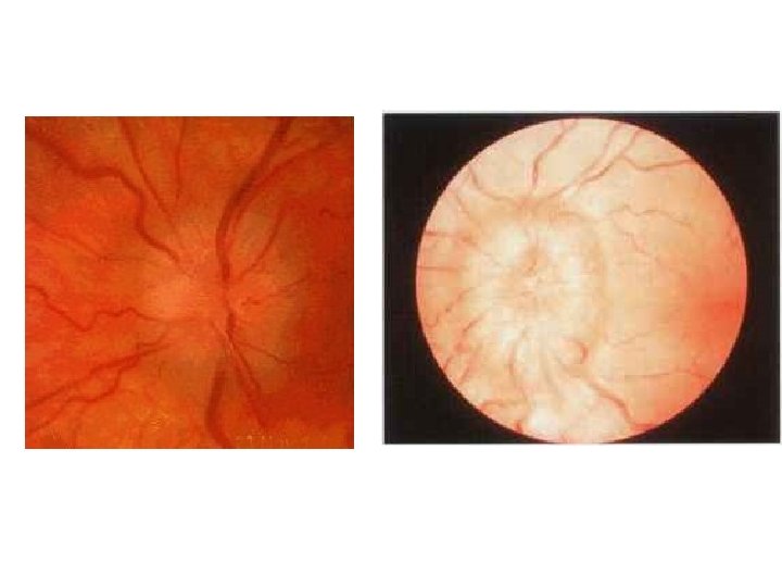
Methyl alcohol amblyopia • Mild disc oedema • Markedly narrowed blood vessels • Bilateral optic atrophy
Eye Pain RAPD Key findings CRAO No Yes Pale retina, cherry-red spot CRVO No +/- Blood and thunder / “Ketchup” fundus RD No +/- May have localized field defect, cloudy veil. But suspect on history AION No Yes Swollen pale disc, signs of temporal arteritis Optic Neuritis Yes Painful EOM, young female pt
Urgency Can wait till AM? ED Treatment CRAO CALL IMMEDIATELY Only if subacute (Many days old) Orbital massage Lower the IOP CRVO CALL when convenient Yes, wait ASA RD CALL IMMEDIATELY At their discretion Bed rest supine Eye shield AION CALL if TA, severe sx, uncertain dx, can wait if not TA Yes, wait Steroids if TA Optic Neuritis CALL Yes, for ophth AVOID oral steroids