
SUBTROCHANTERIC FEMUR FRACTURES Dr Sudhir Rawat Associate Professor
SUBTROCHANTERIC FEMUR FRACTURES Dr. Sudhir Rawat Associate Professor SBKS MIRC
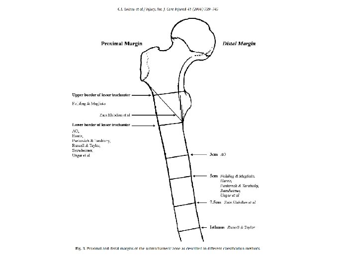
Subtrochanteric fractures involve an area between the lesser trochanter and the isthmus of the diaphysis of the femoral shaft CLASSIFICATION OF SUBTROCHANTERIC FEMORAL FRACTURES C. L. LOIZOU *, I. MCNAMARA, K. AHMED, G. A. PRYOR, M. J. PARKER INJURY, INT. J. CARE INJURED 41 (2010) 739– 745 .
Epidemiology • 10 -34% of all hip fractures Velasco and Comfort • 63% of subtrochanteric fractures in 51 - 70 year olds • Only 24% in those 17 - 50 years old Michelson et al • 14% of hip fractures in patients older than 50 are subtrochanteric fractures
Mechanism of Injury young and healthy individuals- high-energy trauma • elderly population - osteoporotic fall • pathological fractures • periprosthetic fractures

Mechanism Most subtrochanteric fractures are caused by -Direct lateral trauma -Axial loading (with failure in subtrochanteric region).
Low energy High energy • Minimal comminution • Spiral pattern more common • Osteoporotic bone • High comminution involving much of the proximal femur • Significant soft tissue damage
Natural History • High-energy trauma • The other life-threatening conditions must be adequately handled before managing the definitive treatment of the subtrochanteric fracture. • The concept of damage control in acute management
Diagnosis History • Elicit mechanism (low versus high energy) • Preinjury status: preexisting hip pain and constitutional symptoms to rule out pathologic fracture • Ambulatory status/pre-injury functional status/occupation • Chest pain, shortness of breathe, light headedness, dizzyness, palpitations, headache
Physical Exam ABC’s in high energy injuries Pain and apprehension Swollen thigh, bruising Shortened extremity, externally rotated Inability to actively flex/extend hip and pain with passive range of motion • Neurovascular compromise is very rare • Secondary Survey! • • •
Radiographic Exam Full-length femur views in AP and lateral planes • • • Note inner and outer diameter of medullary canal Curvature of femoral shaft Preexisting deformities or implant devices AP Pelvis Neck shaft angle of unaffected side
AP and Lateral of the hip • Determine if fracture line extends into piriformis fossa, medial cortex, etc. • Bone scan/MRI for occult fractures
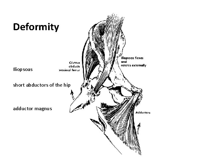
Deformity Iliopsoas short abductors of the hip adductor magnus
Biomechanical Considerations • The fractures occur typically at the junction between trabecular bone and cortical bone where the mechanical stress across the junction is highest in the femur • frequent fracture comminution in this region, due to changes in the structure and the mechanical environment.
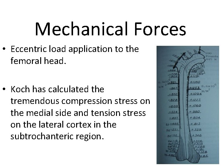
Mechanical Forces • Eccentric load application to the femoral head. • Koch has calculated the tremendous compression stress on the medial side and tension stress on the lateral cortex in the subtrochanteric region.
Biomechanical Considerations • The healing of the fracture is also special, as the highenergy trauma induces more vascular insults to the bone. • Marked displacement of the fracture fragments proximally and distally due to muscular forces.
Classification A review of the literature identified 15 different classification methods for subtrochanteric femoral fractures. There was no agreement between the different classifications regarding the proximal and distal border or for those fractures, which traverse anatomical boundaries Classification of subtrochanteric femoral fractures C. L. Loizou *, I. Mc. Namara, K. Ahmed, G. A. Pryor, M. J. Parker Department of Orthopaedics, Peterborough District Hospital, Thorpe Road, Peterborough, PE 3 6 DA, UK
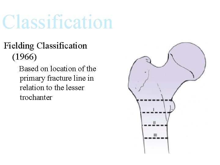
Classification Fielding Classification (1966) Based on location of the primary fracture line in relation to the lesser trochanter

Classification Seinsheimer Classification Based on number of major bone fragments and the location and shape of fracture lines
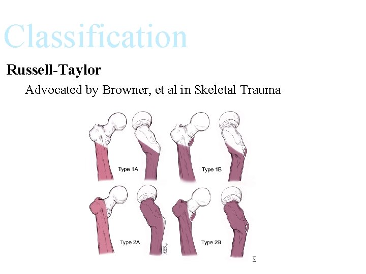
Classification Russell-Taylor Advocated by Browner, et al in Skeletal Trauma

AO classification

So what does Classification matter? Key factors to look for: • Is the medial cortex intact? • Is the greater troch/piriformis fossa involved? • Is it a reverse obliquity type pattern?
Treatment Subtrochanteric fractures result in: • • Shortening of involved limb Varus positioning of the femoral head and neck This creates a functionally weakened abductor group. May cause abductor lurch.
The goals of management are: • • • Restoration of normal length Restoration of normal rotation Correction of femoral head and neck angulation The most common error is reduction in varus Leads to increased stress on implant Diminished effectiveness of the abductors
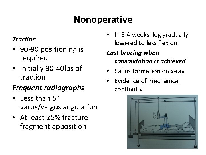
Nonoperative Traction • 90 -90 positioning is required • Initially 30 -40 lbs of traction Frequent radiographs • Less than 5° varus/valgus angulation • At least 25% fracture fragment apposition • In 3 -4 weeks, leg gradually lowered to less flexion Cast bracing when consolidation is achieved • Callus formation on x-ray • Evidence of mechanical continuity
Complications • DVT, PE, pressure sores, pneumonia, pin tract infections • Shortening, varus/valgus deformity, peroneal nerve palsy Velasco and Comfort reported that 11/22 patients had unsatisfactory results with traction
Operative • Avoids complications of prolonged bed rest and hospitalization • Accurate reduction, anatomic reduction, or anatomical alignment • Early surgery reduces blood loss and pain • Early mobilization
Open Anatomical reduction and rigid fixation with interfragmentary compression • Excessive dissection • Bone grafting posteromedially to enhance fracture healing and posteromedial stability • mechanical failure due to the long lever arm and nonunion are relatively common. Banan H, Al-Sabti A, Jimulia T, et al. The treatment of unstable, extracapsular hip fractures with the AO/ASIF proximal femoral nail (PFN)—our first 60 cases. Injury 2002
Closed Anatomical realignment: length and rotation must be corrected • Closed reduction and internal fixation • Load sharing device
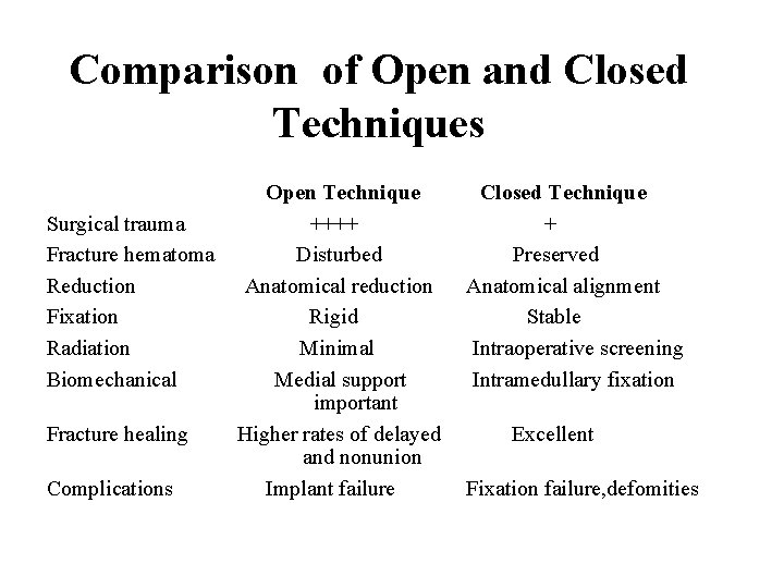
Comparison of Open and Closed Techniques Open Technique Closed Technique Surgical trauma ++++ + Fracture hematoma Disturbed Preserved Reduction Anatomical reduction Anatomical alignment Fixation Rigid Stable Radiation Minimal Intraoperative screening Biomechanical Medial support Intramedullary fixation important Fracture healing Higher rates of delayed Excellent and nonunion Complications Implant failure Fixation failure, defomities
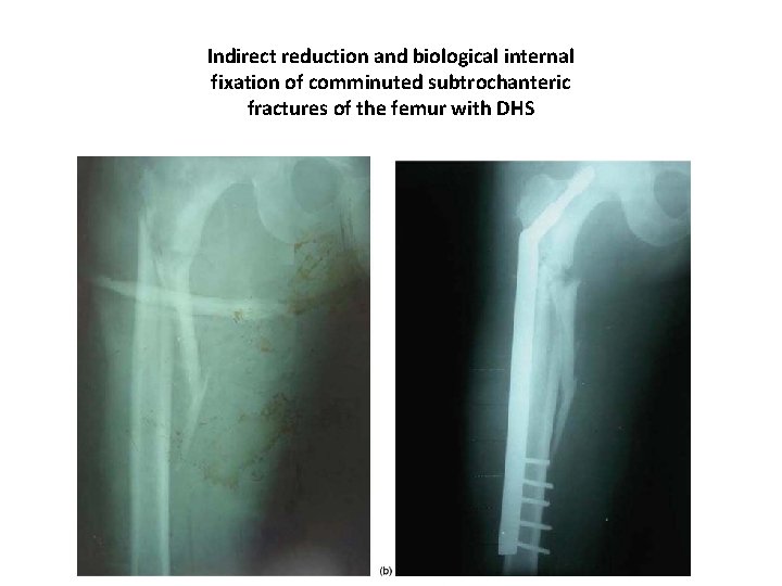
Indirect reduction and biological internal fixation of comminuted subtrochanteric fractures of the femur with DHS
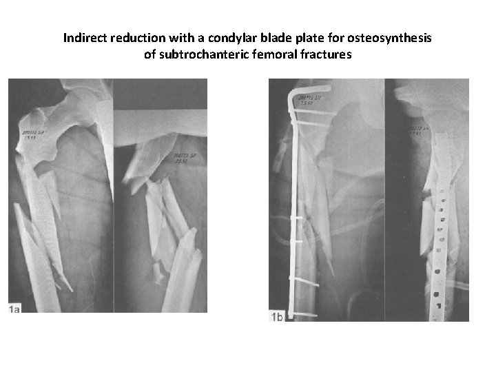
Indirect reduction with a condylar blade plate for osteosynthesis of subtrochanteric femoral fractures
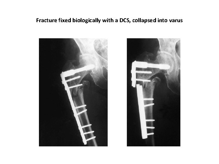
Fracture fixed biologically with a DCS, collapsed into varus
• Using the DCS, the results were good in the young patients with high-energy injuries. However, the implant failure rate is high in elderly patients who suffer low-energy fractures. The DCS should not be used if weight bearing cannot be minimized in this group
• Extramedullary fixation depends on screw fixation of a plate to the lateral cortex, so the mechanical stability of fixation would be problematic. • Moreover, due to the risk of varus collapse and a low union rate, extramedullary fixation is not recommended for complex fractures Sanders R, Regazzoni P. Treatment of subtrochanteric femur fractures using the dynamic condylar screw. J Orthop Trauma 1989
The advantages of intramedullary devices • less extensive exposure • fewer biomechanical stresses with medial movement of the lever arm and earlier weightbearing. Borens O, Wettstein M, Kombot C, et al. Long gamma nail in the treatment of subtrochanteric fractures. Arch Orthop Trauma Surg 2004; 124(7): 443 -447. Cheng MT, Chiu FY, Chuang TY, et al. Treatment of complex subtrochanteric fracture with the long gamma AP locking nail: a prospective evaluation of 64 cases. J Trauma 2005;
Types of Intra Medullary nails Centromedullary (standard ILN) Contained within the medullary canal Condylocephalic nail (Enders) Inserted into the femoral condyle and extended in retrograde fashion into the femoral head/neck Cephalomedullary (Recon Nail) Contained within the medullary canal. Also screw or nail devices that can be inserted into the femoral head and neck

GAMMA NAIL
failures with the Gamma nail • due to a lack of rotational stability of the proximal fragment and/or malpositioning of the neck screw. • The major complications associated with Gamma nail fixation, in fracture of the femoral shaft, have been reported to occur in 2% to 11% of cases Leung KS, So WS, Shen WY, et al. Gamma nails and dynamic hip screws for pertrochanteric fractures: a randomised prospective study in elderly patients. J Bone Joint Surg Br 1992
• PFN overcomes the limitations associated with the Gamma nail. • provides rotational stability to the proximal fragment with two proximal locking hip screws and reduces the incidence of cut-out. 21. Ramakrishnan M, Prasad SS, Parkinson RW, et al. Management of subtrochanteric femoral fractures and metastases using long proximal femoral nail. Injury 2004
• The clinical results of PFNA-long in the treatment of Subtrochanteric femoral fractures have been encouraging, with no mechanical failures of the implant • although technical difficulties are encountered with the insertion of proximal locking screws
• PFNA improves the biomechanical properties and yields better purchase of the helical blade in the femoral head. • The blade has been biomechanically proved to provide increased rotational stability with compaction of cancellous bone around the blade, and better resistance to varus collapse
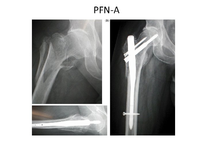
PFN-A
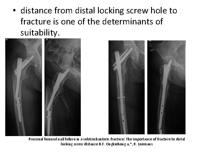
• distance from distal locking screw hole to fracture is one of the determinants of suitability. Proximal femoral nail failure in a subtrochanteric fracture: The importance of fracture to distal locking screw distance B. F. Ongkiehong a, *, R. Leemans
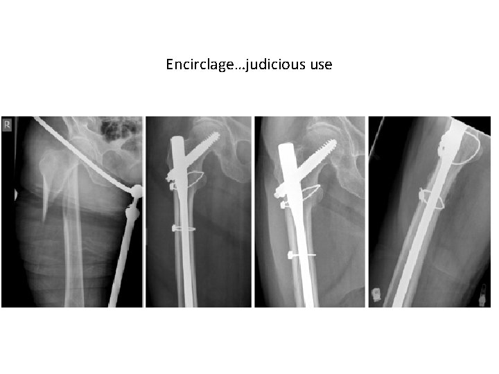
Encirclage…judicious use
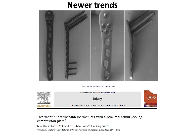
Newer trends
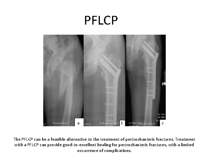
PFLCP The PFLCP can be a feasible alternative to the treatment of pertrochanteric fractures. Treatment with a PFLCP can provide good-to-excellent healing for pertrochanteric fractures, with a limited occurrence of complications.
CASE 1 • 21 YR OLD MALE • RTA • COMUNITED SUBTROCH. EXTENDING TO PY. FOSSA. • NO OTHER INJURY.
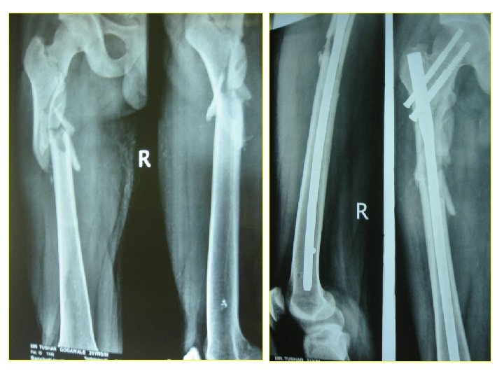
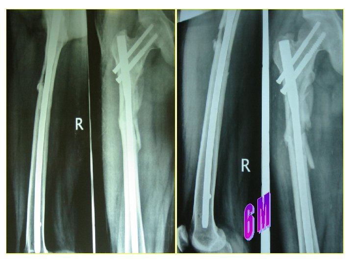
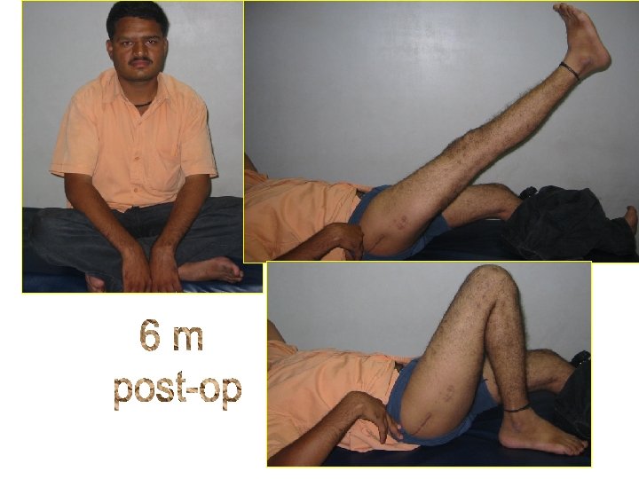
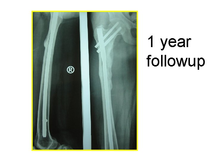
1 year followup
CASE 2 • 40 yr old female lecturer. • Fall from height. • Communited subtroch. • Fragment facing out. • Closed or open reduction?

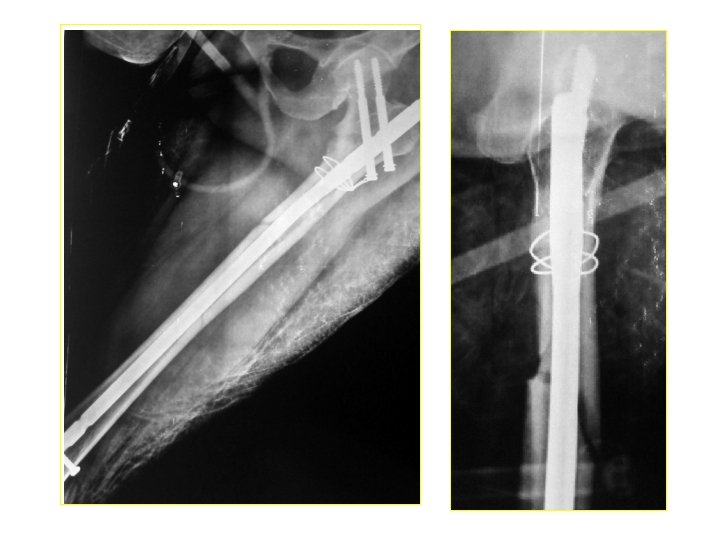
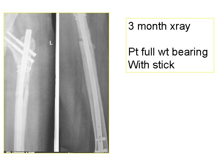
3 month xray Pt full wt bearing With stick
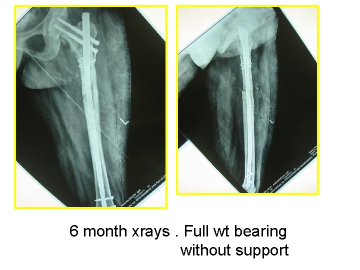
6 month xrays. Full wt bearing without support
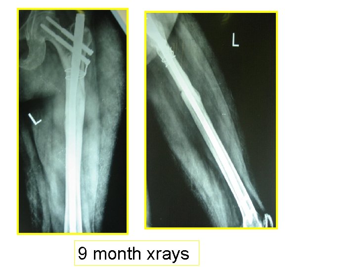
9 month xrays

CASE 3 PRE OP POST OP
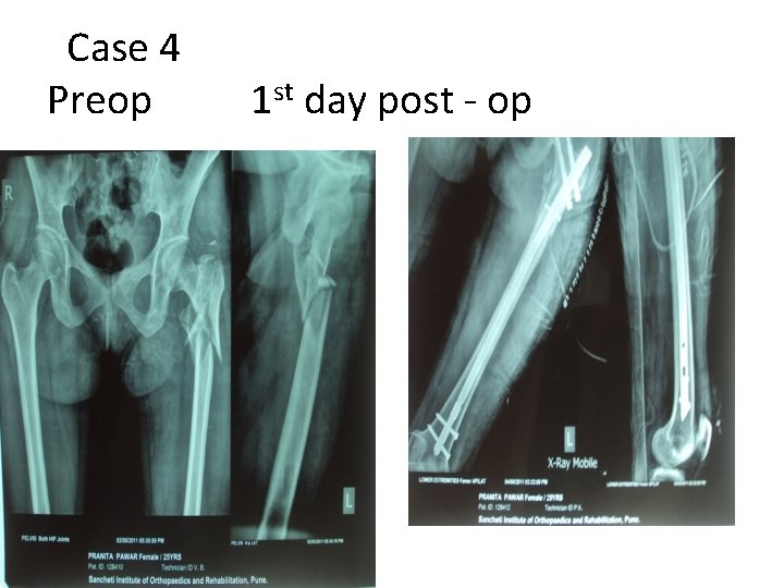
Case 4 Preop 1 st day post - op
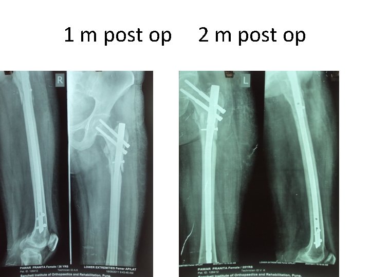
1 m post op 2 m post op
• Reports of atypical femoral fractures in patients on bisphonate therapy has generated both considerable interest and major anxiety as a possible clinical manifestation of the old and unproven “frozen bone” concept, in which treatment-induced suppression of bone turnover is believed to decrease bone strength
Conclusion • Subtrochanteric femur fractures can be challenging to treat in Orthopaedic practice. • Current literature supports intramedullary fixation over surface implants. • Technical problems with instrumentations may be encountered. • Intramedullary implants are biomechanically superior.

THANK YOU……

use of open reduction where necessary to avoid varus malreduction to avoid such complications, particularly as open reduction was not associated with a higher complication rate in this series. The study supports the use of cephalo-medullary nailing for subtrochanteric fractures with a union rate of 95%.
• Extramedullary fixation –disadvantages • extensive surgical dissection, • longer operative time, • more intraoperative blood loss, and • insertion difficulty. • mechanical failure due to the long lever arm and nonunion are relatively common. Banan H, Al-Sabti A, Jimulia T, et al. The treatment of unstable, extracapsular hip fractures with the AO/ASIF proximal femoral nail (PFN)—our first 60 cases. Injury 2002
• The MSP in the biaxial dynamisation mode had a low rate of failure in trochanteric fractures but an unacceptably high rate when used in the biaxial dynamisation mode in subtrochanteric fractures
• distance from distal locking screw hole to fracture is one of the determinants of suitability. Proximal femoral nail failure in a subtrochanteric fracture: The importance of fracture to distal locking screw distance B. F. Ongkiehong a, *, R. Leemans
- Slides: 69