SUBMANDIBULAR REGION Presented by Dr Sushma Tomar Associate

SUBMANDIBULAR REGION Presented by : - Dr. Sushma Tomar Associate Professor Department of

Introduction • The region under cover of body of mandible. • Extends between mylohyoid lines above and hyoid bone below.

v Suprahyoid muscles. Deep Structures v Extrinsic muscles of the tongue. v Salivary glands • Submandibular and Sublingual. v Arteries • Facial and Lingual. v • • • Nerves Lingual. Hypoglossal. Glossopharyngeal. v Ganglion • Submandibular.

Key Muscle • Hyoglossus

• 4 Muscular Planes I Digastric and Stylohyoid. II Mylohyoid. III Geniohyoid, Hyoglossus and Styloglossus. IV Genioglossus and a part of superior constrictor of pharynx.

Submandibular Gland

Introduction • Size- about the size of a walnut.

PARTS 2 • Larger superficial part • Smaller deep part. v Two parts are continuous with each other around the posterior border of the mylohyoid muscle.

Superficial Part Presenting parts 2 Ends— Ø Anterior Ø Posterior 3 Surfaces— Ø Inferior Ø Lateral Ø Medial
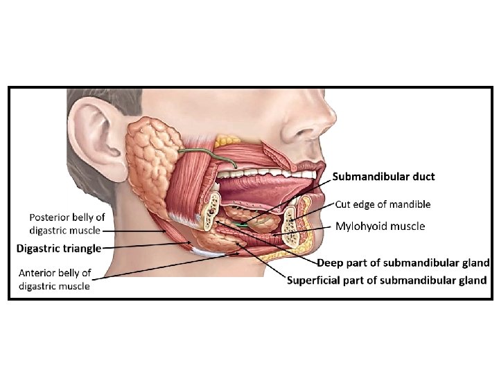

Ends • Anterior end extends upto the anterior belly of digastric muscle. • Posterior end extends upto stylomandi bular ligament which separates the submandibular gland from parotid gland.

Ends contd… • Posterior end presents a groove for the ascending limb of facial artery.
 Relations: v Covered by: • Skin • Superficial fascia •")
Inferior surface (Superficial surface) Relations: v Covered by: • Skin • Superficial fascia • Deep cervical fascia v Crossed by facial vein and cervical branch of facial nerve, under cover of platysma. v Submandibular lymph nodes, beneath the investing layer of deep cervical fascia.

Lateral Surface Relations: • Submandibular fossa of the mandible • Medial pterygoid muscle close to its insertion • Facial artery v Facial artery loops downward and forward between the bone and the gland, and winds around the lower border of body of the mandible at the antero inferior angle of the masseter to reach the face.

Medial Surface • Its relations are divided into three parts: anterior, middle and posterior. Anterior part: • Rests on mylohyoid muscle; separated by the mylohyoid vessels and nerves, and submental branch of the facial artery.

Medial Surface contd… Intermediate part: Rests on: • Hyoglossus muscle • Lingual nerve • Submandibular ganglion • Hypoglossal nerve

Medial Surface contd… Posterior part : related to • Styloglossus muscle • Stylohyoid ligament • Glosso pharyngeal nerve • Wall of pharynx

Deep part • Extends forward in the interval between the mylohyoid and hyoglossus upto the posterior end of sublingual salivary gland.

Relations of Deep part Laterally Mylohyoid Medially Hyoglossus Above Lingual nerve and submandibular ganglion Below Hypoglossal nerve accompanied by a pair of veins.
 • ~ 5 cm long. • Emerges at the anterior")
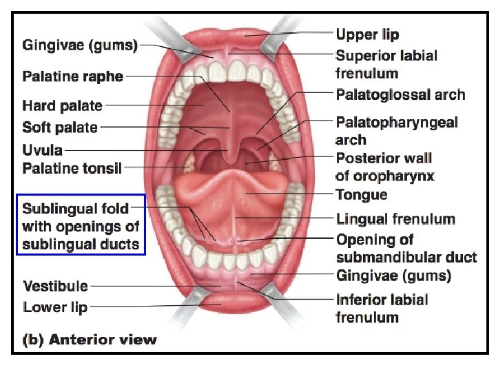
Submandibular duct (Wharton’s duct) • ~ 5 cm long. • Emerges at the anterior end of deep part. • Runs forward on the hyoglossus between lingual and hypoglossal nerves. • Crossed by lingual nerve, near the anterior border of hyoglossus. • Continues forward between sublingual gland genioglossus muscle. • Opens into the oral cavity on the summit of sublingual papilla at the side of frenulum of tongue.

Nerve Supply

Clinical Correlates v Ø • • v Formation of calculi. It is more common in submandibular gland its duct for two reasons: Secretion is more viscid. Duct takes a tortuous and upward course (against gravity). Skin incision for removal of calculus or tumour from submandibular gland 4 cm below the angle of mandible. Bimanual Palpation of Submandibular gland-

• Sublingual Gland It is the smallest of the three large salivary glands. • Mostly mucous in nature. SHAPE-almond shaped. WEIGHT 3 to 4 gm LOCATION • In the floor of the mouth between the mucous membrane and mylohyoid muscle. • Lodges in the sublingual fossa of the mandible.

Ducts of Sublingual Gland • The gland possesses about 8 to 20 ducts: • Most of the ducts (ducts of Rivinus) open separately in the floor of the mouth on the summit of the sublingual fold. • Some ducts from the anterior part of the gland unite to form the sublingual duct (duct of Bartholin). • Duct of Bartholin opens into the submandibular duct.

Sublingual Gland contd… Arterial supply: • The gland is supplied by the sublingual and submental arteries. Lymphatic drainage: • The lymphatics drain into submental and submandibular nodes.
 • It is a para sympathetic ganglion. • A relay")
Submandibular Ganglion (Langley’s Ganglion) • It is a para sympathetic ganglion. • A relay station for secretomotor fibers of submandibular and sublingual salivary glands. CONNECTIONSTopographically with the lingual nerve. Functionally with the facial nerve (through its chorda tympani branch).

Submandibular Ganglion contd… LOCATIONv On outer surface of hyoglossus muscle. v It is suspended from the lingual nerve by two roots: • Posterior. • Anterior.

Submandibular Ganglion contd… ROOTS- 3 • Parasympathetic. • Sensory. Parasympathetic or motor root: • Derived from lingual nerve. • The preganglionic fibres arise from superior salivatory nucleus in the pons. • Preganglionic fibres pass successively through the facial, chorda tympani and lingual nerves and reach the submandibular ganglion. • Preganglionic fibres relay in submandibular ganglion.

Parasympathetic or motor root contd… • Postganglionic fibres directly supply the submandibular gland by five or more branches. • Some postganglionic fibers join the lingual nerve through the anterior root and supply the sublingual gland.

Submandibular Ganglion contd… Sympathetic Root: • It is derived from a plexus around the facial artery which is formed by post ganglionic sympathetic fibres. • Post ganglionic sympathetic fibres arise from superior cervical sympathetic ganglion of the sympathetic trunk. • These fibres pass through the submandibular ganglion without relay. • These fibres supply the blood vessels of submandibular and sublingual salivary glands.

- Slides: 32