SUBMANDIBULAR GLAND Vanishri S Nayak Mixed gland with

SUBMANDIBULAR GLAND -Vanishri S. Nayak

Mixed gland with predominantly serous type 10 -20 gm Situated in Digastric triangle J-shaped Superficial & deep part-Mylohyoid

Submandibular gland: § Ø Ø Location: occupies most of the posterior part of the submandibular triangle. Location of parts: Superficial: larger and lies on the mylohyoid muscle directly underneath the superficial layer of cervical fascia. Deep: folds around the posterior border of the mylohyoid muscle to lie deep in the sublingual space between the mylohyoid muscle and the hyoglossus and genioglossus muscles.

Superficial part 2 ends & 3 surfaces Anterior end-Anterior belly of Digastric Posterior end-Stylomandibular ligament

Submandibular gland: Relations v v v Inferior Surface: Platysma Cervical branch of facial nerve Deep fascia Facial vein Submandibular lymph nodes

Submandibular gland: Relations Lateral Surface: v Submandibular fossa of mandible v Insertion of medial pterygoid v Facial artery

Submandibular gland: Relations v v v v § v v v Medial Surface: Anterior part Mylohyoid, nerve & vessels Medial Surface: Middle part Hyoglossus Styloglossus Lingual nerve Submandibular ganglion Hypoglossal nerve Medial Surface: Posterior part Styloglossus Stylohyoid ligament Glossopharyngeal nerve Wall of pharynx Stylohyoid Posterior belly of digastric

Deep part Lateral-Mylohyoid Medial-Hyoglossus Above-lingual n. & submandibular ganglion Below-Hypoglossal n.

submandibular duct Length: 5 cm Beginning: from the deep part of this gland Course & Termination: passes forward and medialward to open in the sublingual caruncle at the side of the lingual frenulum.

Submandibular gland Arterial Supply: facial artery Venous drainage: facial vein Lymphatic drainage: submandibular lymph nodes.

Submandibular gland: Nerve Supply Innervation of submandibular gland: Sympathetic nerves from the superior cervical sympathetic ganglion reach the submandibular gland via the facial plexus along the facial artery. Parasympathetic innervation comes from the chorda tympani branch of the facial nerve (VII). The chorda tympani gives rise to the submandibular ganglion, and the gland is innervated by postganglionic parasympathetic fibers from this ganglion.
")
figure 2 - stone formation in submandibular region (stone circled in red)

Submandibular ganglion: Situation On upper part of hyoglossus muscle

Submandibular ganglion: Situation On upper part of hyoglossus muscle above deep part of submandibular gland

Submandibular ganglion: Situation n On upper part of hyoglossus muscle above deep part of submandibular gland between lingual nerve & submandibular duct. Suspended from lingual nerve by 2 short branches

n. Component parts: TOPOGRAPHICALLY: LINGUAL NERVE FUNCTIONALLY: FACIAL NERVE PARASYMPATHETIC Superior salivatory nucleus–> Facial nerve –> Chorda tympani nerve –> lingual nerve SYMPATHETIC Superior cervical Sympathetic ganglion –> Plexus around facial & lingual arteries SENSORY Through lingual nerve Relay in ganglion No relay in ganglion
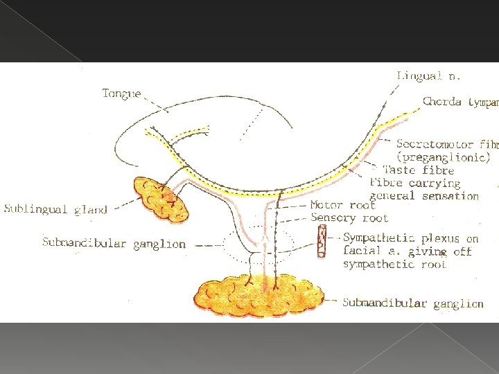

Submandibular ganglion : Branches & Supply Parasympathetic fibres: 1. To submandibular gland: through 5 or 6 branches from ganglion 2. To sublingual & anterior lingual gland: reenter lingual nerve through anterior root & travel distal part of lingual nerve Sympathetic fibres: vasomotor fibres to submandibular & sublingual nerve

THANK U
- Slides: 19