Submandibular gland Surgical Anatomy Tumors Surgery Dr Amit

Submandibular gland: Surgical Anatomy Tumors Surgery Dr Amit Gupta Associate Professor Dept Of Surgery

Surgical Anatomy • Composed of a superficial part and a deep part. • Superficial part: lies in the submandibular triangle between two bellies of digastric muscle • Deep part: lies below and lateral to the line of the submandibular duct, in the floor of the mouth, above and deep to the mylohyoid.
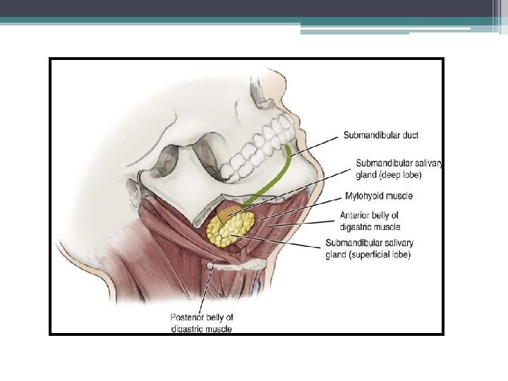

• Borders – Lateral – proximal half of the mandible. – Posterior – anterior to but near the low anterior margin of the parotid gland. – Inferior – approaches the level of the hyoid bone. – Majority of gland lies over the external surface of the mylohyoid muscle. – Lateral to and abuts the lingual and hypoglossal nerve and is medial to the marginal mandibular and cervical branch of the facial nerve. • Drains through Wharton’s duct in anterior floor of the mouth • Lymphatic Drainage Level IB

Submandibular ganglion is attached to the nerve at this point and is also within the sheath of the gland Submandibular duct lies beneath the lingual nerve as it emerges from the upper pole of the gland. Lingual nerve lies first between the duct and the deep part of the gland, then crosses medially beneath the duct to ascend on the hyoglossus to supply the tongue. In some subjects a posterior sublingual gland drains into the submandibular duct Bartholin's major sublingual gland may drain via A single duct into the submandibular duct or by a separate open

Salivary Gland Tumors

Epidemiology • Relatively uncommon – 2% of head and neck neoplasms • Diverse histopathology Distribution • Parotid: 80% overall; 80% benign; 25% malignant • • Submandibular: 15% overall; 50% benign; 43%malignant • Sublingual/Minor: 5% overall; 40% benign; 65%malignant

• Preponderance of benign tumors in women • Malignant tumors exhibit an equal sex distribution • Patients with benign tumors are younger (mean age: 46 years) compared with those with malignant tumors (mean age: 54 years) • Trend to an older age for submandibular and minor salivary gland locations

Risk factors • Nutrition: low intake of vitamins • Smoking • Irradiation • Ultraviolet exposure (controversial)

Molecular targeting Recent interest in molecular targeting of salivary gland malignancies Molecular markers • EGFR ▫ Overexpression in all histologic subtypes • HER 2 ▫ Ductal carcinoma • C-kit ▫ Adenoid cystic Although there is overexpression of these molecular markers, the rates of true genetic mutation is much lower

Clinical Presentation • Painless, rapidly enlarging mass • Indolent period >10 years • Pain is more frequently associated with malignant disease Clinical features suggesting a malignant tumour: • • • Rapid growth rate Pain Facial nerve palsy Childhood occurrence Skin involvement Cervical adenopathy

WHO classification of salivary gland tumors

Morphology • Pleomorphic adenomas: originate from the intercalated duct cells and myoepithelial cells • Oncocytic tumors: originate from the striated duct cells • Acinic cell tumors: originate from the acinar cells • Mucoepidermoid & squamous cell carcinomas: develop in the excretory duct cells.
 scans • Magnetic resonance")
Diagnostic Work-Up • History /Physical Examination • Computed tomography (CT) scans • Magnetic resonance imaging (MRI) (T 1 -weighted images are excellent to assess the margins, deep extent, and patterns of infiltration because the (fatty) background of the gland is hyperintensive) Perineural invasion may be evaluated with both CT and MRI • PET CT Scan Potential role for staging and management in salivary gland carcinomas • Fine-needle aspiration cytology

Axial CT scan showing left submandibular gland lesion

MRI scan showing right submandibular gland lesion

PET CT scan showing right submandibular gland lesion
")
Treatment • Surgery • Radiotherapy • Chemotherapy (not efficacious)

Surgery • Mainstay of treatment • Submandibular Gland excision with level Ib dissection recommended • Radical resection is indicated with tumors that invade the mandible, tongue, or floor of mouth

Submandibular Triangle Dissection • Scalpel proceeds through the skin in a curvilinear incision from just off the midline anteriorly to just below the earlobe posteriorly • Flap is elevated deep to platysma muscle • Surgeon must be aware of the course of the marginal branch of the facial nerve. • It is important to identify this nerve and mobilize it anteriorly and posteriorly.

Incision site for submandibular triangle dissection Incision for combined neck dissection Skin flap raised superiorly with platysma muscle left in place

Skin flap raised superiorly with platysma muscle Marginal branch of the facial nerve and its relationship with the facial vessels

Detail of the submandibular triangle with the submaxillary gland resected

Management of Neck

Management of the N 0 neck • Tumors >4 cm • High grade histology Management of the N+ neck • Ipsilateral MRND for clinically or radiographically positive nodes • Incidence of multilevel node involvement

Postoperative Radiotherapy Indications • T 3 -4 tumors • Close or positive margin • Incomplete resection • Bone involvement • Perineural invasion • Positive nodes • Recurrent cancer

Prognosis • Survival of patients with submandibular cancers is inferior to that of parotid cancers • Perineural invasion: independent prognostic factor for distant metastases or DFS • Impairment of function of the facial nerve : poor DFS • Acinic cell and (low-grade)mucoepidermoid cancer: best prognosis • Undifferentiated & squamous cell cancer: worst prognosis
- Slides: 27