Subclavian artery Subclavian artery B Brachiocephalic trunk C


Subclavian artery

Subclavian artery B= Brachiocephalic trunk C= Common carotid A S= Subclavian A Outer border of the first rib S Right C B C Arch S Begin End parts Axillary artery
Left subclavian artery: from the arch")
SUBCLAVIAN ARTERY Beginning: different on both sides: 1)Left subclavian artery: from the arch of aorta. 2) Right subclavian artery from the brachiocephalic artery. End; at the outer border of the 1 st rib and continues as axillary artery. Course: - It is divided by the scalenus anterior muscle into 3 parts: * 1 st part: medial to scalenous anterior. * 2 nd part: behind scalenous anterior. * 3 rd part: lateral to scalenous anterior.
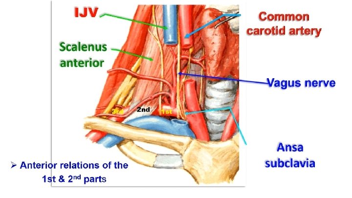

Ø Anterior relations of the first part • 3 superficial structures: skin, superficiăl fascia (containing platysma muscle) and deep fascia. • 3 muscles: (a) Sternomastoid. (b) Sternohyoid. (c) Sternothyroid. • Veins (Internal jugular vein) • Artery (Common carotid artery) • Nerves: (a) vagus nerve. (b) Ansa subclavia (is a branch from the middle cervical sympathetic ganglia forms a loop around the 1 st part of subclavian artery to join the inferior cervical sympathetic Ganglion). • Left phrenic nerve and Thoracic duct anterior to the left artery Ø Anterior relations of the second part • Scalenus anterior muscle and subclavian vein
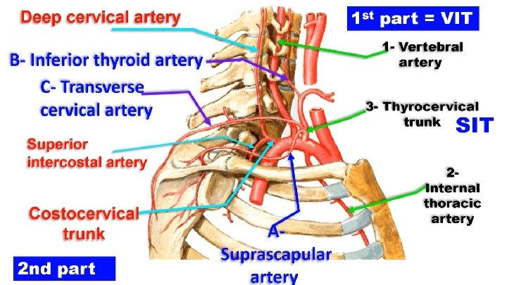
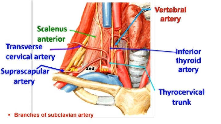

• Branches of subclavian artery 1 - 1 st part: gives 3 branches (VIT) (1) Vertebral artery. (2) Internal thoracic (mammary) artery. (3) Thyrocervical trunk (SIT = Suprascapular - Inferior thyroid – Transverse cervical) 2 -2 nd part: costocervical trunk divided into a) Superior intercostal artery; to the 1 st and 2 nd posterior Intercostals arteries. b) Deep cervical artery: ascends to back of neck to anastomoses with descending cervical Branch of occipital artery (site of anastomosis between carotid and subclavian system). 3 - 3 rd part: no branches, but occasionally gives dorsal scapular artery if deep branch of transverse cervical artery absent

Vertebral artery
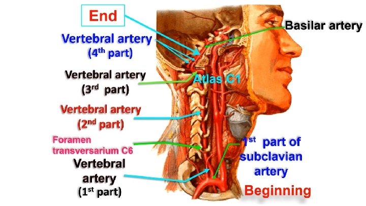
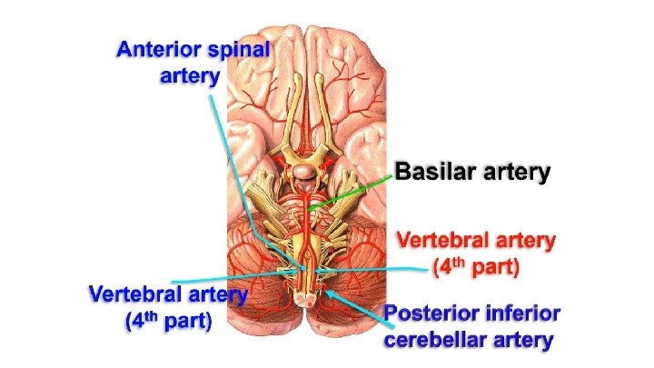

Vertebral artery Course and relations: divided into 4 parts: 1 st part: ascends along the medial border of scalenus anterior to the foramen transversarium of C 6. 2 nd part: ascends in the foramina transversaria of the upper 6 cervical vertebrae. 3 rd part: in the suboccipital triangle. 4 th part: enter the cranial cavity through foramen magnum then united together to form basilar artery in the basilar sulcus on the anterior surface of the pons. Branches -The 2 nd part: radicular branches to the spinal cord. -The 4 th part: (1) Anterior spinal artery. (2) Posterior spinal artery. (3) Posterior inferior cerebellar artery. (4) Medullary branches to the medulla oblongata.

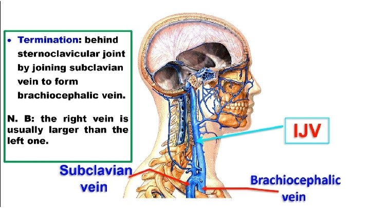
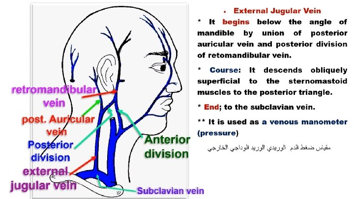
Ø Subclavian vein - It is the continuation of the axillary vein at the outer border of the first rib - It passes superficial to the scalenus anterior muscle - It joins with the internal jugular vein to form brachiocephalic vein -Tributaries A- External jugular vein B- Dorsal scapular vein C- Thoracic duct opens at the junction of left subclavian vein with left internal jugular vein

Carotid artery

Upper border of Thyroid cartilage Right Common carotid brachiocephalic trunk C 3 C 4 End Left Common carotid artery Arch of aorta Begin
 Internal carotid")
At the bifurication of common carotid artery Carotid sinus Baroreceptor (pressure receptor) Internal carotid artery Carotid body Chemoreceptors (innervated by IX & X)

External carotid artery

Superficial temporal artery End Behind neck of mandible inside the parotid gland Maxillary artery External carotid artery
 Beginning: one of the 2 terminal")
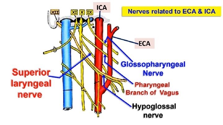
• EXTERNAL CAROTID ARTERY (E. C. A) Beginning: one of the 2 terminal branches of the common carotid artery at the upper border of thyroid cartilage (disc between C 3 & C 4) Termination: it ends inside parotid gland by dividing into maxillary and superficial temporal arteries Nerves related to ECA & ICA • One nerve passes superficial to ICA & ECA, Hypoglossal nerve. • One nerve passes deep to ICA & ECA, Superior laryngeal nerve • Three pharyngeal structures between ICA and ECA - Glossopharyngeal nerve. - Stylopharyngeus muscle. - Pharyngeal branch of vagus nerve.
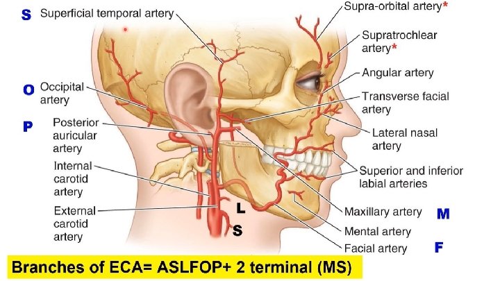

Superficial temporal artery Posterior auricular artery Occipital artery A 1 artery Ascending from medial pharyngeal artery External carotid artery OP 2 arteries from behind 2 terminal branches Maxillary artery MS Facial artery Lingual artery Superior thyroid artery 3 arteries from SLF front

internal carotid artery
 • Beginning: one of the 2")
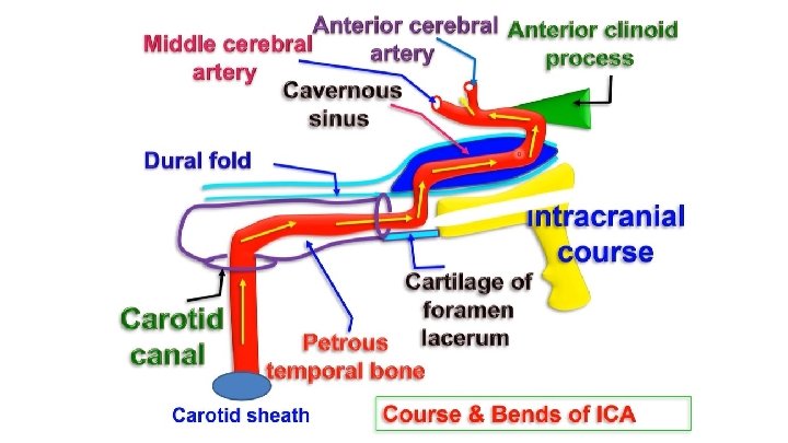
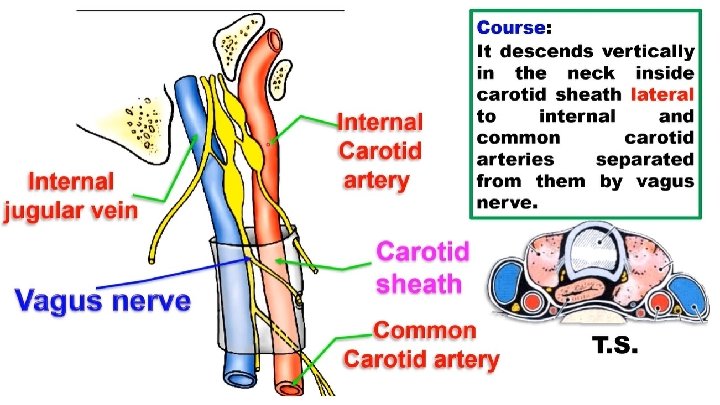
• INTERNAL CAROTID ARTERY (I. C. A) • Beginning: one of the 2 terminal branches of C. C. A at the upper border of thyroid cartilage (disc between C 3 & C 4) • Termination: below the base of the brain in the cranial cavity by dividing into anterior and middle cerebral arteries. • Course: its course is dividing into 4 parts: (1) Cervical part: ascends inside the carotid sheath. (2) Intrapetrous part: in carotid canal inside petrous part of temporal bone. (3) Intracavernous part: inside the cavernous sinus. (4) Intracranial part: terminal part of the artery - The ICA has bends that damp down the pulsation and give more a regular stream of blood for brain

• Branches of ICA A- Cervical part: no branches in the neck. B- Branches in the carotid canal: • (1) Caroticotympanic artery to the middle ear cavity (vertigo). • (2) Artery to the pterygoid canal. C- Branches within the cavernous sinus: • (1) Cavernous branches. • (2) Superior and inferior hypophyseal arteries to pituitary gland. D- Branches Outside the cavernous sinus: (1) Ophthalmic artery. (2) Anterior choroidal artery. (3) Posterior communicating artery. (4) Anterior cerebral artery. (5) Middle cerebral artery.

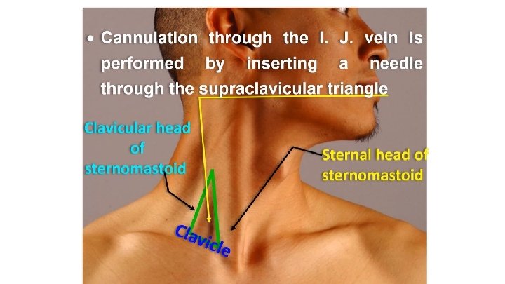
Internal Jugular vein
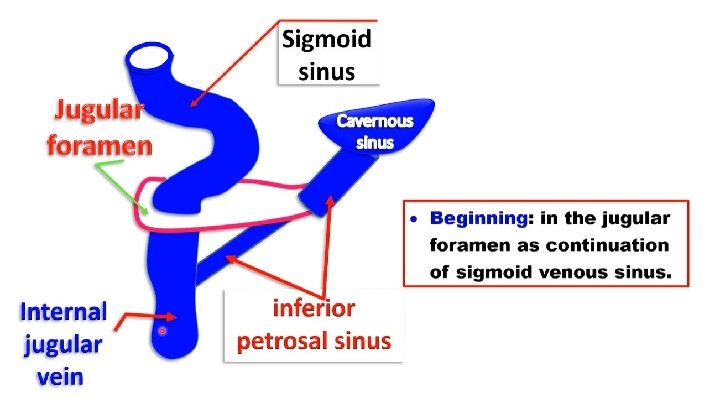

Common facial vein Superior thyroid vein Middle thyroid vein Sigmoid sinus Inferior petrosal sinus Lingual vein Tributaries IJV (SLFO-P-SIM)

Th ank Qu you est ion s I/Azzam - 2004
- Slides: 35