STURMS CONOID REDUCED EYE OPTICAL ABERRATIONS Dr Vidya

STURM’S CONOID, REDUCED EYE, OPTICAL ABERRATIONS Dr. Vidya Hegde

Specific learning objectives At the end of the class, students should be able to understand ü Sturm”s conoid and its importance ü Reduced eye and its application ü Also about optical aberrations in the eye
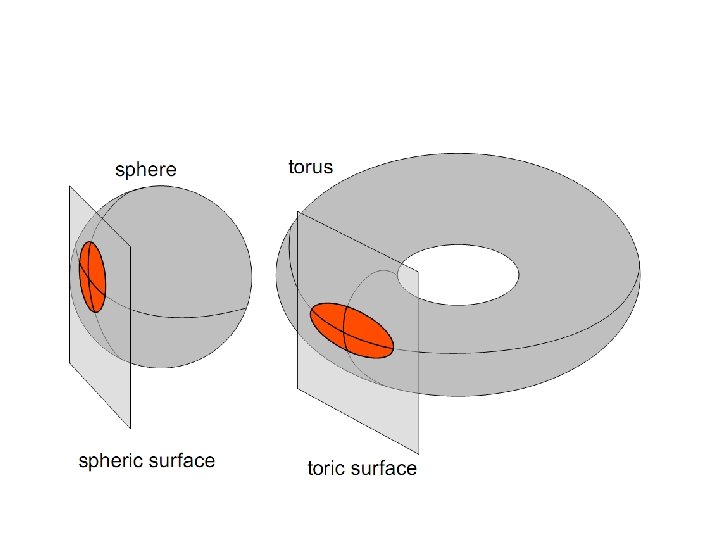

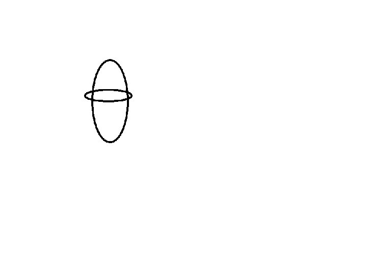
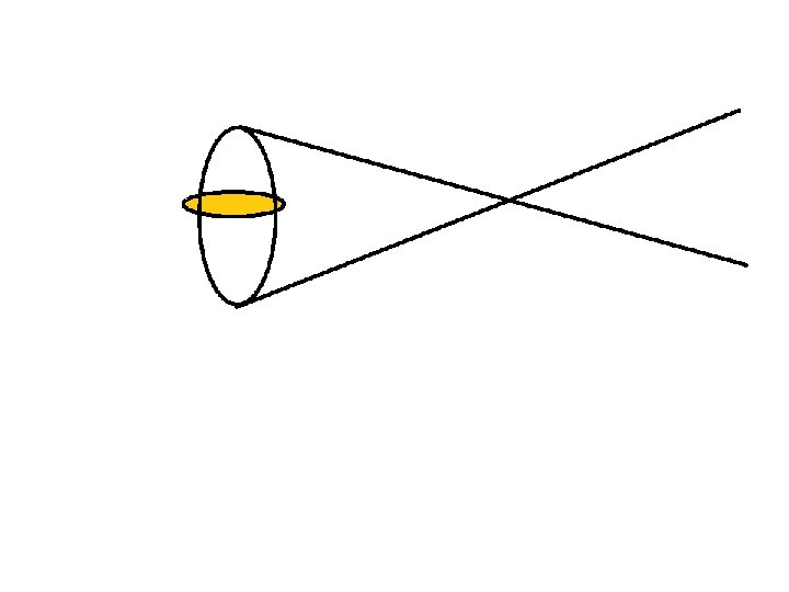
STURM’S CONOID • Configuration of rays refracted through a toric surface – STURM’S CONOID • For a toric surface- light focuses in 2 lines at right angles to each other. • The interval between 2 line foci – STURM INTERVAL


A B C D E Focal Interval F G
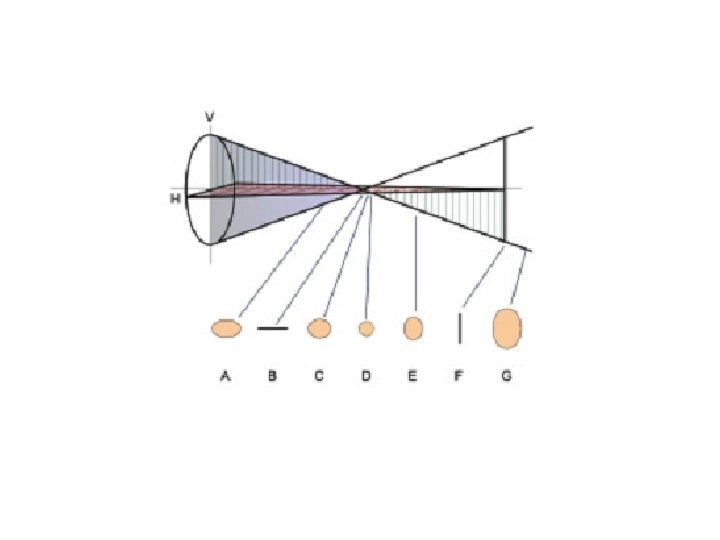

STURM’S CONOID • An image that falls on the retina can be thought of as being made up of many dots. • If an ametropic eye , is not optically corrected, then the image will consist of many blur circles instead of sharp dots. The more out of focus the image is, the larger the circles are. If an astigmatic eye is not optically corrected, the blur circles will be distorted into ellipses.

SIGNIFICANCE OF STURM’S CONOID • Forms the basis for classification of astigmatism. • The location of the circle of least confusion is equal to the spherical equivalent of the prescription.

SCHEMATIC EYE • • • Optics of eye- Homocentric lens system. Six cardinal points – Two principal foci (F 1 and F 2) Two principal points (P 1 and P 2) Two nodal points (N 1 and N 2) • Total dioptric power – 58. 64 D

REDUCED EYE • By Listing and Donder • • Single Principal point – 1. 5 mm Single Nodal point – 7. 2 mm Anterior Focal point – 15. 7 mm Posterior Focal point – 24. 13 mm Anterior Focal length – 17. 2 mm Posterior Focal length – 22. 63 mm Total Diopteric Power – 60 D

SIGNIFICANCE OF REDUCED EYE • • Designing the instuments Making calculations Localizing a foreign body Derive formulae for intraocular lens power

OPTICAL ABERRATIONS • Imperfections or lapses in the optical system • Though they normally exist to a small degree, functionally they are immaterial

NATURAL MECHANISMS • Cutting off peripheral rays by iris • High refractive index of the core of nucleus of the lens than that of the peripheral cortex • Low sensitivity of the peripheral retina • Stiles Crawford effect

DIFFRACTION OF LIGHT Bending of light caused by • Edge of an aperture or • Rim of a lens Even a perfect lens will not focus light to a point due to diffraction

DIFFRACTION OF LIGHT • The actual pattern of a diffracted image point produced by the pupil is a series of concentric dark and bright rings. • At its centre is a bright spot known as the Airy Disc. • Diffraction blur increases with the small size of the pupil

SPHERICAL ABERRATIONS • Spherical lens refracts peripheral rays more strongly than paraxial rays • As a result the incoming rays do not come to a point focus

SPHERICAL ABERRATIONS Factors contributing to diminution of spherical aberrations in human eye are 1. Lens is flatter at the periphery than the centre. 2. Central portion of the lens has greater density and curvature than the periphery. 3. Iris blocks the peripheral rays and allows only paraxial rays

CHROMATIC ABERRATIONS Occurs due to to the fact that the RI of any transparent medium varies with the wavelength of the incident light. emmetropic eyehypermetropic for red and myopic for blue & green

CHROMATIC ABERRATIONS • emmetropic eyehypermetropic for red and myopic for blue & green. • This forms the basis of the duochrome test in subjective refraction.

CHROMATIC ABERRATIONS Chromatic aberrations in the eye is minimised by • Yellow rays form most sharply defined images on the retina. • Fovea lacks blue cones. • Narrow spectral sensitivity band of long & medium wavelength cones.
- Slides: 23