Study of muscles blood supply of forelimb in

Study of muscles & blood supply of forelimb in ox

Muscles n Muscles are structures formed by the bundles of muscles cell in the form of fibers and possess the property of contraction on stimulation. n n n They are derived from mesenchymal cells. The three type muscles 1) skeletal muscles 2) smooth muscles 3) cardiac muscles

Muscles of fore limb These are Categaries into : Muscles of shoulder Girdle Muscles of shoulder Muscles of arm Muscles of forearm and manus

Muscles of shoulder Girdle The forelimb is attached to the body by a set of muscles called muscles of shoulder girdle Ø Trapezius Ø Rhomboideus Ø Brachiocephalicus

Lattismus dorsi Serratus ventralis Omo transversarious Pectoral

Trapezius It is a thin triangular muscle at the upper most part of the fore limb. Its dorsal border extends from the atlas to a considerable part of thoracic region. ü ü ü Origin – Supraspinous processes from the level of 12 th thoracic vertebra to atlas. Insertion – Scapular spine and fascia of shoulder and arm. Blood supply-: Deep cervical branch of costo cervical artery and intercostal artery.

Trapezius

Rhomboideus This muscle is roughly triangular and thick. Laterally it is covered by trapezius. It helps to move the shoulder forward and upward. Origin - Summit of 1 st to 8 th thoracic spine and ligamentum nuchae. v Insertion – Medial surface of the scapular cartilage. Blood supply - Deep cervical branch and dorsal branch of costo cervical artery.

Rhomboides thoracis Rhomboides cervicis

Bracheocephalicus It is a long flat muscle placed diagonally at the side of the neck and extends from the arm to the cephalon. It act to extend the head and neck. It also extends the shoulder when the head and neck are fixed. It is divide into a dorsal part and a ventral part. Origin –Dorsal part originates from occipital bone and ligamentum nuchae and the ventral part originate from temporal bone, wings of atlas and mandible. Insertion – Both dorsal and ventral part unite together and insert to the cranial border of musculospiral groove of humerus. BLOOD SUPPLY : Anterior circumflex , inferior cervical, vertebral and carotid arteries.

Br ac hi oc ep ha lic us
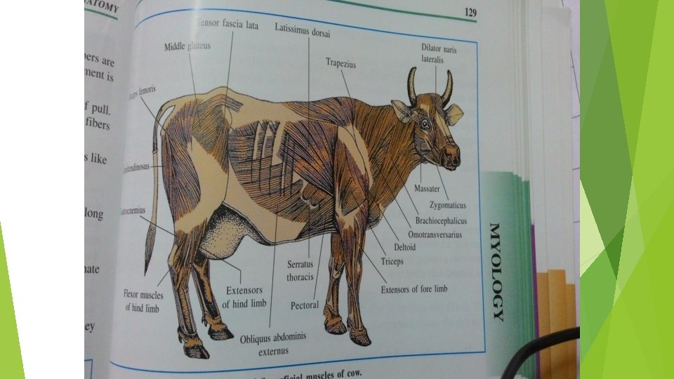

Latissimus dorsi It is a very wide and thin muscle spread at the dorso-lateral aspect of thorax. The muscles is aponeurotic at the origin and becomes thicker and narrower towards the arm. It helps in flexing the shoulder. ü Origin – lumbar and dorsal spine. ü Insertion – At the termination it becomes blended with tendons of teres major, and inserted to the teres tuberosity of humerus. ü BLOOD SUPPLY : Thoracodorsal branch of sub scapular artery, intercostal and lumbar arteries.

Latissimus dorsi

Serratus ventralis It is a serrated fan shaped fleshy muscle, spread on the lateral side of the neck and thorax. The part which covers the neck is known as serratus cervicis and the portion which covers the thorax is known as serratus thosacis. Origin - C 4 to 10 th rib. Insertion - medial scapula and scapular cartilage.

Serratus cervicis Placed partly on the neck and partly on the thoracic wall. It extends from 2 nd cervical vertebra to the 5 th rib. It helps to extend the neck and pull the cervical angle of the scapula cranially. Origin – Transverse processes of 2 nd to 7 th cervical vertebrae and lateral surface of 1 st to 5 th ribs. Insertion – The rough triangular area at the cranio-dorsal part of the medial surface of scapula. BLOOD SUPPLY : Dorsal branch of costocervical artery, intercostal and deep cervical arteries.

Serratus thoracis Thin flat and ventrally presents six prominent digitations. It covers the posterior part of the serratus cervicis. Muscles of both sides together act to rise thorax and individually pull the scapula dorso caudally. In resting condition it acts as a muscles of inspiration. Origin – Lateral surface of 4 th and 9 th rib. Insertion – The rough triangular area at the caudodorsal part of the medial surface of scapula. BLOOD SUPPLY : Intercostal arteries.

Serratus ventralis cervicis Serratus ventralis thoracis

Omo transversarious It is a flat long muscle, extends from atlas to shoulder and remains mostly covered by brachiocephalicus. The muscles pulls the ventral angle of scapula craniodorsally. Origin – Wing of atlas. Insertion – Spine of the scapula. BLOOD SUPPLY : Branches from carotid and inferior cervical arteries.

ns ra ot Om rse ve s riu

Pectoral Superficial pectoral Deep pectoral

Superficial pectoral 1. Anterior superficial pectoral 2. Posterior superficial pectoral Anterior superficial pectoral: It extends from first sternebra to the arm. This is thick and forms prominence at the brisket. It helps in adduction of the limb. Origin – 1 st sternebra Insertion – Ventral part of the crest of humerus in commomn with brachiocephalicus. BLOOD SUOOLY : External thoracic , Internal thoracic and anterior circumflex arteries.

Posterior superficial pectoral Its anterior part is blended with the anterior superficial pectoral. The muscles is thin and extends from sternum to arm. It acts to adduct the limb. Origin – Cranio lateral aspect of sternum. Insertion – Crest or the humerus and fascia of forearm. BLOOD SUOOLY : External thoracic , Internal thoracic and cranial circumflex arteries.

Pectoralis ascendens Pectoralis descendens Pectoralis transversus

Deep pectoral This is a fleshy muscle extends from last sternebra to the shoulder. It adducts and retracts the limb. Origin – Ventral surface of the sternum. Insertion – Medial tuberosity of the humerus and the fascia covering the tendon of the biceps brachi. BLOOD SUPPLY : cranial circumflex, Internal and external thoracic arteries.

Pectoral muscles

Muscles of shoulder Lateral shoulder curvature: Deltoideus Supraspinatus Infraspinatus Teres minor Medial shoulder curvature: Subscapular Teres major Coracobrachialis
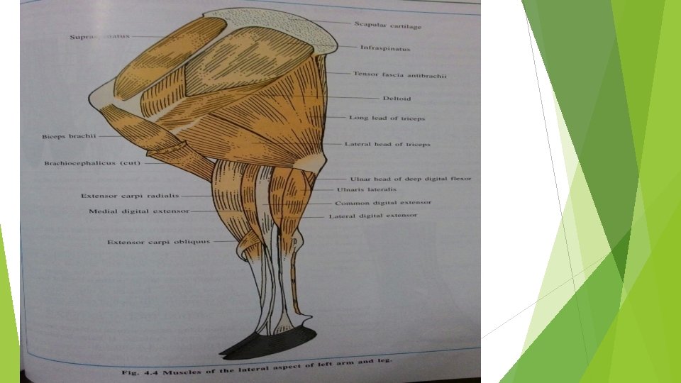

Muscles of arm Brachialis Biceps brachii Triceps brachii Tensor fascia antibrachii Anconeus

Tri c eps bra chi i Brac hiali s Bice p brac s hii
Extensor carpi radialis")
Muscles of forearm and manus Blood supply of Extensor Group muscles 1)Extensor carpi radialis : Brachial artery 2)Extensor carpi obliquus : Interosseos artery 3)Medial digital extensor : Brachial artery 4)Common digital extensor: Brachial artery 5)Lateral digital extensor : Brachial & interossesous. A
Pronator teres : Median artery 2)Flexor carpi radialis")
Blood supply of Flexor Group Muscles 1)Pronator teres : Median artery 2)Flexor carpi radialis : Median artery 3)Flexor carpi ulnaris : Brachial & medial arteries 4)Ulnaris lateralis : Brachial & Interosseous arteries 5)Superficial digital flexor: Median & Interosseous arteries 6)Deep digital flexor : Median & Interosseous arteries 7)Interosseous medius

Diagram of arterial system in cow

Axillary artery Each artery leaves the thoracic cavity by turning round the anterior border of first rib between costal and chondral insertion of scalenus ventralis. Then it bends backward and downward in axilla. It reaches the medial surface of shoulder toward the subscpulaiis and teres major muscles where it continues as brachial miens. Each axillar artery gives rise intrathoracic and extrathoracic branches.

Branches of axillary artery 1. Suprascapular/prescapular artery : - It arises from the axillars artery and supplies to subscapularis and supraspinatus. 2. External thoracic artery : - It is voluminous branch arises near first rib and supplies to pectoral muscle. . 3. Subscapular artery : - It is very large branch which is supposed to be continuation of axillary artery. It supplies to subscapularis, teres major, triceps, infraspinatus and deltoideus.
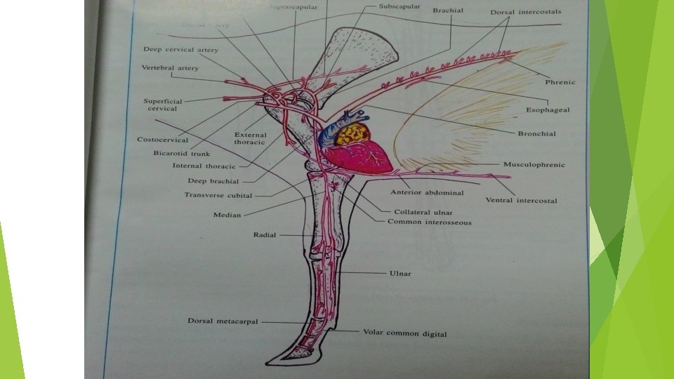
 Thoracodorsal artery : - It supplies to latissitnus dorsi. teres")
Subscapular artery a. ) Thoracodorsal artery : - It supplies to latissitnus dorsi. teres major, serratus thoracic, triceps and tensor fascia antibrachi muscles. b. ) Posterior circumflex or scapulo-humeral artery : It arises above The thoraeodorsal artery between long_ and lateral heads of triceps to which it supplies. It also supplies to shoulder Joint. infraspinatus. teres minor and deltoideus. c. ) Circumflex artery of scapula : - It supplies to infraspinatus. teres minor and supraspinatus.
 Anterior circumflex")
In the arm region axillary artery continued as brachial artery 1. ) Anterior circumflex or prehumeral artery : - It supplies to coracobrachialis, biceps brachia brachiocephalicus and deep pectoral muscles 2. ) Deep humeral or brachial artery : - It supplies to triceps, tensor fascia antibrachi and anconeus muscles. 3. ) Collateral ulnar artery : - It supplies to pectoral and skin over it , triceps, anconeus. ulnar and humeral heads of flexor carpi ulnar, ulnar head of deep digital flexor and superficial digital flexor. 4. ) Collateral radial artery : - It supplies to biceps, brachialis. extensor muscles and humerus,
")
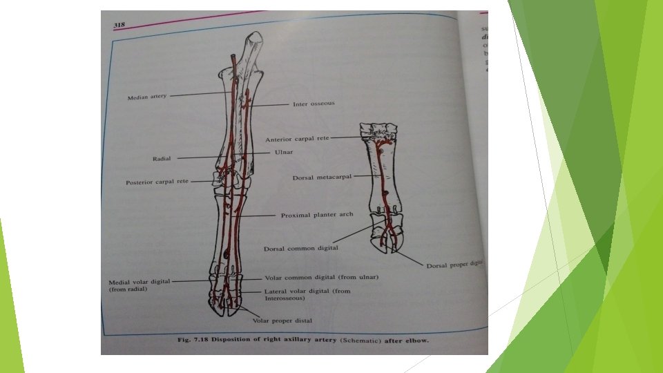
Median artery It divides into radial and ulnar arteries. Collateral branches : 1. ) Large and small muscular branches supplies to pronator teres, flexor carpi radialis and ulnanris superficial and deep digital flexors. 2. ) Common interosseous artery : - It is a large branch which anastomoses with branches of the deep humeral artery. It detaches branches to humeral and radial divisions of deep digital flexor. It passes through the proximal interosseous space and is continued down as anterior interosseous artery. While passinio through mterosseous space it elves nutrient artery to radius and ulna.

Ø Anterior interosseous artery : - It runs down in the groove between the radius and ulna. Ø It continues forward and assist in the formation of anterior carpal arterial network. Ø It gives a branch near distal interosseous space which passes backward and assist in the formation of posterior arterial network and is then continued as postero-external deep metacarpal artery. Ø This artery runs downward and joins with the middle deep metacarpal artery to assist in the formation of deep volar arch.

Radial artery : - It descends downward on the back side and continued as postero-internal deep metacarpal artery. It gives following branches. 1. ) One branch assist in the formation of the posterior arterial network. It crosses the lower part of the radius and comes on the anterior face. passes downward anastomoses with the branches of the anterior interosseous artery to form the anterior carpal arterial network. 2. ) The other branch is posterior deep middle metacarpal artery which forms the deep volar arch and runs through the proximal foramen of large metacarpus and divides into two. One of the branch forms the anterior carpal arterial network and other joins with anterior metacarpal artery. The postero-intenal deep metacarpal artery descends on the posterointernal face of the metacarpus and assist to form superficial volar arch.

Ulnar artery : - It runs downward in the metacarpal region as posterior superficial large metacarpal artery which continued as posterior common digital artery. The posterior lateral proper digital arteries are two in number. ( 1. ) Postero-internal proper digital artery : - It is the continuation of the postero-internal metacarpal artery. (2. ) Postero-external artery. : - It arises from deep volar arch or may be the continuation of the postero-external deep metacarpal artery. Each of them detaches a branch to the rudimentary digit. It then reaches the bulb of heel and anastomoses with the artery of volar cushion.

Posterior common digital artery : - it is the continuation of posterior superficial large metacarpal artery and passes downward in the interdigital space at the level of lower extremity of the first phalanx and it divides into posterior proper digital arteries for each digit. Artery of the volar cushion : - It arises from the proper digital arteries and supplies to the volar cushion; sensitive sole, coronary cushion and sensitive laminae. Anterior metacarpal artery : - It passes downward in the anterior longitudinal groove of the large. metacarpal bone and unite with the perforating metacarpal artery and it is continued on the anterior common digital artery.

Anterior common digital artery : - It receives a communicating branch from the posterior common digital artery and divides into two anterior proper digital arteries, one for each digit which supplies in its course to the first and second phalanx, tendon, fascia. skin and hone and in the foot to the coronary cushion and lamina Tissue

Veins of the forelimb : - The axillar veins are the other root of the anterior vena cava. 1. Digital veins a. Anterior digital vein b. Postero proper digital vein. c. Postero-internal proper digital vein. d. Postero-external proper digital vein. 2. The digital veins are continued by five metacarpal veins. a. Anterior superficial metacarpal vein. b. Postero superficial metacarpal vein. c. Postero-internal deep metacarpal vein. d. Postero-external deep metacarpal vein. e. Postero-middle deep metacarpal vein.

Veins of the forearm and arm : Radial vein : - It is upward continuation of posterior branch of postero-internal deep metacarpal vein and joint with the internal and external ulnar vein and continued as median vein. Near the medial condole of humerus it continued as brachial vein which receives the blood from common interosseous vein and muscular branches. It terminates below and behind the shoulder joint and continued as axillary vein. It crosses the lower part of anterior border of first rib and joins jugular vein. It receives the blood from external thoracic, suprascapular and subscapular veins.

Superficial veins : Cephalic vein : - It is formed on the inner face of lower third of radius by union of anterior superficial metacarpal vein and anterior division of postero-intenal deep metacarpal vein and joins with the median cephalic branch given by median vein at the upper part of groove formed by brachiocephalicus and anterior superficial pectoral.

Thank you
- Slides: 49