STRUKTUR DNA Drs Sutarno MSc Ph D Komponen

 adalah rantai nukleotida yang panjang (poly nucleotide) yang")


")


 is in blue,")


:")


- Slides: 28
STRUKTUR DNA Drs. Sutarno, MSc. , Ph. D.
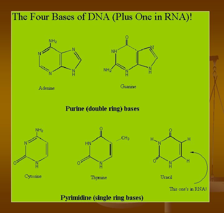
Komponen DNA n Deoxyribonucleic acid (DNA) adalah rantai nukleotida yang panjang (poly nucleotide) yang terdiri dari: n Deoxyribose (suatu pentosa, yaitu gugusan gula yang terdiri dari 5 atom karbon (C). n Fosfat (Phosphoric Acid) n Basa nitrogen (nitrogenous bases) yang terdiri dari (Purin: Adenine dan Guanine, atau Pyrimidin: Cytosine dan Thymine)
Nukleotida
Nukleotida
Nukleotida
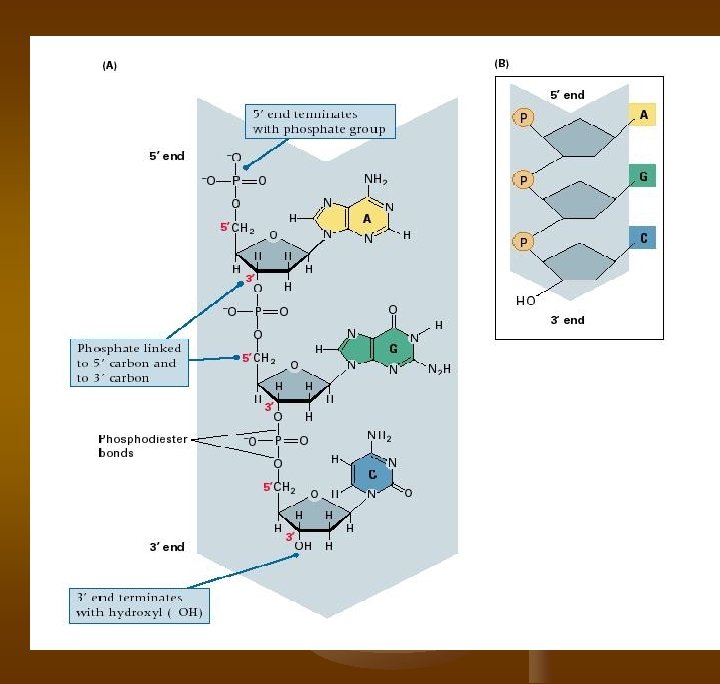
Gugus Gula n Pada nukleotida, atom-atom pada gugusan gula baik yang berupa deoksiribosa seperti pada DNA maupun ribosa seperti pada RNA diberi nomor: 1', 2', 3’, 4’, 5'. n Atom-atom pada komponen gula suatu nukleotida menghubungkan basa nitogen dan gugus fosfat. Atom karbon no 1 berikatan dengan N dari suatu basa purin, atau N suatu basa pirimidin. Sedangkan gugus OH (hydroxyl) pada C no 5' digantikan oleh suatu ikatan dengan gugus fosfat (ester). n
n n n DNA terdiri dari dua rantai polynukleotida berpasangan yang berpilin membentuk suatu helix, yang sering disebut double helix. Masing-masing rantai polynukleotida merupakan polymer linier yang dibentuk oleh monomer-monomer (deoxynucleotida) yang antara monomer satu dengan yang lainnya dihubungkan oleh ikatan phosphodiester Ikatan phosphodiester ini menghubungkan karbon no 3' ribosa dari suatu deoxynucleotida dengan karbon no 5' pada ribosa suatu ribose of deoxynucleotida yang berdekatan.
IKATAN PHOSPHODIESTER (Phospho diester bonds)
DNA RANTAI TUNGGAL Gambar: suatu potongan rantai tunggal asam nukleat
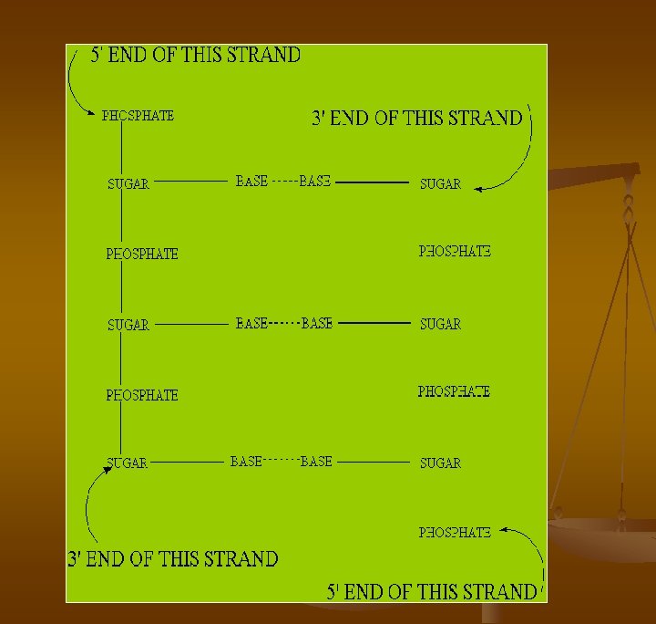
n n The sugar / phosphate backbone is on the outside while the organic bases project into the inside of the double helix. By convention a polynucleotide is read from the 5' end to the 3' end. The orientations of the two strands are antiparallel : their 5' - 3' directions are opposite. The two strands are held together by the energy of many hydrogen bonds. The base pairing is regular and precise.
The four nucleotides
The two sugar backbones are outlined in dark grey deoxyadenylate (A) is in blue, deoxythymidylate (T) is in green, deoxyguanylate (G) is in red, deoxycytidylate (C) is in orange
Base pairing of G and C Figure: Base pairing. The hydrogen bonds between the NH (blue) and O (red) are in green.
Base pairing of A and T Figure: Base pairing. The hydrogen bonds between the NH (blue) and O (red) are in green.
p. CATG GTACp
Double helix DNA n The Watson and Crick model of a double helix structure provides an answer to the regularity of the composition in bases and its physiological properties (replication in the cell). It is confirmed by diffraction data. n The base composition is variable, but in all cases the amount of adenine is equal to the amount of thymine (A=T). In the same manner, C=G. Consequently A+C= T+G. n E. coli has a single circular DNA molecule of 4, 600, 000 base pairs. The total length is 1. 4 mm. n In man, the DNA molecule in a diploid cell, if fully extended, would have a total length of 1. 7 metres. If you unwrap all the DNA you have in all your cells, you could reach the moon. . . 6000 times! n
The Double Helix
n n Pasangan dari pita 3’--> 5’ adalah pita 5’ --> 3’. Double helix akan stabil bila A -- T, G – C, Chargaff (1955), hidrolisis DNA >> A/T, G/C ~ 1
Struktur Double Helix n n n n Struktur Double Helix (Watson – Crick, 1953): mengandung dua rantai polinukleotida berpilin kanan pada pusat aksis Dua rantai DNA berorientasi anti parallel: 3’-5’ berpasangan 5’--3’ Basa nitrogen berpasangan dg ikatan hidrogen: A = T, G = C. Basa nitrogen Setiap satu putaran helix sempurna sepanjang 34 A (3, 4 nm), masing 2 rantai mengandung 10 basa. Di sepanjang aksis terjadi pergantian antara lekuk ‘major grooves’ yg lebih besar dan ‘minor grooves’ yang lebih kecil Diameter dari heliks adalah 20 A (2 nm) Struktur double heliks
Denaturation of DNA Denaturation: conversion of ds. DNA to two strands of ss. DNA n When the H bonds break that join the 2 strands in ds. DNA, they all tend to break simultaneously. . . thus, the ds. DNA "melts" into two strands Agents which "denature" DNA: n a. Increase the temperature past the "melting" temperature: Tm b. Increase the p. H to above about 11. 3 c. Decrease the ionic strength n The Tm is the "midpoint" in the melting reaction. . . Tm is proportional to %(G+C), due to 3 H-bonds between C and G but only 2 H-bonds between A and T n
Renaturation of DNA n n n Renaturation: joining of two DNA strands of complementary sequence to form ds. DNA 1. Reverse of Denaturation. . . but requires that the two DNA strands have complementary sequence, i. e. permit A joining to T, C joining to G, along the entire length of both DNA strands or chains. 2. Renaturation is also called Hybridization 3. Can also occur between DNA and RNA, to form a DNA: RNA hybrid, or between two RNA strands of complementary sequence. Also note that purine/pyrimidine bp fit "just right" so that they occupy the same space compare the other possible schemes
Structure relates to Function There are 3 ESSENTIAL REQUIREMENTS of a genetic material: 1. It must be able to replicate accurately. n Strands separate. . . each serves as a "template" for synthesis of a new strand T dictates A, A dictates T, C dictates G, G dictates C 2. Maintain Genetic Information It must have the capacity to carry all the information needed to direct organisation and metabolic activities of the cell.
3. It must be capable of having an OCCASIONAL mutation or change in information. n Mutagenesis: any change in the Nucleotide Sequence of a DNA molecule Base Sequence changes Spontaneously at Low Probability Provides for: Genetic Variation. . . to adapt evolutionarily to new environments Transitions vs Transversions n Transitions: Pyrimidine <--> Pyrimidine; or Purine <--> Purine AT --> GC, TA --> CG, n Transversions: Purine <---> Pyrimidine AT --> TA (NOT the same!), AT --> CG, TA --> GC,
Similarities and Differences Between DNA and RNA
What's in a name n n n D Deoxyribo : the pentose does not have any oxygen in position 2. Compare a deoxyribose with a ribose. N Nucleic: these molecules were first found in the nucleus of the cell , before being found in mitochondria, chloroplasts (of plant cells), and in the cytoplasm of prokaryotes. A Acid: only two of the three acid groups of the phosphoric acid are used to form the DNA chain. The third one gives the phosphoribo-backbone an acidic property.