
Structures of the Eye and Sheep Eye Dissection

- Slides: 25
Structures of the Eye and Sheep Eye Dissection Prep
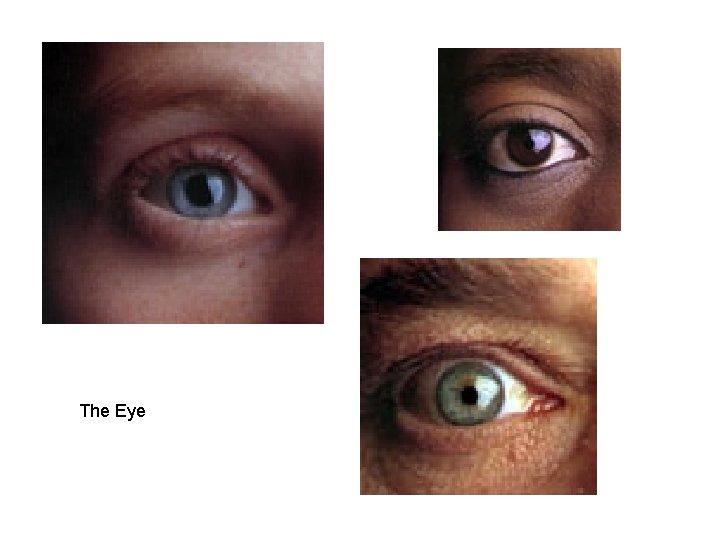
The Eye
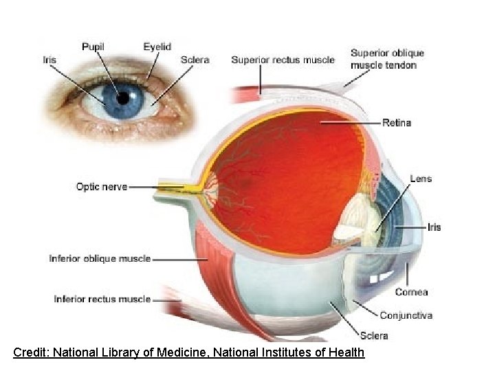
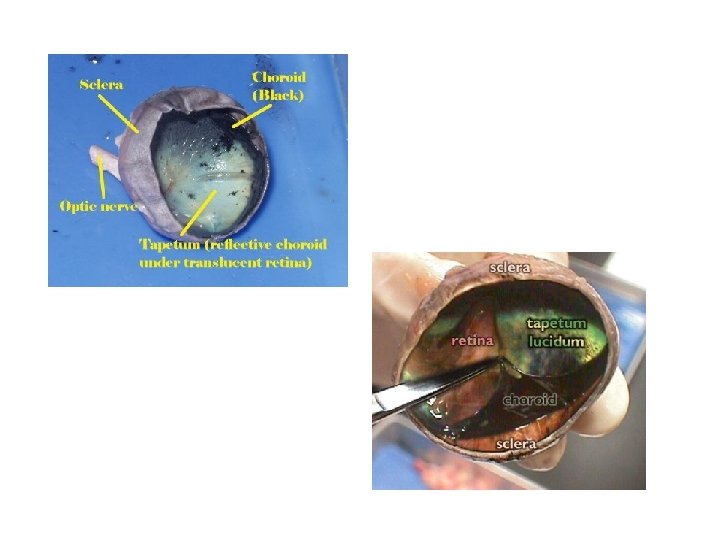
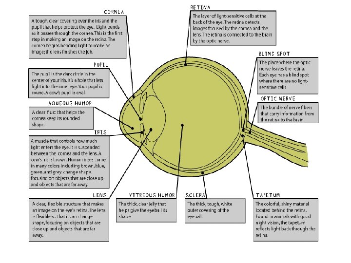
• Eye: sense organ that receives light and focuses it; sending impulses to the brain • 3 Main Parts – Sclera: Outer Layer of Eye tough white outer tissue of the eye – Choroid: Middle Layer of Eye – Retina—inner layer of the eye
Credit: National Library of Medicine, National Institutes of Health
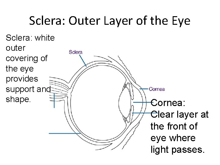
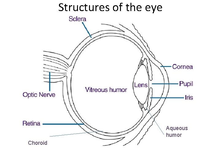
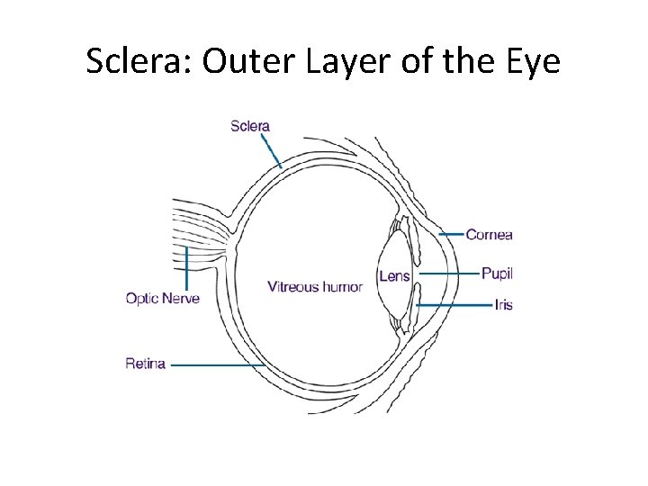
Sclera: Outer Layer of the Eye Sclera: white outer covering of the eye provides support and shape. Cornea: Clear layer at the front of eye where light passes.
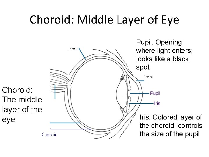
Choroid: Middle Layer of Eye Pupil: Opening where light enters; looks like a black spot Choroid: The middle layer of the eye. Iris: Colored layer of the choroid; controls the size of the pupil
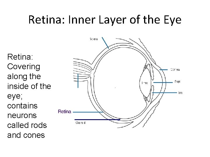
Retina: Inner Layer of the Eye Retina: Covering along the inside of the eye; contains neurons called rods and cones
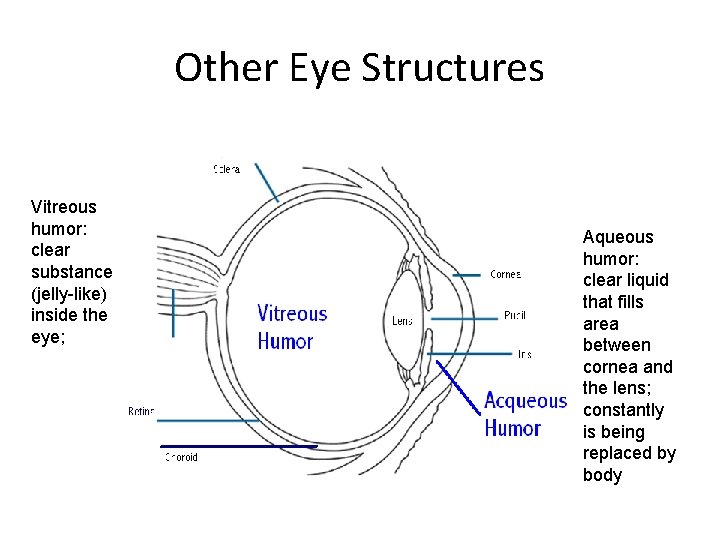
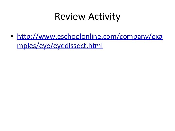
Other Eye Structures Vitreous humor: clear substance (jelly-like) inside the eye; Aqueous humor: clear liquid that fills area between cornea and the lens; constantly is being replaced by body
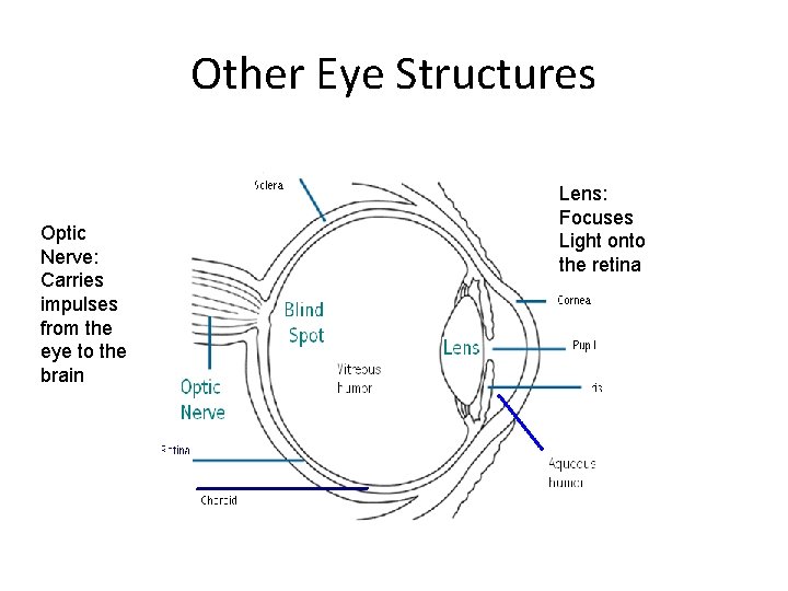
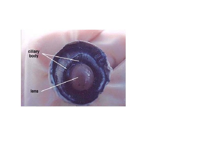
Other Eye Structures Optic Nerve: Carries impulses from the eye to the brain Lens: Focuses Light onto the retina
Structures of the eye Aqueous humor Choroid
Sheep Eye Dissection
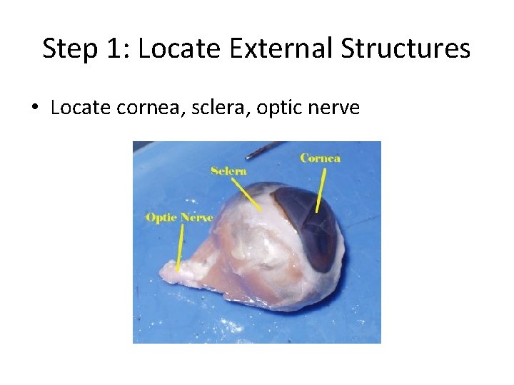
Step 1: Locate External Structures • Locate cornea, sclera, optic nerve
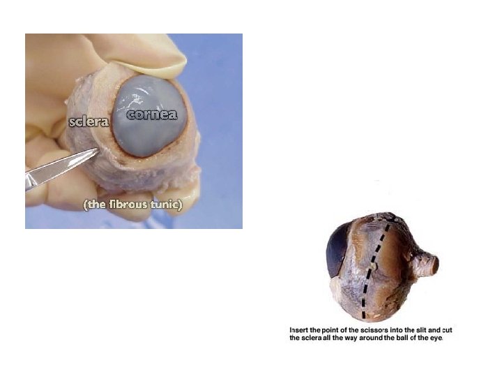
Step 2: Begin Dissection • Using your scalpel cut into the eye at the junction of the sclera and the cornea • Make incision with the scalpel and then cut around the cornea with the scissors
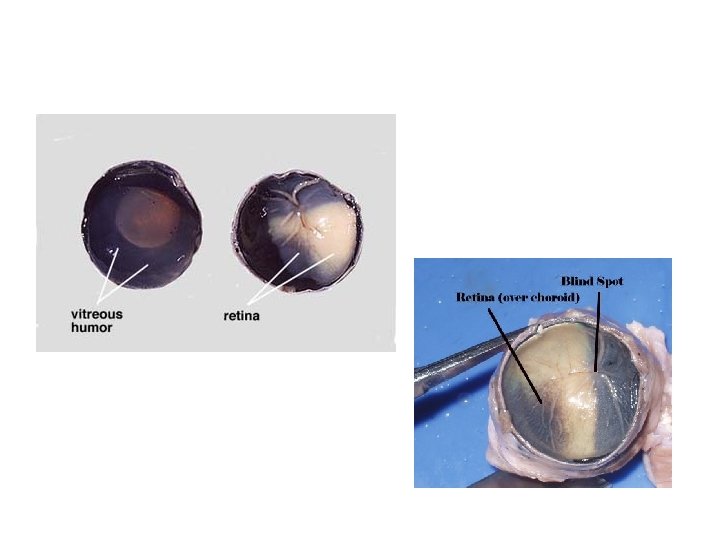
Step 3: Separate eye and identify structures • Separate the front of the eye and set aside • Identify the structures of the back of the eye – Aqueous humor (watery liquid) – Victreous humor (jelly-like liquid) – Retina/Blind Spot (where optic nerve attaches) – Lens (looks like marble)
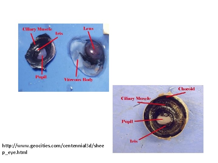
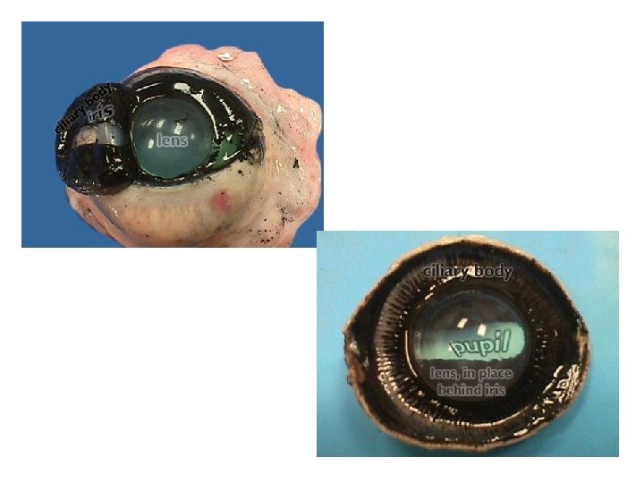
Step 4: Identify structures of front section of eye • Identify – Cornea (clear covering at front of eye) – Iris (colored section of eye) – Pupil (opening through iris)
http: //www. geocities. com/centennial 3 d/shee p_eye. html
Review Activity • http: //www. eschoolonline. com/company/exa mples/eyedissect. html
Sclera: Outer Layer of the Eye