Structure of skeletal muscle Dr Kalpana B Assistant

Structure of skeletal muscle Dr. Kalpana. B Assistant professor Physiology

By the end of class student should. . • Understand the structural organization of skeletal muscle fibers • Name the contractile and regulatory proteins and its role in muscle contraction. • Draw the diagram of sarcotubular system and give its role in muscle contraction.

SKELETAL MUSCLE q Long cylindrical cells q Many nuclei per cell q Striated q Voluntary

CARDIAC MUSCLE q Branching cells q One or two nuclei q Striated q Involuntary

SMOOTH MUSCLE q Fusiform cells q One nucleus per cell q Nonstriated q Involuntary

SKELETAL MUSCLE • Human body contains over 400 skeletal muscles and constitutes 40 – 50% of total body weight • They are attached to the skeleton by tendons • The tendons transmit the muscle force to the bone • Skeletal muscle causes the skeleton to move at joints

• Skeletal muscle contains muscle bundles or fascicles • Fascicles consists of muscle fibers • Structural unit of muscle-myocyte • Cell membrane-sarcolemma • Cytoplasm-sarcoplasm

• A single muscle e. g. biceps contains approx 1000 muscle fibres. • These fibres run the whole length of the muscle • Muscle fibres are joined together at the tendons
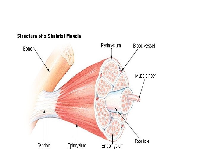

Structure of Skeletal Muscle: Connective Tissue Covering • Epimysium – Surrounds entire muscle • Perimysium – Surrounds bundles of muscle fibers • Fascicles • Endomysium – Surrounds individual muscle fibers

• Muscle fiber 10 to 80μ in diameter each and is composed of 1000 s of myofibrils • Sarcoplasm is filled with myofibrils • Myofibrils-myofilaments-thick and thin filaments. • Striations-alternate dark and light bands • Each myofibrils is made up of unitssarcomere-different muscle proteins
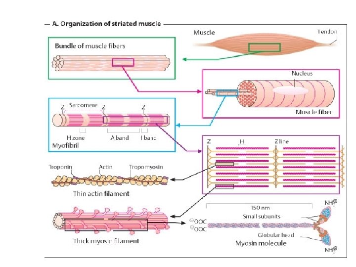

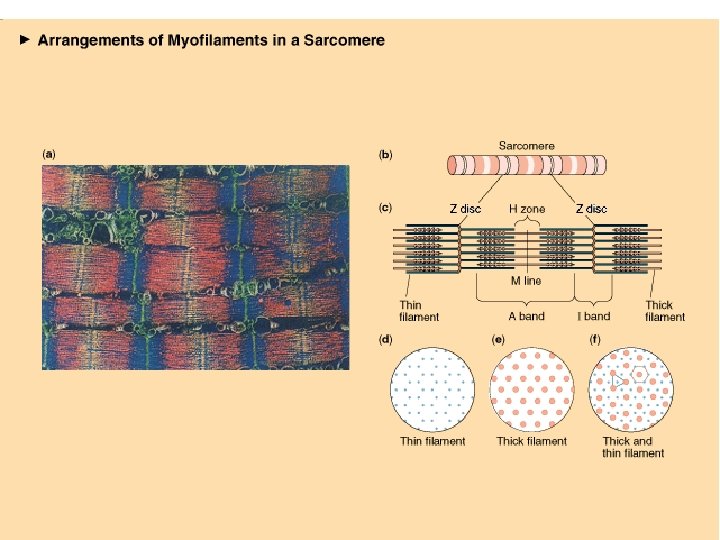
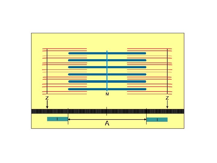
• Sarcomere- between two z-lines. • Structural and functional unit of myofibril • Cross striations due to alternate dark and light bands • Light band - Isotropic band - I band Thin filament-actin filament • Dark band - Anisotropic band - A band Thick filament-myosin filament • • • H zone - lighter zone in A band Z line - in the center of I band M line - in the center of A band
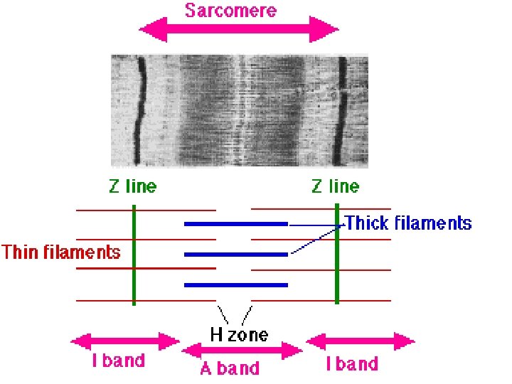

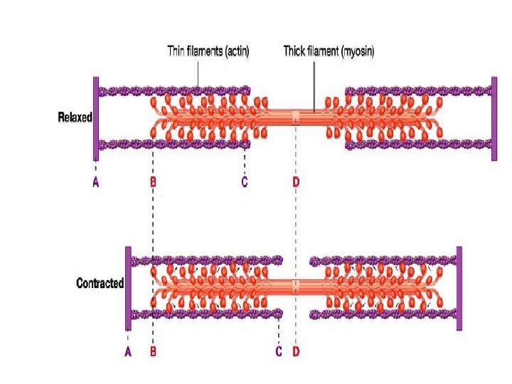
Sarcomere: functional unit of striated muscle


• Sarcomere • Distance between two Z lines. • Whole of A band one half of I band on either side of A band. • At rest the sarcomere length is 2. 5 µm • At contracted state its length is 1. 6 µm • At relaxed state its length is 3. 5 µm

Muscle proteins 1. Contractile proteins-myosin and actin 2. Regulatory proteins-troponin and tropomyosin 3. Attachment proteins-titin, alpha actinin and dystrophin
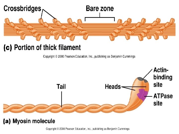
 Myofilament • • Many elongated myosin molecules. Single filament contains roughly 300")
Myosin (Thick) Myofilament • • Many elongated myosin molecules. Single filament contains roughly 300 myosin molecules Molecule consists of two heavy chain and four light chain. Myosin heads 1. Can bind to active sites on the actin molecules to form cross-bridges. (Actin binding site) 2. Attached to the rod portion by a hinge region that can bend and straighten during contraction. 3. Have ATPase activity: activity that breaks down adenosine triphosphate (ATP), releasing energy. Part of the energy is used to bend the hinge region of the myosin molecule during contraction
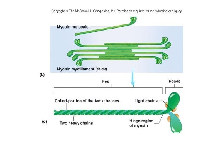

• Has a diameter of 10 -11 nm and a length of 1. 6 micron • Located in the middle of sarcomere-A band. • Cross bridges project from the lateral sides in a helical fashion • Myosin filament is surrounded by 6 actin filaments.
 1. F (fibrous) actin-thin-4 -5 nm Actin in diameter")
Thin Filament: • • (Thin) 1. F (fibrous) actin-thin-4 -5 nm Actin in diameter 2. Tropomyosin Myofilaments 3. Troponin Two strands of fibrous (F) actin form a double helix which is made up of globular molecules of G-actin. F- actin formed by polymerization of G actin - each of which has a myosin-binding site Tropomyosin: an elongated protein winds along the groove of the F actin double helix. Covers the binding site of actin where myosin comes in contact.

TROPONIN: Globular units located at intervals along the tropomyosin molecule. Has 3 components • Troponin C- contain binding site for calcium • Troponin T – binds the troponin component to tropomyosin • Troponin I –inhibit the interaction of myosin with actin.

Attachment proteins • Titin-extends from Z line to M line • Stabilize the position of thick filament • Alpha actinin-anchors thin filament to Z line • Dystrophin –Links thin filaments to internal membrane proteins in sarcolemma.
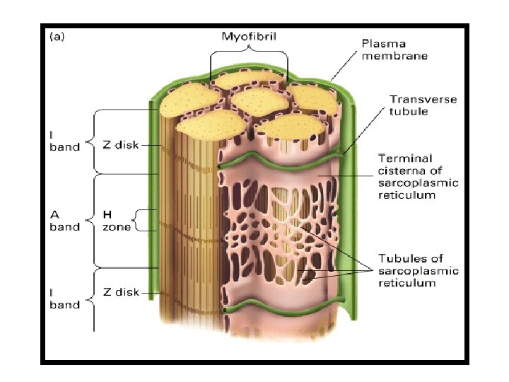

SARCOTUBULAR SYSTEM The myofibrils are surrounded by some important membranous structures which appear in the form of vesicles and tubules. These structures are together called Sarcotubular system • T-tubules (Transverse Tubules) • L-tubules (Sarcoplasmic Reticulum)

T –tubule: • They are inwardly directed extensions of the sarcolemma into the muscle fibers at the junction between A and I bands. • Each Sarcomere has 2 tubules. • Its function is the rapid transmission of the action potential from the cell membrane to all the fibrils in the muscle.

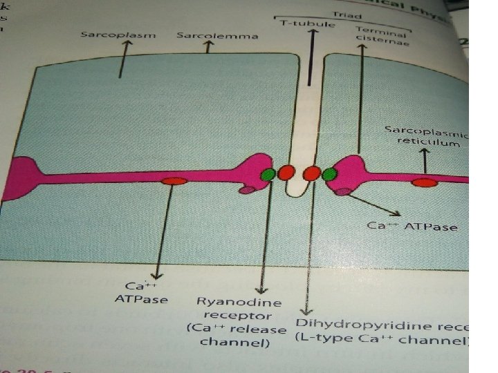
Longitudinal Sarcoplasmic Reticulum • Longitudinally on the either side of tubular system • Vesicles /sacs- Terminal cisternae. • Stores a large quantity of calcium ions-calsequestrin. • Rich in glycogen • Triggers the process of muscle contraction • Depolarization of T – tubules activate the L- tubule via dihyropyridine receptors- release of Ca 2+ from Ltubules via Ryanodine receptor. • Transverse tubule with 2 terminal cisterns – TRIAD

Sarcomere Completely Contracted

Summary
 G K Pal")
References • Comprehensive Textbook of Medical physiology (Vol 2 first edition) G K Pal • Text book of medical physiology (Vol 2 6 th edition) A K Jain • Essentials of medical physiology (6 th edition) K Sembulingam and Prema Sembulingam AEJ 36
- Slides: 36