Structure of Bone Cartilage Sanjaya Adikari Department of

Structure of Bone & Cartilage Sanjaya Adikari Department of Anatomy
")
Connective tissue • Cells producing extracellular material • Other cells (immune cells, fat cells) • Extracellular material • Ground substance – matrix of organic material • Fibres

Connective tissues

Cartilage • A semi-rigid tissue • Variable amounts of collagen and elastic fibres • Three types – Hyaline cartilage (consist of collagen fires) – Elastic cartilage (consist of collagen and lot of elastic fires) – Fibrocartilage

Cartilage Blood vessels diffusion Nerves Perichondrium cells

Chondroblasts Lacuna/ space Chondrocytes
 – Appositional")
Cartilage… • Grow by – Interstitial growth (chondrocytes dividing & forming clusters) – Appositional growth (chondroblasts differentiating into chondrocytes) • Adults have a limited distribution of cartilage • Newborns have more extensive distribution

Hyaline Cartilage • Most common type • Found in, nasal septum, larynx, tracheal rings, costal cartilages and articular surfaces of sinovial joints • Newborns have more extensive distribution since they act as templates for most of the long bones

Elastic Cartilage • Found in, external ear, external auditory canal, epiglottis and wall of Eustachian tubes. • Differ from hyaline cartilage by having numerous elastic cartilage bundles in the matrix

Hyaline cartilage Elastic cartilage

Fibrocartilage • Found in, intervetebral discs, some articular cartilages and pubic symphysis. • Features appear intermediate between cartilage and dense fibrous connective tissue • There alternating layers of hyaline like cartilage and thick layers of collagen fibres.

Fibrocartilage

Bone • A specialized connective tissue • Extracellular components are mineralized - rigid • Supporting and protective functions • Function as a calcium ion store • Growth and resorption throughout life - dynamic
 – Osteocytes: maintain bone")
Bone…… • Cells – – Osteoblasts: secrete extracellular matrix (osteoid) – Osteocytes: maintain bone matrix – Osteoclasts: remove bone matrix • Extracellular matrix – – glycoprotein ground substance, collagen fibres, mineral component by calcium hydroxyapatite
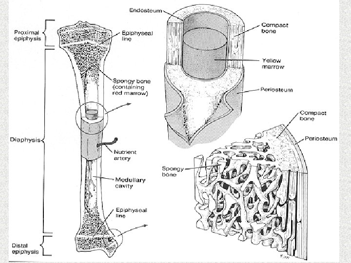
 red marrow epiphysis")
periosteum epiphysis growth plate endosteum diaphysis cavity (contains yellow marrow) red marrow epiphysis

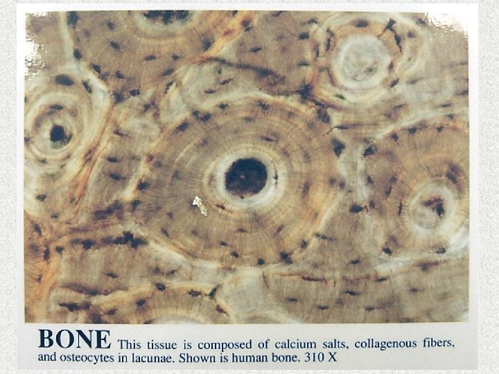
Microscopic structure • Haversian canals with vessels and nerves • They are arranged parallel to the long axis • Connected by canals running across, called • Bone cells are arranged around Haversian canals in circles • The cells lay down bone in circles called lamellae

Microscopic structure Cortical bone Haversian system Volkmann’s canal

Canaliculi
 New Haversian system")
Cortical bone Interstitial system (old Hav Sys) New Haversian system

Woven bone immature bone. Random organization of fibrous elements Bone Compact bone Lamellar bone mature bone forms walls of the shaft & a thin layer around the epiphysis of long bones Cancellous bone found in the epiphysis or around the medullary cavity

Resorption Apositional growth Increased calcium ions + Calcitonin _ Osteoblast Osteoclast + PTH + Reduced calcium ions Periosteum
- Slides: 23