Structure Function of Immune System Cells and tissues


 Lymphoid system Lymphoreticular cells Secondary (Peripheral) Thymus Bone marrow Lymph")


 which include")


 • Peyer’s patches")
")


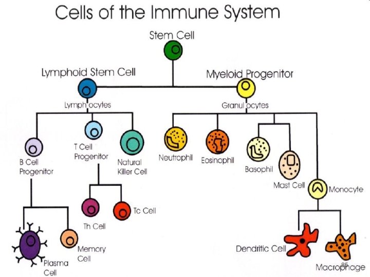
 Lymphoid stem cell (progenitor) B cell progenitor (pro-B")


 CD")

 Suppressor cells (CD 8")
 • Double the size of")


- Slides: 44
Structure & Function of Immune System
Cells and tissues of the immune system • Lympho – reticular system is a complex organization of cells of diverse morphology • Distributed widely in different organs and tissues of the body. • The cells of the system are responsible for the development of specific immunity.
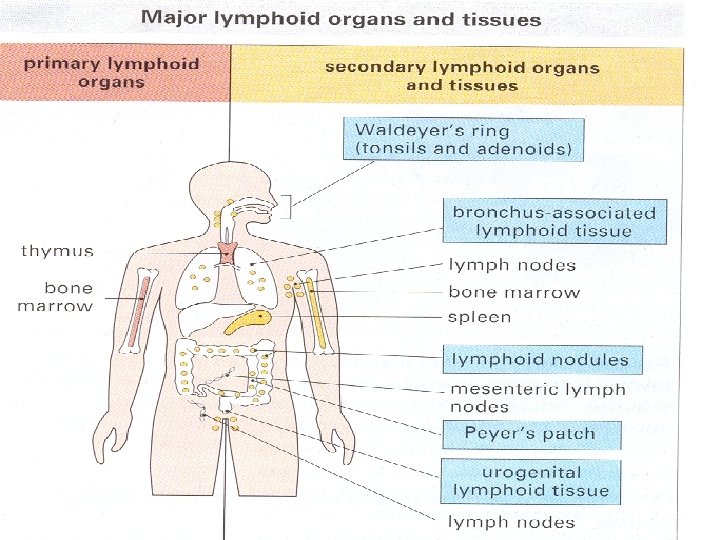
Lymphoid organs Primary (Central) Lymphoid system Lymphoreticular cells Secondary (Peripheral) Thymus Bone marrow Lymph node (Encapsulated) Lymphoid Reticular Plasma spleen MALT (Un-encapsulated) Phagocytic cells MALT: Mucosa Associated Lymphoid Tissue
Primary Lymphoid organs • Major sites for lymphopoiesis • The lymphocytes, develop, differentiate proliferate and mature into functional cells. • In mammals T cells mature in thymus and ‘B’ cells in fetal liver cells and Bone marrow. • It is in the primary lymphoid organs the lymphocytes acquire the antigen receptors to fight with the antigenic challenge subsequently during life.
Primary Lymphoid Organ • Major site of lymphocyte development. • In mammals, T lymphocytes mature in thymus whereas B lymphocytes mature in the liver or bone marrow. • Birds have specialised sites of B cell maturation called bursa of Fabricus.
Secondary lymphoid organs • Spleen, Lymph node, mucus associated lymphoid tissue (MALT) which include tonsils and payer's patches. • Here the lymphocytes interact with lymphocytes and accessory cells (macrophage both phagocytic and antigen presenting) and also with antigen. • The immune response is generated and disseminated in secondary lymphoid tissue.
Secondary Lymphoid Organs • Lymph nodes • Spleen • Accumulation of lymphoid tissues like • MALT (mucosa associated lymphoid tissue) • GALT (gut associated lymphoid tissue) • BALT (bronchus associated lymphoid tissue)
Thymus • • Bilobed organ Present in mediastinum Consists of Cortex and Medulla 70 g in infants and 3 g in adults • Reaches maximal size at birth, continues to grow till 12 th year and progressively involutes at puberty • Lymphocyte proliferation in thymus is not dependent on antigenic stimulus.
Structure • Two lobes, numerous septa divide the organ into lobules. • Outer cortex and inner medulla. • Cortex: crowded with actively proliferating small lymphocytes. • Medulla: Epithelial cells and mature lymphocytes. Hassall’s corpuscles
Functions • Production of thymic lymphocytes • Lymphocyte proliferation. But only 1% leave thymus. • Lymphocyte acquire ‘Thy’ Ag • Lymphocytes conditioned in thymus are called thymus (T) dependent or T cells. • Immunological competence of lymphocytes
• T lymphocytes are selectively seeded into peripheral lymphatics like white pulp of spleen, paracortical areas of LNs, and around central arterioles. • Thymus dependent areas.
Thymus deficiency • Di. George syndrome: Congenital aplasia of thymus • Runt disease: Deficiency of CMI. Seen in neonatally thymectomised mice
Bone marrow • Site of origin of all lymphocytes • Site of ‘B-Cell’ origin & development • A Selection process – eliminates B cell with self reactive Ab Receptors
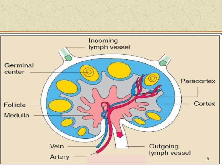
Lymph nodes • Present along the course of lymphatic channels. • Surrounded by a capsule. • Consists of outer cortex & inner medulla. • Cortex: primary lymphoid follicles (accumulation of lymphocytes)
Cortex • Within follicles on antigenic stimulus, germinal centres develop, called secondary follicles. • Dendritic macrophages to capture and process antigens are present in follicles • Medulla: lymphocytes, plasma cells & macrophages arranged as Medullary cords. • Cortical follicles & medullary cords rich in B cells. • Paracortical area rich in T cells.
Functions • LNs help in proliferation & circulation of T & B cells. • LNs enlarge following local antigenic stimulus.
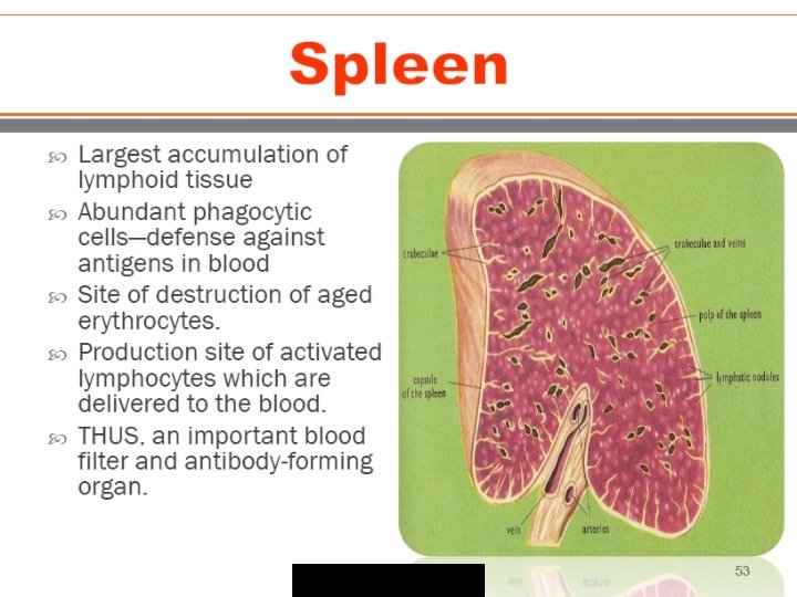
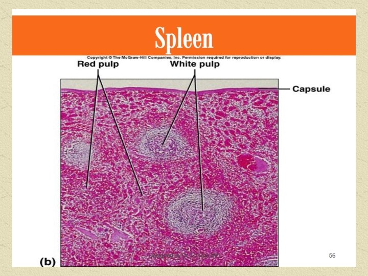
Spleen • Largest lymphoid organ. • Abundant phagocytic cells- Defense against antigens in blood • Consists of white pulp & red pulp. • Production site of activated lymphocytes & delivered to blood • Blood filter and Ab forming organ.
White pulp and Red pulp • Capsule forms trabeculae and divides spleen into compartments. • Along the trabeculae, Splenic artery descends and branches to form central arterioles. • Arterioles surrounded by sheath of lymphoid tissue (White Pulp) • Central arterioles descend down into red pulp rich in red cells (Red Pulp) • Peri arterial lymphoid collection in white pulp is called Malpighian corpuscles or follicles. Rich in T cells.
MALT • Contains lymphoid & phagocytic cells. • Both B & T cells present. • Secretory Ig. A is predominant.
GALT • Gut Associated Lymphoid Tissue • Tonsils, adenoids (Waldeyer’s ring) • Peyer’s patches • Lymphoid aggregates in appendix & small intestine
Cells of the Acquired Immune System • Lymphocytes • Antigen presenting cells (APCs)
Subsets of Lymphocytes • 2 subsets : T cells & B cells • T cells are thymus derived. • B cells are bone marrow derived. • B cells : ability to synthesize proteins called Immunoglobulins (Igs). • Each Ig can bind specifically & with high affinity to a particular Ag.
B Cell Development • Maturation-Stem cells to mature, naïve B cells • Activation-Ag binding; initiation of cell changes • Differentiation-Cell division and changes into effector B cells (plasma cells) and memory B cells
B Cell Maturation Hematopoietic stem cells(HSC) Lymphoid stem cell (progenitor) B cell progenitor (pro-B cell) Pre-B cell Immature B cell naïve B cell Mature
B Cell Activation Triggered by combining with Ag Two types of Ags that activate B cells • Thymus dependent Ag e. g. soluble proteins • Thymus independent Ag • Type 1, e. g. LPS • Type 2, e. g. capsular polysaccharides
Functions of B cells • Chief cell type in humoral immunity. • Function as Ag Presenting Cells. • Secrete lymphokines & other factors that influence the growth and activities of other immunologically important cells.
LYMPHOCYTES • Small, round cells present in the peripheral blood, lymphoid organs and in many other tissues. • Constitute 20 -45% of leucocytes in the peripheral blood. • Major cell types in lymphoid organs. • 1012 lymphocytes in human body & only 1% of it is present in the blood.
T cell development T cell precursors ( yolk sac, fetal liver & BM) CD 7+ pro T cells Migrate to thymus (acquires CD 2) Synthesize CD 3 & become pre T cells Immature T cells Mature T cell
T cells • 2 most imp subsets are distinguished by the surface proteins CD 4 & CD 8. • A mature, functional T lymphocyte express only one of the 2 proteins & are designated as CD 4+CD 8 - or CD 4 -CD 8+. 1. CD 4+ Helper T cells 2. CD 8+ Cyotoxic T cells.
T cell types Regulatory cells- Helper cells (CD 4 cells) Suppressor cells (CD 8 cells) - balanced activity results in optimal response. - over activity of helper T cells & low suppressor cell activity – Autoimmunity - decreased helper T cell activity & increased suppressor T cell activity – Immunodeficiency Effector T cells – cytotoxic T cells - Delayed type hypersensitivity (DTH) cells - Mixed lymphocyte reactivity cells (MLR) cells
Null cells • Also called large granular lymhocytes (LGL) • Double the size of small lymphocytes • Three types - natural killer cells - Ag dependent cytotoxic cells (ADCC) - Lymphokines activated killer (LAK) cells
Natural killer cells • Cytotoxicity towards various target cells mainly tumor & virus infected • Cytotoxicity not Ab dependent or MHC restricted • Important in immune surveillance & natural defence against virus infected & malignant mutant cell
MAJOR HISTOCOMPATIBILITY COMPLEX Its discovery was based on transplantation experiments First work done by GORER in 1930 on the Ag responsible for allograft rejection in inbreed mice Snell, Dausset and Benacerraf was awarded the Nobel Prize for their work on MHC and the genetic control of immune system
This is cell surface Ag that evoke immune response to an incompatible host resulting in allograft rejection The H 2 Ag system are found to be the major histocompatibility Ag for mice This Ag is coded for by a closely linked multiallelic cluster of genes called MHC
HLA COMPLEX Alloantigens present on surface of leucocyte in man are called human leucocyte Ag (HLA) and set of genes coding for them is called the HLA complex
HLA COMPLEX • Gene for this complex are located on chromosome 6 • It consists of three separate cluster of gene - Class I: A, B & C loci - Class II: DR, DQ, & DP loci - Class III: Genes for C 2, C 4, Properdin factor B, Heat shock proteins & TNF α and β
Typing of HLA • Microtoxicity • Mixed leucocyte reaction • Primed lymphocyte typing
Application • Transplantation: used for testing compatibility between recipient and potential donor • Disputed Paternity • Anthropological studies • Genetic predisposition of disease • Ankylosing Spondylitis, Insulin Dependent Diabetes, Multiple Sclerosis, Myasthenia Gravis, Rheumatoid Arthritis, Systemic Lupus Erythromatosis, Narcolepsy