STRUCTURE FUNCTION OF HEMOGLOBIN DR AMINA TARIQ BIOCHEMISTRY

STRUCTURE & FUNCTION OF HEMOGLOBIN DR. AMINA TARIQ BIOCHEMISTRY

GLOBULAR HEME PROTEINS �Heme proteins are a specialized group of proteins that contain heme as a Prosthetic group. Role of heme group is dictated by the environment. �Examples: a) a. Cytochromes b) b. Catalase c) c. Hemoglobin d) d. Myoglobin

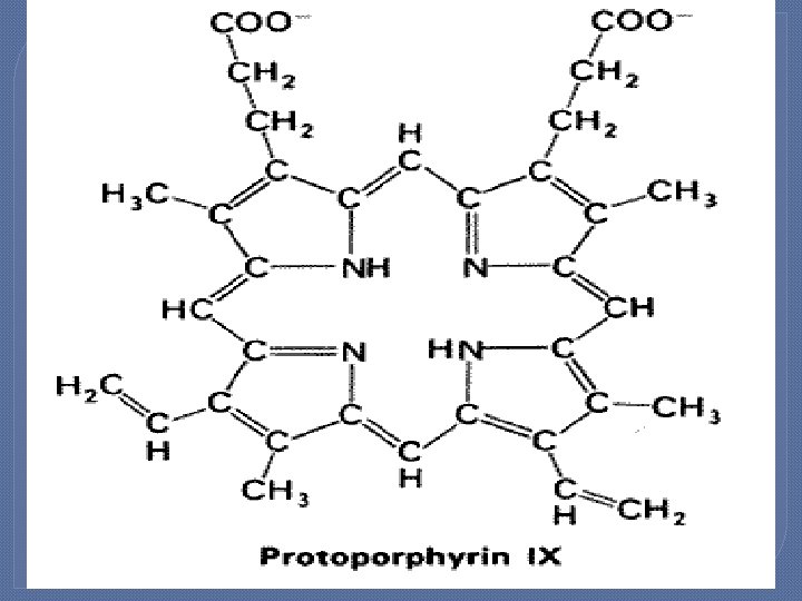
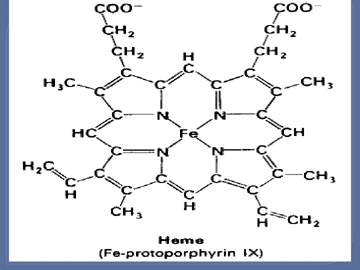
Structure and bonding �A Heme group is a flat ring molecule containing carbon, nitrogen and hydrogen atoms, with a single Fe 2+ ion at the center. Without the iron, the ring is called a Porphyrin. �In a heme molecule, the iron is held within the flat plane by four nitrogen ligands from the porphyrin ring.

HEMOGLOBIN �Hemoglobin is the protein that carries oxygen from the lungs to the tissues and carries carbon dioxide from the tissues back to the lungs. .

�The oxygen-carrying protein hemoglobin was discovered in 1840.

�Hemoglobin's reversible oxygenation was described a few years later. �In 1959 Max Perutz determined the molecular structure of hemoglobin by X-ray crystallography. This work resulted in his sharing with John Kendrew the 1962 Noble prize in chemistry.

STRUCTURE OF HEMOGLOBIN �Hemoglobin molecule consists of four polypeptide chains: �Two alpha chains, each with 141 amino acids and �Two beta chains, each with 146 amino acids.

�The protein portion of each of these chains is called "globin". �The α and β globin chains are very similar in structure. In this case, α and β refer to the two types of globin.

the α and β globin chains contain primarily α helix secondary structure with no β sheets. �Both
 which, in")
α or β globin chain folds into 8 α helical segments (A-H) which, in turn, fold to form globular tertiary structures. �Each �The folded helices form a pocket that holds the working part of each chain, the heme.
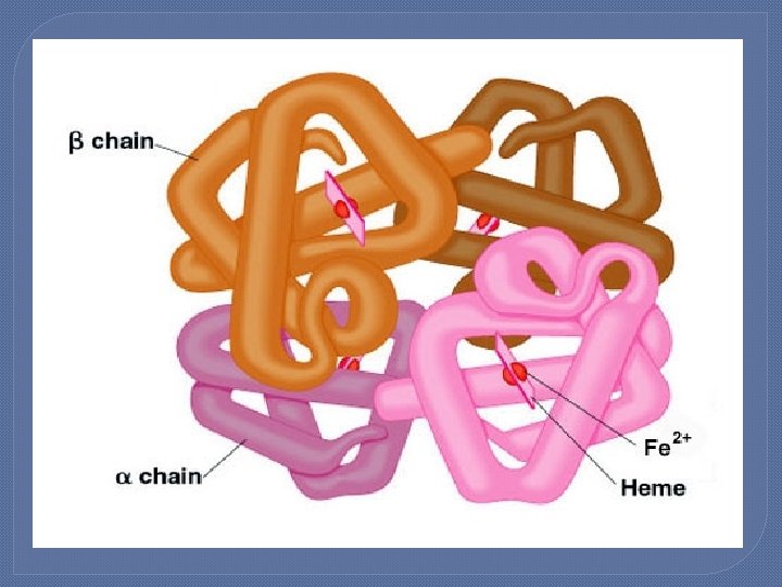
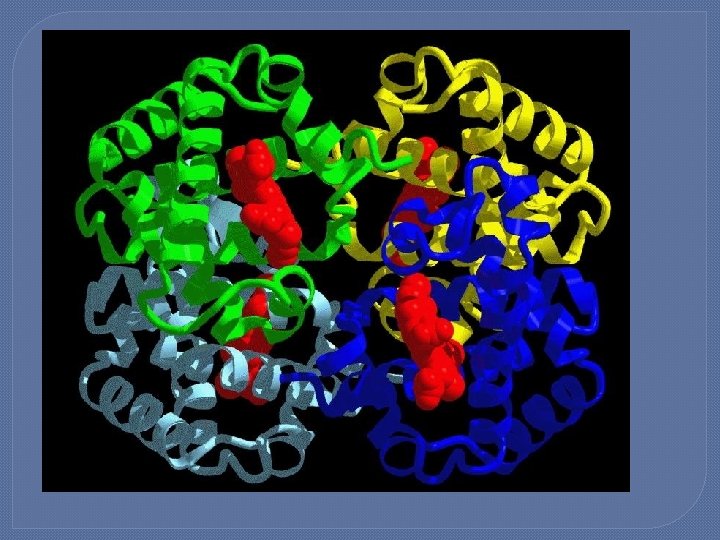

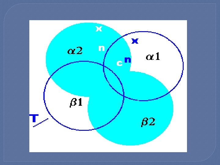
Quaternary structure of Hb �Hb molecule is a tetramer. It is composed of two identical dimers (αβ)1 and (αβ)2. Polypeptides of α and β chain are held together by hydrophobic interactions. �Polypeptides between αβ dimers are held by ionic and hydrogen bonds.
 which has a")
�There are two alternative structures of hemoglobin; the relaxed structure (R) which has a greater oxygen affinity, and the tense structure (T) which has lower affinity for oxygen.

�T-Form: The deoxy form of Hb is called the tense form. Dimers interact through ionic and hydrogen bonds. These restrict the movement of the chains. This form has got low oxygen affinity.

�R- Form: The oxy form of Hb is called Relaxed form. The binding of oxygen to the Hb causes the rupture of the ionic and hydrogen bonds. This leads to increase movement of the polypeptide chains. This form has got high affinity for oxygen.

�The change between the T and R structures is the result of a rotation of 15 degrees between the two alpha-beta dimers.

Allosteric Effects �In order to function most efficiently, hemoglobin needs to bind to oxygen tightly in the oxygen-rich atmosphere of the lungs and be able to release oxygen rapidly in the relatively oxygen-poor environment of the tissues.

�Essentially, hemoglobin is an allosteric protein that has more than one shape and can undergo conformational changes in its structure based on environment conditions.

�The ability of hemoglobin to take up oxygen molecules in the lungs and then release them in the tissues is regulated by several factors both within the hemoglobin molecule itself and through external chemical factors.

�The ability of hemoglobin to reversibly bind oxygen is affected by : � 1. p. O 2 � 2. p. H � 3. p. CO 2 � 4. 2 3 BPG

1. Heme-heme interactions �One of the biggest regulators of the oxygen affinity of the hemoglobin is the presence of oxygen itself.

�In the lungs where the oxygen levels are high, the hemoglobin has a higher affinity for oxygen and this affinity increases disproportionately with the number of molecules it already has bound to it

�In other words, after the oxyhemoglobin binds one molecule of oxygen its affinity for oxygen increases until the hemoglobin is fully saturated.

�In the same way, the deoxyhemoglobin has a lower affinity for oxygen and this affinity decreases disproportionately with the number of molecules it already has bound.

�Thus, the loss of one oxygen molecule from the deoxyhemoglobin lowers the affinity for the remaining oxygen. This regulation is known as Cooperativity.

� Cooperativity is essential to the functioning of the hemoglobin because it allows the oxyhemoglobin to carry the maximum amount of oxygen to the tissues and then allows the deoxyhemoglobin to release the maximum amount of oxygen into the tissues.

�Net effect is that the affinity of Hb for the last oxygen bound is 300 times greater then its affinity for the first oxygen bound. �This effect is called heme- heme interaction.

Dissociation Curve: �It is sigmoidal in shape for Hb. �This means that the subunits cooperate in binding oxygen. �It shows that binding of an oxygen molecule at one heme group increases the affinity of the remaining heme groups for oxygen.

�It is hyperbolic in shape for myoglobin. �Myoglobin reversibly binds a single molecule of oxygen. �Oxygenated and deoxygenated forms exit in equilibrium. �Myoglobin is designed to release the oxygen in muscles in response to oxygen demand.

2. Bohr Effect: �When CO 2 is released into the blood from the tissues it acidifies the blood by increasing the concentration of hydrogen ions.

�This lowering in p. H causes the oxygen affinity of the hemoglobin to decrease, which is known as the Bohr effect.

�The molecular basis behind the Bohr effect is that the T structure of hemoglobin binds hydrogen more readily than the R structure, so under a condition of low p. H (high hydrogen ion concentration) the T structure, which has a decreased oxygen affinity, dominates.

�Bohr effect is because of the ionizable groups plus histidine side chain. �When the conc of ions increase these groups are charged and are able to form ionic bonds. These bonds stabilize the T form and so the affinity for oxygen decreases.

�Bohr effect causes the shift to the right in the oxygen dissociation curve.

Sources of Protons: a. Hydrogen ions and CO 2 more in the metabolically active tissues. b. Organic acids produced during anaerobic metabolism ( role of carbonic anhydrase).

Mechanism of Bohr effect: �Bohr effect reflects that the deoxyform of Hb has got a greater affinity for protons than oxy. Hb. �It is because of the ionizable groups.

�When conc of protons increases these groups become charged and form ionic bonds. �These bonds stabilize the deoxyform of Hb and produces a decrease affinity for oxygen.

�Bohr effect shifts the curve to the right.

3. 2, 3 – Bisphoglycerate : � 2, 3 - bisphoglycerate is an allosteric effector that changes the oxygen affinity of hemoglobin by binding to the hemoglobin itself.

�It decreases the oxygen affinity of hemoglobin by binding to deoxy form. This stabilizes the taut structure. Hb. O 2 + 2, 3 BPG Hb-2, 3 BPG + O 2

� 2, 3 BPG binds to a pocket formed by two beta- globin chains in the center of the tetramer. �Pocket has got positively charged amino acids that forms ionic bonds with 2, 3 BPG. �When O 2 binds then 2, 3 BPG is expelled.

� 2, 3 BPG causes the shift to the right in the oxygen dissociation curve.
 and")
� 2, 3 BPG levels are increased in chronic hypoxia (high altitudes, COPD) and chronic anemia. � 2, 3 BPG absent in fetal Hb. �Role in transfused blood (acid citrate dextrose)(Inosine hypoxanthine ribose).

� 2, 3 BPG is essential for the normal transport function of Hb. Storing of blood in acid citrate medium decreases 2, 3 BPG and the affinity of Hb for oxygen increases.

�Fetal Hb has got a gamma globin chain, this has got less positive charge so does not bind 2, 3 BPG. �This allows the Hb. F to facilitate the transfer of oxygen from the maternal blood to the fetal blood.

4. Binding of CO 2 : �CO 2 has a similar effect on the hemoglobin, but instead of binding to the heme molecule like oxygen, CO 2 binds to the N-terminus of the alpha globin molecule.

�The CO 2 binds better to the globin in the T structure, so the release of oxygen in the tissues by the T structure of hemoglobin facilitates the uptake of CO 2.

�CO 2 is mostly transported in the form of bicarbonate. �Some is transported by Hb. It is called carbamino hemoglobin or carbamate, because it is attached to the uncharged α amino groups.

Ligand binding �Besides the oxygen ligand which binds to hemoglobin in a cooperative manner, hemoglobin ligands also include competitive inhibitors such as carbon monoxide (CO) and allosteric ligands such as carbon dioxide (CO 2).

Competitive �Hemoglobin's oxygen-binding capacity is decreased in the presence of carbon monoxide because both gases compete for the same binding sites on hemoglobin, carbon monoxide binding preferentially in place of oxygen.
 (for")
�The binding of oxygen is affected by molecules such as carbon monoxide (CO) (for example from tobacco smoking, car exhaust and incomplete combustion in furnaces). CO competes with oxygen at the heme binding site.

�Hemoglobin binding affinity for CO is 200 times greater than its affinity for oxygen, meaning that small amounts of CO dramatically reduce hemoglobin's ability to transport oxygen.

�When hemoglobin combines with CO, it forms a very bright red compound called carboxyhemoglobin, which may cause the skin of CO poisoning victims to appear pink in death, instead of white or blue.

�When inspired air contains CO levels as low as 0. 02%, headache and nausea occur; if the CO concentration is increased to 0. 1%, unconsciousness will follow. In heavy smokers, up to 20% of the oxygenactive sites can be blocked by CO.
, sulfur monoxide (SO),")
�In similar fashion, hemoglobin also has competitive binding affinity for cyanide(CN), sulfur monoxide (SO), nitrogen dioxide(NO 2), and sulfide(S 2 -), including hydrogen sulfide (H 2 S). All of these bind to iron in heme without changing its oxidation state, but they nevertheless inhibit oxygenbinding, causing grave toxicity.

Allosteric �Carbon dioxide occupies a different binding site on the hemoglobin. Carbon dioxide is more readily dissolved in deoxygenated blood, facilitating its removal from the body after the oxygen has been released to tissues undergoing metabolism. This increased affinity for carbon dioxide by the venous blood is known as the Haldane effect.

�Through the enzyme carbonic anhydrase, carbon dioxide reacts with water to give carbonic acid, which decomposes into bicarbonate and protons: �CO 2 + H 2 O → H 2 CO 3 → HCO 3ˉ + H⁺

�Hence blood with high carbon dioxide levels is also lower in p. H (more acidic). Hemoglobin can bind protons and carbon dioxide which causes a conformational change in the protein and facilitates the release of oxygen.

Hb. O 2 + H⁺ Hb. H + O 2
- Slides: 63