Structure Bacteria produce a single endospore internally The


Structure Bacteria produce a single endospore internally. The spore is sometimes surrounded by a thin covering known as the exosporium, which overlies the spore coat. The spore coat, which acts like a sieve that excludes large toxic molecules like lysozyme, is resistant to many toxic molecules and may also contain enzymes that are involved in germination.

In Bacillus subtilus endospores, the spore coat is estimated to contain more than 70 coat proteins, which are organized into an inner and an outer coat layer. The cortex lies beneath the spore coat and consists of peptidoglycan. The core wall lies beneath the cortex and surrounds the protoplast or core of the endospore. The core contains the spore chromosomal DNA which is encased in chromatin-like proteins known as SASPs (small acid-soluble spore proteins), that protect the spore DNA from UV radiation and heat. The core also contains normal cell structures, such as ribosomes and other enzymes, but is not metabolically active.
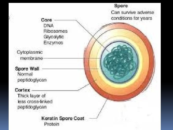

Up to 20% of the dry weight of the endospore consists of calcium dipicolinate within the core, which is thought to stabilize the DNA. Dipicolinic acid could be responsible for the heat resistance of the spore, and calcium may aid in resistance to heat and oxidizing agents. However, mutants resistant to heat but lacking dipicolinic acid have been isolated, suggesting other mechanisms contributing to heat resistance are also at work Small acid-soluble proteins (SASPs) are found in endospores. These proteins tightly bind and condense the DNA, and are in part responsible for resistance to UV light and DNA-damaging chemicals.

Visualising endospores under light microscopy can be difficult due to the impermeability of the endospore wall to dyes and stains. While the rest of a bacterial cell may stain, the endospore is left colourless. To combat this, a special stain technique called a Moeller stain is used. That allows the endospore to show up as red, while the rest of the cell stains blue. Another staining technique for endospores is the Schaeffer-Fulton stain, which stains endospores green and bacterial bodies red. The arrangement of spore layers is as follows: Exosporium Spore coat Spore cortex Core wall

Location The position of the endospore differs among bacterial species and is useful in identification. The main types within the cell are terminal, subterminal, and centrally placed endospores. Terminal endospores are seen at the poles of cells, whereas central endospores are more or less in the middle. Subterminal endospores are those between these two extremes, usually seen far enough towards the poles but close enough to the center so as not to be considered either terminal or central. Lateral endospores are seen occasionally.
 central endospore; (2, 3, 5) terminal endospore; (6)")
Variations in endospore morphology: (1, 4) central endospore; (2, 3, 5) terminal endospore; (6) lateral endospore

Formation and destruction Under conditions of starvation, especially the lack of carbon and nitrogen sources, a single endospore forms within some of the bacteria through a process called sporulation When a bacterium detects environmental conditions are becoming unfavourable it may start the process of endosporulation, which takes about eight hours. The DNA is replicated and a membrane wall known as a spore septum begins to form between it and the rest of the cell. The plasma membrane of the cell surrounds this wall and pinches off to leave a double membrane around the DNA, and the developing structure is now known as a forespore.

Calcium dipicolinate, the calcium salt of dipicolinic acid, is incorporated into the forespore during this time. The dipicolinic acid helps stabilize the proteins and DNA in the endospore. Next the peptidoglycan cortex forms between the two layers and the bacterium adds a spore coat to the outside of the forespore. In the final stages of endospore formation the newly forming endospore is dehydrated and allowed to mature before being released from the mother cell.

The cortex is what makes the endospore so resistant to temperature. The cortex contains an inner membrane known as the core. The inner membrane that surrounds this core leads to the endospore's resistance against UV light and harsh chemicals that would normally destroy microbes Sporulation is now complete, and the mature endospore will be released when the surrounding vegetative cell is degraded.

Endospores are resistant to most agents that would normally kill the vegetative cells they formed from. Unlike persister cells, endospores are the result of a morphological differentiation process triggered by nutrient limitation (starvation) in the environment; endosporulation is initiated by quorum sensing within the "starving" population Most disinfectants such as household cleaning products, alcohols, quaternary ammonium compounds and detergents have little effect on endospores.

quorum sensing bacteria produce and release chemical signal molecules called autoinducers Gram-positive and Gram-negative bacteria use quorum sensing communication circuits to regulate a diverse array of physiological activities. These processes include symbiosis, competence, antibiotic production, motility, sporulation, and biofilm formation.
- Slides: 13