Structure and function of the mammalian skeleton Todays


- Slides: 23
Structure and function of the mammalian skeleton
Today's Objective • The skeleton
The skeletal system consists of: – Bones – Cartilage – Ligaments The skeletal system allows movement and provides support and protection as well as a storage area, and a production site for red blood cells
Skeletal Functions • Support – Rigid bone for weight bearing – Cartilage for flexible support - ear • Protection – Hard bone protects underlying organs – chest to protect lungs and heart • Movement – Muscles are attached by tendons – Joints (where two bones meet and come together) allow movement. Bones are attached to each other by ligaments • Storage – Storage of certain minerals (especially calcium) • Blood Cell Production – Occurs in the bone marrow
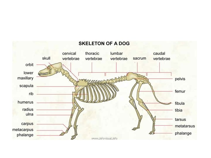
The skeleton is divided into sections: • Axial Skeleton – This forms the straight line of the body – this is the vertebral column – the spine / skull / ribs / sternum • Appendicular Skeleton – These are bones that are attached to the axial skeleton. These are the forelimbs, hind limbs, pelvis • Splanchic Regions – Os penis – bone that develops in soft tissue
Axial Skeleton
Atlas Cervical Vertebrae Axis Thoracic Vertebrae Lumbar vertebrae Sacrum Coccygeal Verterae Axial Skeleton
The Forelimb
Scapula Humerus Radius Ulna Carpals Metacarpals The Forelimb
The Hindlimb
Pelvis femur phalanges The Hindlimb Tibia Fibula Tarsals
Other Bones
Cranium Maxilla Mandible Rib Cage Other Bones
Atlas Cervical Vertebrae Axis Lumbar vertebrae Thoracic Vertebrae Mandible Sacrum Coccygel Vertebrae Scapula Pelvis Humerus femur Rib Cage Radius Tibia Ulna Fibula Carpals Tarsals Metacarpals
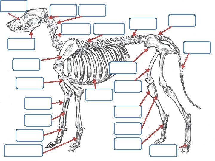
Your homework • You are required to remember the anatomy of the skeleton!
Joints • Joints are placed where two or more bones meet • There are 3 types of joints: – A- Fibrous – B- Cartilaginous – C- Synovial
Fibrous Joints • There are no movement • Found in the skull Cartilaginous Joints • Slight movement • Found between vertebrae, between ribs , sternum and pelvis Synovial Joints Free movement Found in appendicular skeleton The end of the bone in a synovial joint have a thin layer of cartilage, and fluid lubricant within the joint capsule named synovial fluid. Found in the knee joint, hip joint
Synovial Joints
Muscles Location Smooth Cardiac Skeletal Wall of hollow organs, vessels, respiratory passageways Wall of heart Attached to bones Involuntary Voluntary Cell Characteristics Control Action
Next Week • Your assessment on the skeletal system!
QUIZ • http: //www. vet. osu. edu/assets/flash/educati on/outreach/games/skeleton. html