
Structure and Function of the Eye SPE 516
- Slides: 31
+ Structure and Function of the Eye SPE 516
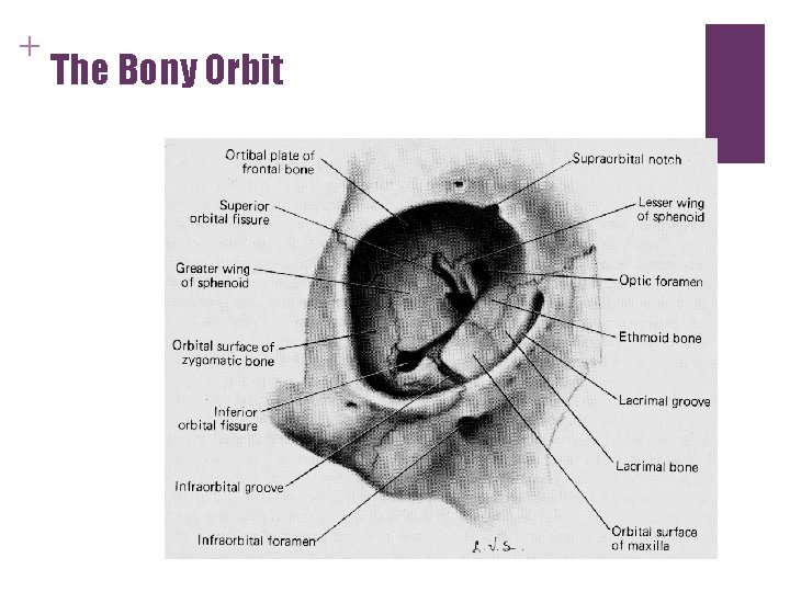
+ The Bony Orbit
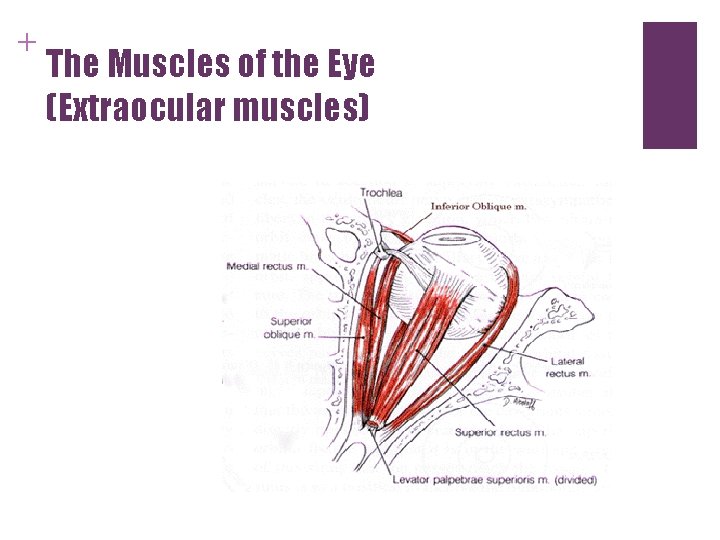
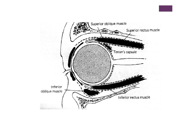
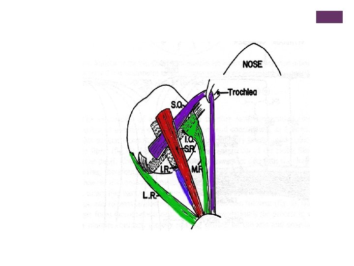
+ The Muscles of the Eye (Extraocular muscles)
+ Superior and Inferior rectus n Superior rectus n Attached to the eye at 12 o’clock n Moves the eye up. n Inferior rectus n Attached to the eye at 6 o’clock n Moves the eye down.
+ Lateral Rectus n Also called the external rectus n Attaches on the temporal side of the eye n Moves the eye toward the outside of the head (toward the temple)
+ Medial Rectus n Also called the internal rectus n Attached on the nasal side of the eye n Moves the eye toward the middle of the head (toward the nose)
+ Superior Oblique n Attached high on the temporal side of the eye. n Passes under the Superior Rectus. n Moves the eye in a diagonal pattern -- down and in. n Travels through the trochlea
+ Inferior Oblique n Attached low on the nasal side of the eye. n Passes over the Inferior Rectus. n Moves the eye in a diagonal pattern -- up and out.
+ The Nerves That Control the Muscles of the Eye n Third Cranial Nerves n Fourth Cranial Nerves n Sixth Cranial Nerves
+ Lids and Lashes n Main function is the protection of the eye. n They also help to distribute tears which wash and lubricate the eyes.
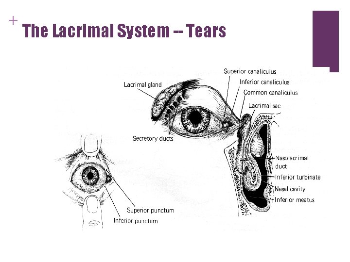
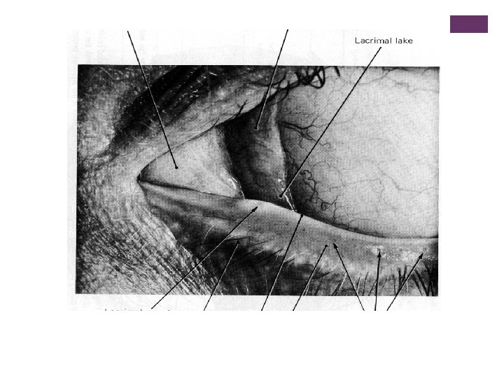
+ The Lacrimal System -- Tears
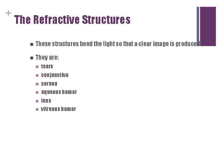
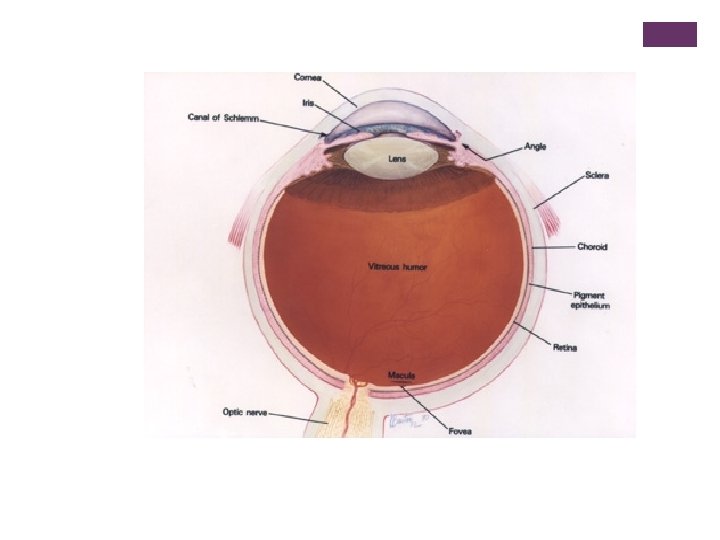
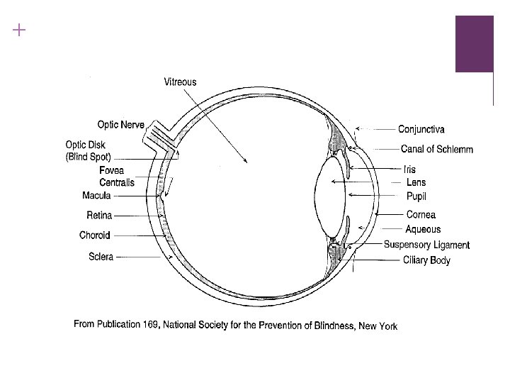
+ The Refractive Structures n These structures bend the light so that a clear image is produced. n They are: n tears n conjunctiva n cornea n aqueous humor n lens n vitreous humor
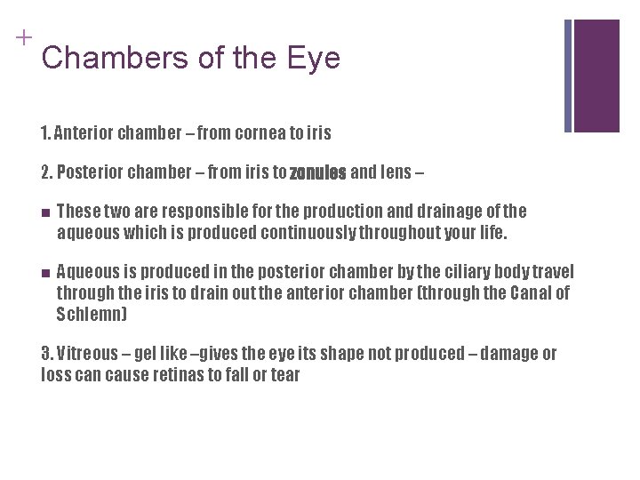
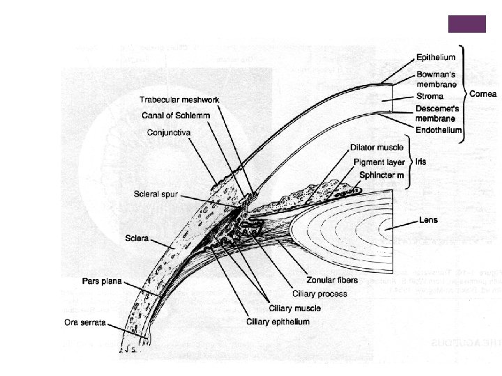
+ Chambers of the Eye 1. Anterior chamber – from cornea to iris 2. Posterior chamber – from iris to zonules and lens – n These two are responsible for the production and drainage of the aqueous which is produced continuously throughout your life. n Aqueous is produced in the posterior chamber by the ciliary body travel through the iris to drain out the anterior chamber (through the Canal of Schlemn) 3. Vitreous – gel like –gives the eye its shape not produced – damage or loss can cause retinas to fall or tear
+ Layers of the eye n Sclera and Cornea n uveal tract n n Choroid n Iris n Ciliary body retina
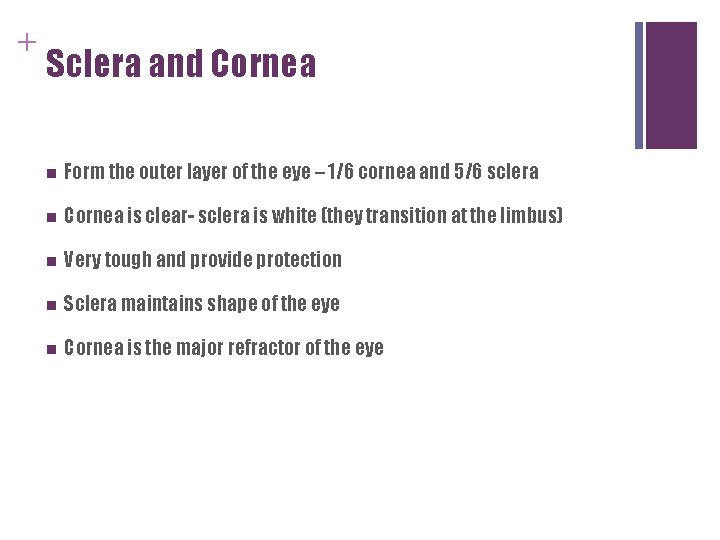
+ Sclera and Cornea n Form the outer layer of the eye – 1/6 cornea and 5/6 sclera n Cornea is clear- sclera is white (they transition at the limbus) n Very tough and provide protection n Sclera maintains shape of the eye n Cornea is the major refractor of the eye
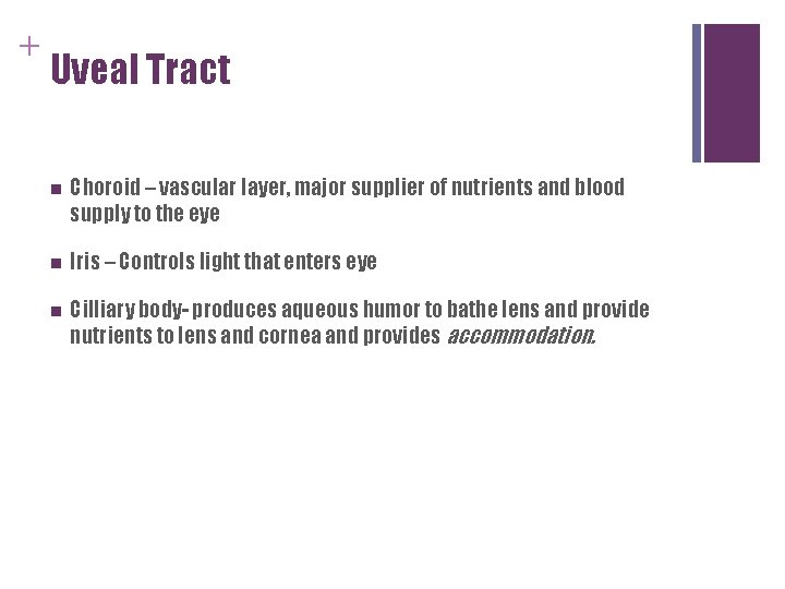
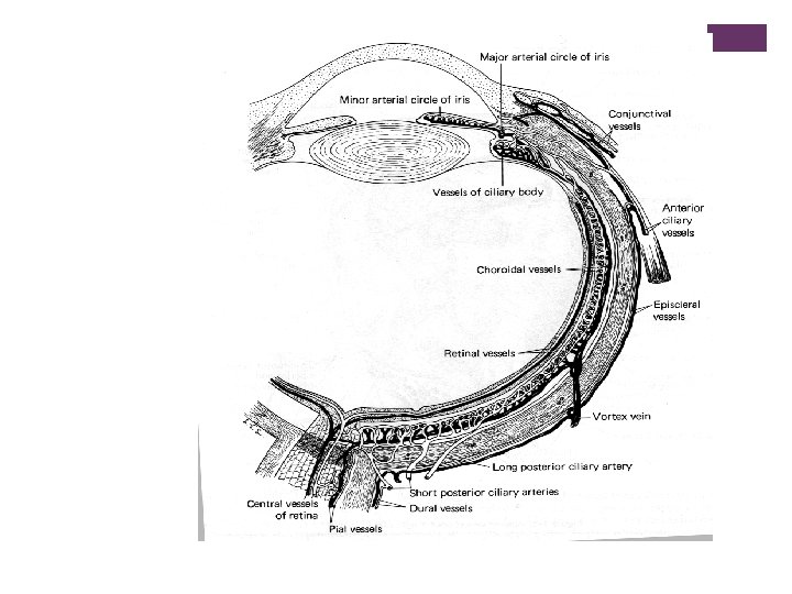
+ Uveal Tract n Choroid – vascular layer, major supplier of nutrients and blood supply to the eye n Iris – Controls light that enters eye n Cilliary body- produces aqueous humor to bathe lens and provide nutrients to lens and cornea and provides accommodation.
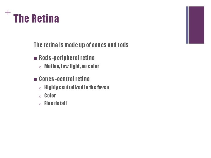
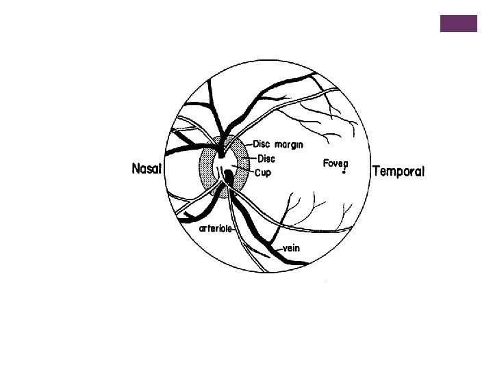
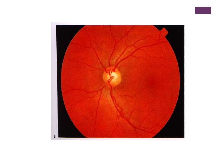
+ The Retina The retina is made up of cones and rods n Rods -peripheral retina o n Motion, low light, no color Cones -central retina o Highly centralized in the fovea o Color o Fine detail
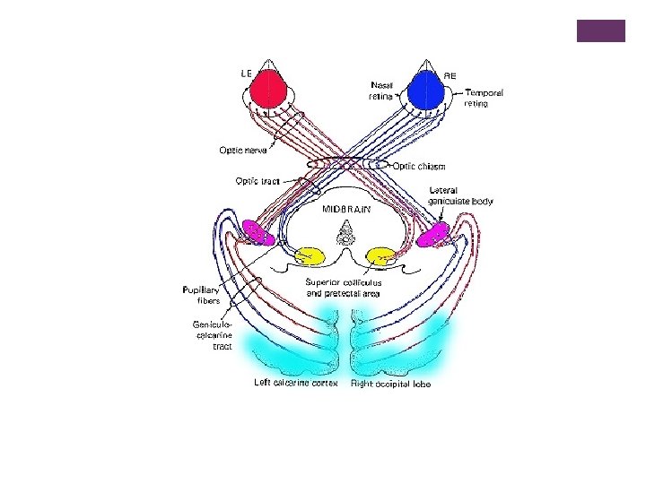
+ The Optic Pathway n Begins at the optic nerve. n Impulses cross and partially split at the optic chiasm. n After the chiasm, it becomes the optic tract. n Lateral geniculate bodies (sensory way stations) n Some fibers go to the colliculus (located in the mid brain)
+ Optic Pathway (cont. ) n The other fibers fan out into the visual cortex which is located at the top and back of the brain.
+ Vision and the Brain n n Primary visual cortex (Striated Cortex) -n spatial organization of a scene n shapes of objects n brightness and shading of parts of objects Secondary visual cortex (Prestriated Cortex) -n pattern recognition
+ The Brain and Vision (cont) n n Temporal Lobes n center for visual learning n recognition by sight Midbrain -- Limbic sector n n emotional responses to visual stimuli Midbrain -- Superior Colliculus -n guides visual attention