Structure and Function of Muscle ANSC 3404 Objectives

Structure and Function of Muscle ANSC 3404 Objectives: Study the structures of muscle and the mechanism of muscle contraction.

Muscle Types SKELETAL SMOOTH CARDIAC METHOD OF CONTROL VOLUNTARY INVOLUNTARY BANDING PATTERN STRIATED NON-STRIATED NUCLEI/CELL MULTI SINGLE

Cardiac Muscle

Smooth Muscle

Skeletal Muscle

Muscle Cross Sections Showing Bundles of Myofibers FAT CELL BLOOD VESSEL

Cross Section of Muscle Fibers NUCLEI

Myofiber

Red and White Fibers in Muscle

Fiber types

The Blood Supply for Myofibers

Connective Tissues

Position of Mysiums in Muscle Perimysium Epimysium Endomysium ENDO = within PERI = around EPI = upon

• Endomysium from muscle not aged • Endomysium after cooler aging (28 D At 4 o. C)

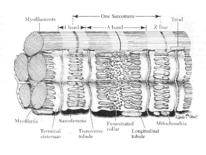
The Sarcoplasmic Reticulum • Sarcoplasmic reticulum – T-tubule • Calcium Storage • Required for contraction

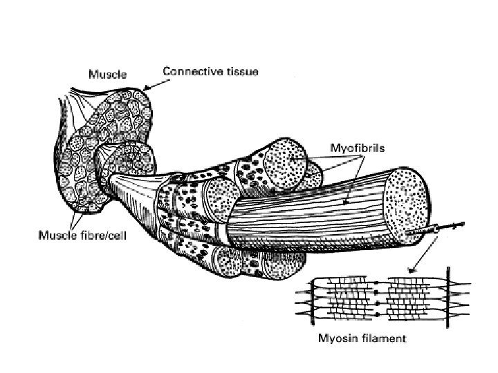
Structure of Muscle
")
Structure of Muscle (Cont)

Sarcomere • Functional unit of a muscle • Runs from z-line to z-line – Actin – Myosin

Myosin Filament

Actin Filament

Muscle Structure

Critical Contractile Proteins

REVIEW

Fat Structures

A D I P O S E T I S S U E ADIPOCYTE BLOOD VESSEL ADIPOCYTES
 I. M. =")
Fat Layers and Depots I. F. = Inter-fasicular or intramucular (marbling) I. M. = Intermuscular (seam fat) PR. = Perinephric or Perirenal (fat around the kidneys)

FAT CELLS Adipoblasts develop at widely varying rates in different parts of the body.

Adipogenesis • Adipoblasts – 20 microns in diameter • Adipocytes – 120 micron in diameter – 300 micron in obese • Cellular make-up – 95% of cytoplasm is lipid – Remainder primarily nucleus

A D I P O C Y T E

HOW ARE ADIPOCYTES FORMED? ONCE RECRUITED & FILLED, AN ADIPOCYTE IS EASIER TO FILL AGAIN THAN IF NOT FILLED BEFORE When filled with lipid, it is a mature adipocyte Adipose cells begin to accumulate

Muscle Contraction

Introduction • Overall structure of muscle is designed for contraction and relaxation, which leads to movement and locomotion. • The ability to contract and relax is lost during the transformation of muscle to meat. • Events surrounding this conversion greatly impact meat palatability

Introduction • The biochemical processes that provide energy to the living muscle cause the accumulation of metabolites during harvest – Affects color, WHC, p. H, others • An understanding of muscle contraction is necessary to understand these processes

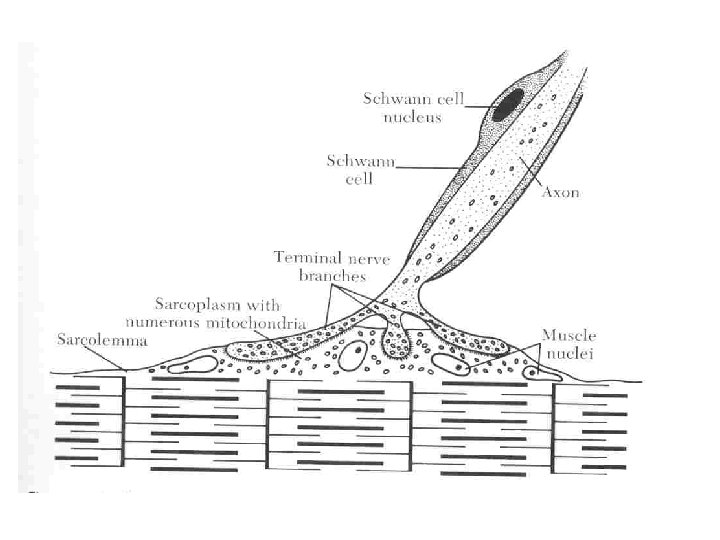
Contraction • Begins with stimuli that arrive at the surface of the muscle fiber at the sarcolemma • Nerve impulse starts in the brain and is transmitted via nerves to the muscle

Transmembrane Potentials • Under resting conditions, an electric potential exists between the inside and outside of the cell – Fluids inside are negative – Fluids outside are positive – Results in a resting membrane potential
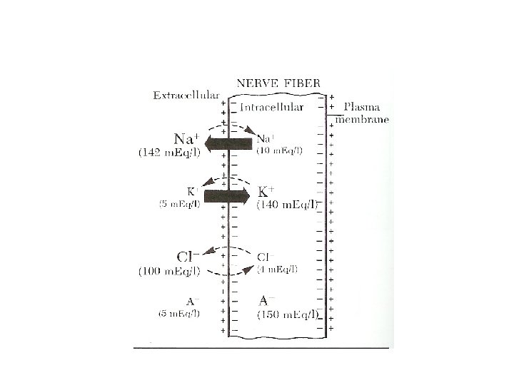

Transmembrane Potentials • Extracellular – Na+ and Cl • Intracellular – K+ and A • Na+ and K+ gradient maintained by a sodiumpotassium pump.

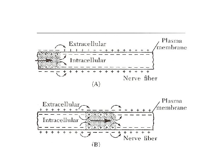
Action Potential • Transmits electric impulse to muscle • Travels along the membrane surface of the nerve fiber by depolarization – Initiated by a dramatic increase in the permeability of Na+ – Na+ rushes into cell to establish equilibrium; however K+ stays in cell causing a change in the net charge inside the cell to positive • Lasts only a millisecond (0. 5 to 1 millisecond) before the permeability to Na+ is changed to resting state

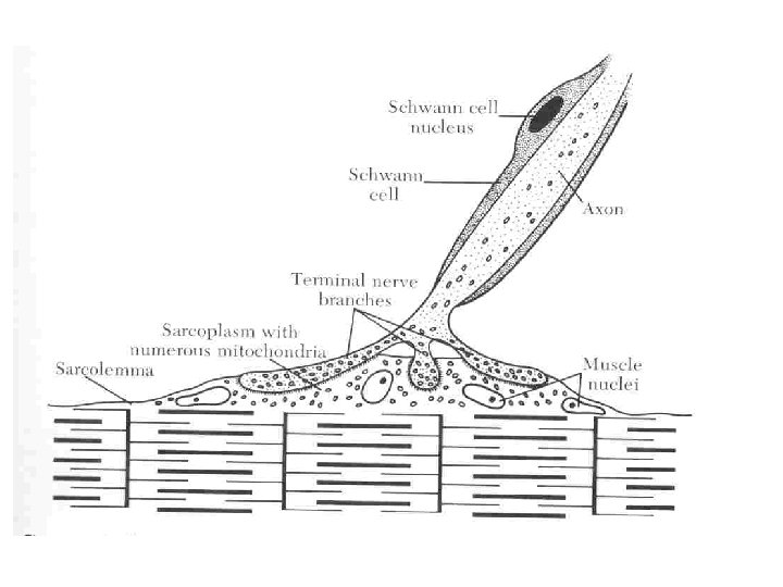
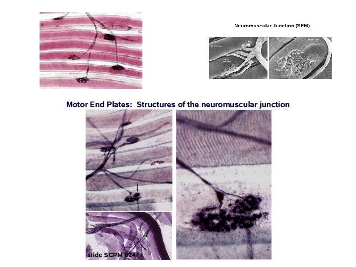
Myoneural Junction • Action potential is not strong enough to elicit a response alone • Uses a chemical transmitter called acetylcholine to be released. – Acetylcholinesterase is quickly released to neutralize the acetylcholine

Muscle Action Potentials • Same as the action potential for nerve fibers • Communicated to the inner muscle cell via the T-tubule system – Action potential transverse a muscle fiber via the ttubules and are ultimately responsible for the release of calcium from the SR
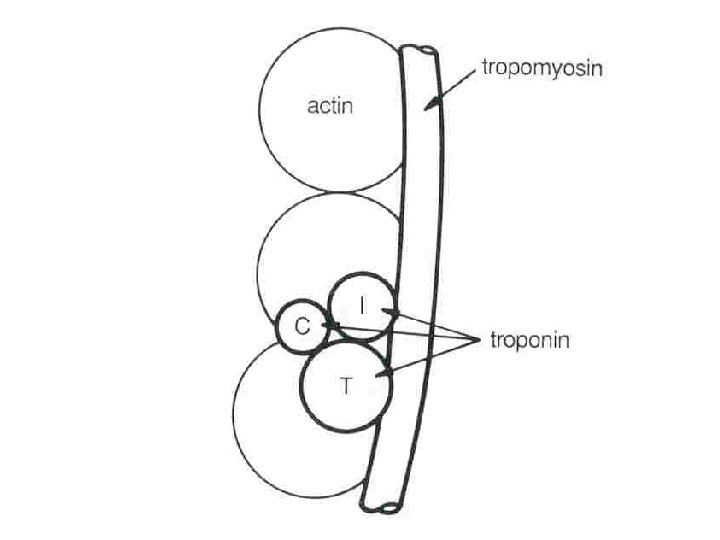
 Tropomyosin")
Sarcomere - Basic contractile unit of the muscle 1. Myosin 2. Actin a) Tropomyosin b) Troponin 3. Z lines

Elements required for muscle contraction and relaxation 1. Acetylcholine and Acetylcholinesterase 2. Calcium 3. Adenosine 5'-triphosphate (ATP) a) Derived from aerobic and anaerobic metabolism

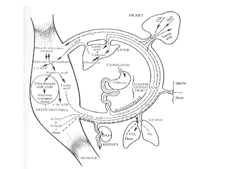
Sources of Energy for Muscle Contraction and Relaxation

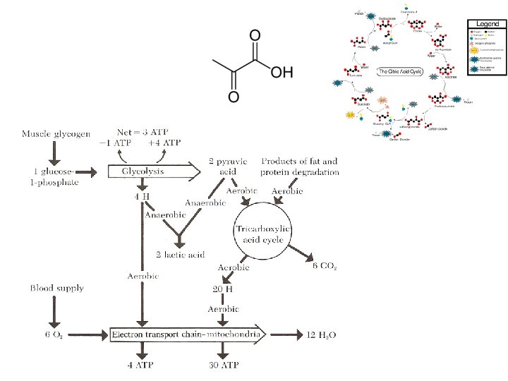
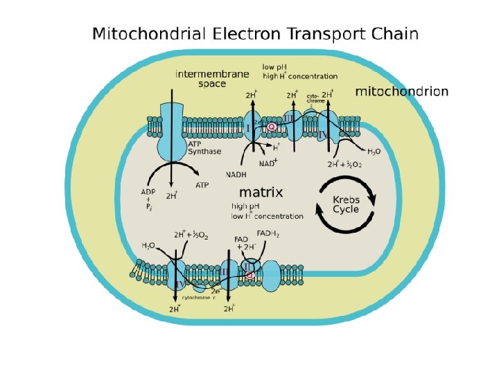
Energy • Aerobic – Glycolysis – TCA cycle – Electron transport chain • Anerobic – Excess Hydrogen is used to reduce pyruvic acid to lactic acid, which permits glycolysis to proceed at a rapid rate – Easily fatigued

Contraction Phase 1. Nerve pulse/impulse transmitted through action potential 2. Acetylcholine is released at neural juncture 3. Action potential transmitted to muscle fiber via the T-tubles to the sarcoplasmic reticulum (SR)

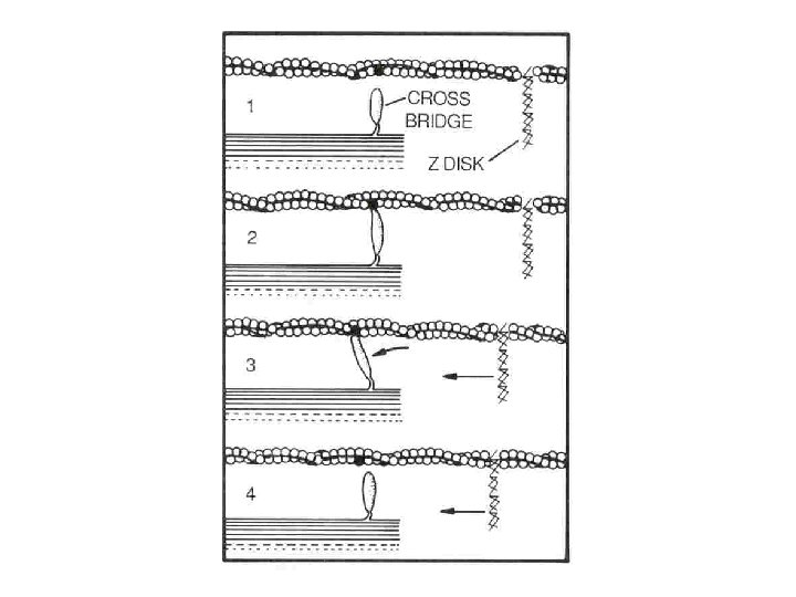
Contraction phase 4. Calcium is released from SR into sarcoplasm 5. Calcium binds to troponin 6. ATP is hydrolyzed (burned) 7. Energy causes a shift in tropomyosin and actin binding site is exposed
 9. ATP")
Contraction phase 8. Actin-myosin cross bridge forms (cross bridge is termed actomyosin) 9. ATP hydrolyzed 10. Myosin head rotates 11. Repeated over and over; filaments slide causing shortening of sarcomere

Contraction

ACTIN A sarcomere contracting Notice that neither filament changes length MYOSIN

http: //www. unmc. edu/Physiology/Mann/ mann 14. html http: //www. blackwellpublishing. com/matt hews/myosin. html http: //entochem. tamu. edu/musclestrucco ntractswf/index. html
 2. Calcium pump activated by SR")
Relaxation phase 1. Acetylcholinesterase is released (neutralizes acetylcholine) 2. Calcium pump activated by SR to sequester calcium 3. Actin-myosin cross bridge terminated 4. Tropomyosin shifts covering the binding site on actin

Relaxation phase 5. Passive sliding of filaments 6. Sarcomere returns to resting state

Be Able To: • Draw a sarcomere showing: Myosin Actin Z Line A Band I Band H Zone M Line • Explain how a muscle contracts and relaxes – starting with a nerve impulse and including the SR role • Explain the role of ATP in muscle contraction and relaxation • (See p. 889 -900 In The Meat We Eat)
- Slides: 64