Structural and Functional Mitral Regurgitation Echo Subleties and

Structural and Functional Mitral Regurgitation: Echo Subleties and Hallmarks Steven A. Goldstein, MD Director, Noninvasive Cardiology Washington Hospital Center Sunday, February 21, 2010

The Mitral Apparatus • Mitral leaflets • Chordae tendinae • Papillary muscles • Mitral annulus • Left ventricle • Left atrium

Mitral Valve Anatomy Dilatation Left atrium Annulus Leaflets Dilatation Calcification Prolapse Redundancy Thickening Perforation Cleft Commissural fusion Chordae tendinae Abnormal insertion Elongation Rupture Thickening/fusion Papillary muscles Ischemia Fibrosis Rupture LV free wall Lateral displacement (ischemia, fibrosis, dilatation)

Mechanisms of Mitral Regurgitation Normal Prolapse Apical Tethering Dilated anulus Restricted PML Ruptured pap muscle

Mitral Valve Repair Mechanisms to be Discussed • Ruptured chordae tendinae • Mitral valve prolapse (MVP) • Ischemic/functional MR

Ruptured cords

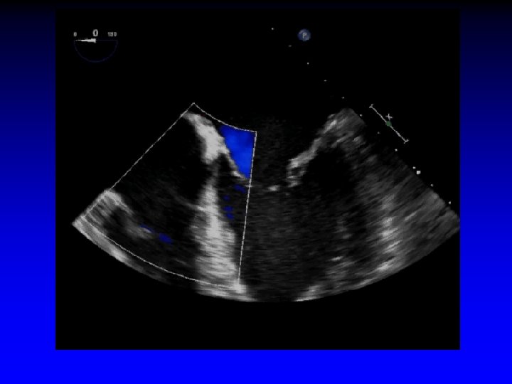
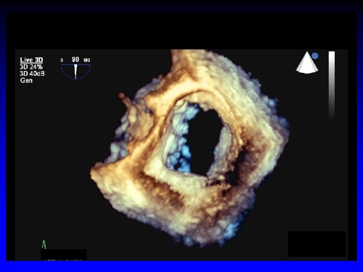
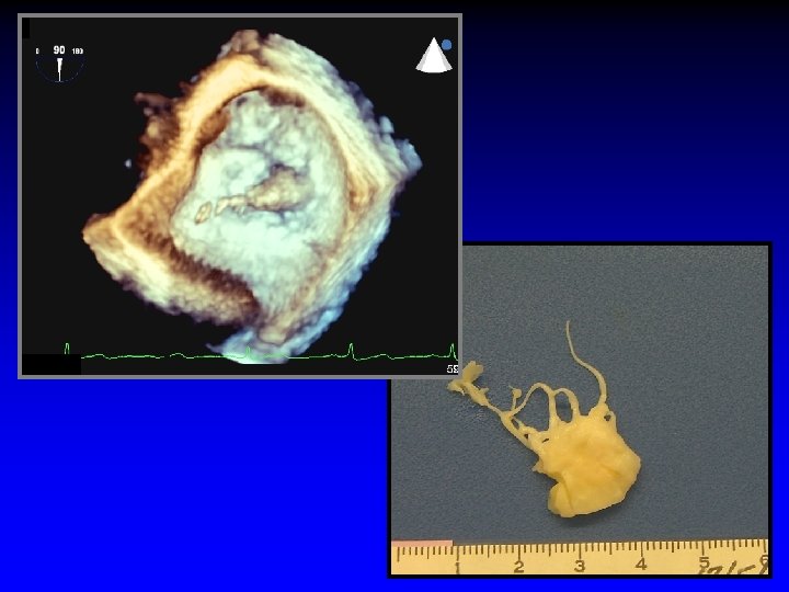
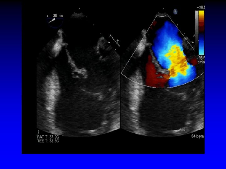
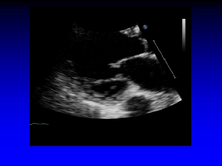
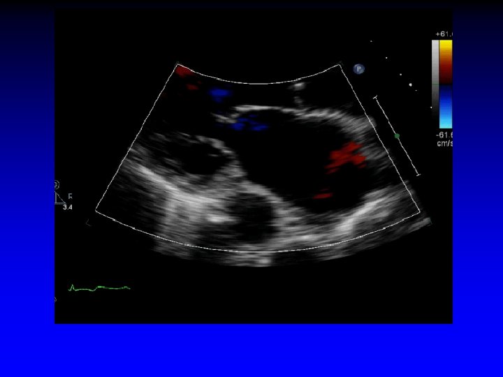
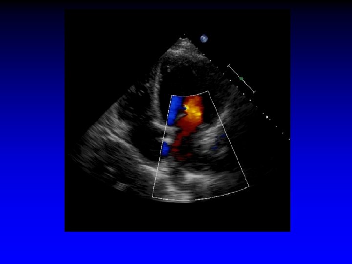
Case 1 Ruptured cords P 2
")
1. 6 cm +. . . + Gastric short-axis view (PSR view)

L-upper pulm vein Systolic flow reversal severe MR

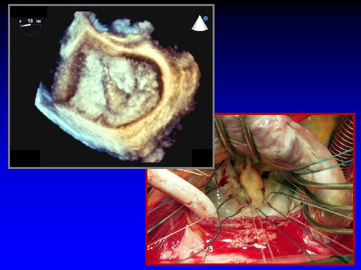
Roadmap for the surgeon
 - affected segment(s) •")
Mitral Valve Repair Information Required by Surgeon • Exact lesion(s) - affected segment(s) • Extent • Degree of calcification (leaflet/annulus) • Dilatation/size of annulus

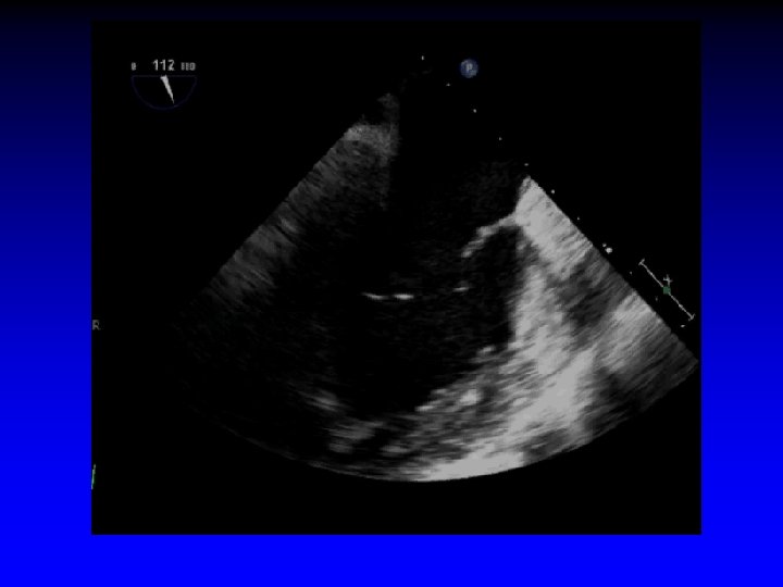
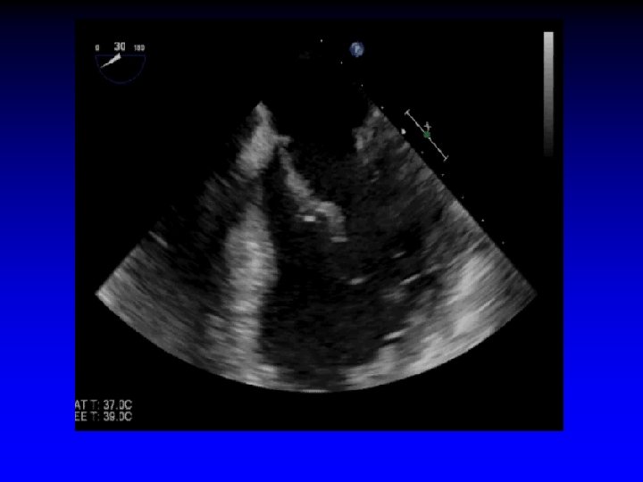
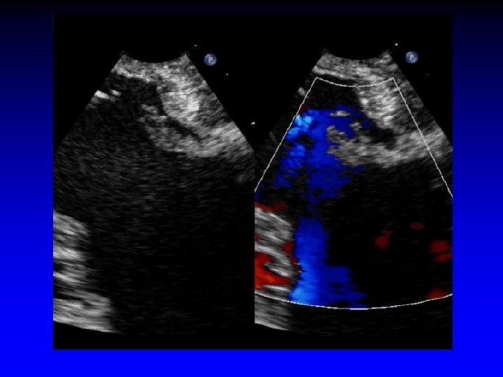
Case 2 Ruptured cord P 2

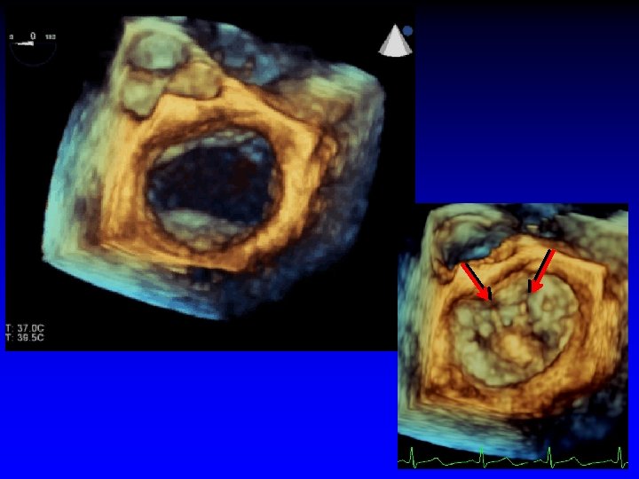
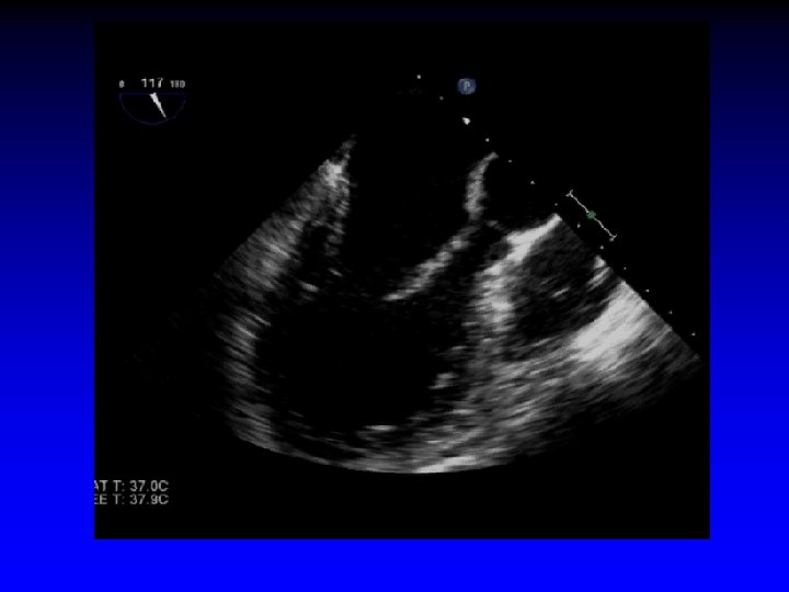
Case 3 Ruptured cords A 2 and A 3

Surgeon’s roadmap A 2 and A 3

MVP

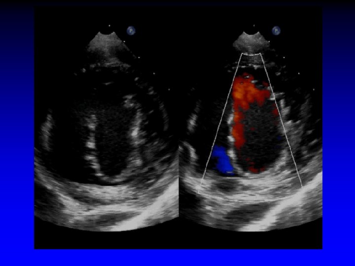
Case 4 Mitral Valve Prolapse

AML

S/P repair

Functional MR

Morphologic Changes in Heart Failure Papillary muscles displaced apically and laterally Bolling J Heart Valve Dis 11: S 28(2002)

Functional Mitral Regurgitation • Global LV dysfunction • Regional LV dysfunction • Sphericity of LV • Excessive pap muscle displacement • Decreased overlap of leaflets • LA enlargement • Loss of systolic mital anular contraction • Increased “tenting” area • Delayed activation of P-M pap muscle (dyssynchrony)

Ischemic Mitral Regurgitation C B A D Mitral leaflets are tented apically Mitral annulus is enlarged (A-B = 45 mm) Mitral coaptation depth is increased (C-D = 13 mm)

Effect of Annular Dilatation on Tethering Area MROP 340
")
Apical Tethering of Mitral Leaflets Basal cord tents anterior leaflet (“seagull sign”)
 Increased tenting area")
Basal cord tents anterior leaflet (“seagull sign”) Increased tenting area

Distance from Mitral Annulus to Mitral Leaflet Coaptation Point Normal Ventricle Spherical Ventricle

Ischemic MR

Ischemic Mitral Regurgitation "Ischemic MR" is a commonly used term, but its definition is not clearcut; Most articles discussing ischemic MR do not even define it !

Ischemic Mitral Regurgitation Definition Moderate to severe MR due to CAD (MI, myocardial ischemia, or resulting LV remodeling) in the absence of primary, preexisting leaflet or chordal pathology

Ischemic Mitral Regurgitation Mechanisms • Dislocation of papillary muscles • Increased tenting length and area • Antero-posterior dilatation of the MV annulus • Kinetics of MV annulus during cardiac cycle • Intraventricular synchrony/dyssynchrony • Atrio-ventricular synchrony

Ischemic Mitral Regurgitation Single Papillary Muscle Involved Local malfunction of LV wall adjacent to a single pap muscle Calafiore Eur Assoc Cardio-Thorac Surg 2005

Abnormal Mitral Valve Coaptation Normal coaptation Abnormal coaptation

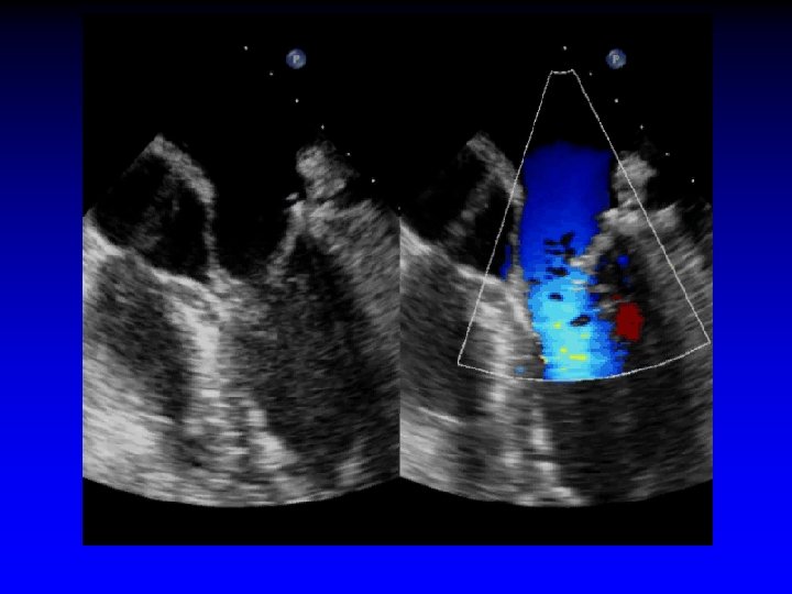
Case 7 Ischemic MR restricted PML


LVIDd = 6. 4 cm Dilated LV; posterior wall thinner than septum

Bent anterior mitral leaflet

Increased tenting area

")
Mitral Annular Calcification (MAC)

Mitral Annulus Calcification Ventricular and Leaflet Extension

Ischemic Mitral Regurgitation Mechanisms • Dislocation of papillary muscles - Papillary muscles dislocated toward apex - Apply traction to chorade - Chordae tendinae lack elasticity • Increased tenting length and area • Antero-posterior dilatation of the MV annulus • Kinetics of MV annulus during cardiac cycle • Intraventricular synchrony/dyssynchrony • Atrio-ventricular synchrony

Ischemic Mitral Regurgitation Mechanisms • Dislocation of papillary muscles • Increased tenting length and area 1. Anterior MI displacement of the 2 pap muscles in the global LV enlargement 2. Posterior MI displacement of the postero-medial pap muscle responsible for an asymmetric tethering • Antero-posterior dilatation of the MV annulus • Kinetics of MV annulus during cardiac cycle • Intraventricular synchrony/dyssynchrony • Atrio-ventricular synchrony

Ischemic Mitral Regurgitation Mechanisms • Dislocation of papillary muscles • Increased tenting length and area • Antero-posterior dilatation of the MV annulus A-P dilatation of the mitral annulus can determine the degree of MR, even if no anomaly of the mitral leaflets exists • Kinetics of MV annulus during cardiac cycle • Intraventricular synchrony/dyssynchrony • Atrio-ventricular synchrony

Ischemic Mitral Regurgitation Mechanisms • Dislocation of papillary muscles • Increased tenting length and area • Antero-posterior dilatation of the MV annulus • Kinetics of MV annulus during cardiac cycle Progress in echo imaging, particularly including 3 D-echo have demonstrated the importance of motion and specific features of kinetics of the annulus • Intraventricular synchrony/dyssynchrony • Atrio-ventricular synchrony

Ischemic Mitral Regurgitation Mechanisms • • Dislocation of papillary muscles Increased tenting length and area Antero-posterior dilatation of the MV annulus Kinetics of MV annulus during cardiac cycle • Intraventricular synchrony/dyssynchrony - Several investigators have demonstrated correlation b/w width of QRS and degree of “functional” MR - Observational data on resynchronization by pacing can improve functional ischemic MR • Atrio-ventricular synchrony
- Slides: 60