Stroke Stroke is a term used to describe

Stroke

• Stroke is a term used to describe an abrupt onset of focal neurologic deficit that lasts at least 24 hours and is presumed to be of vascular origin. • Stroke can be either ischemic or hemorrhagic in origin. • Transient ischemic attacks (TIAs) are focal ischemic neurologic deficits lasting less than 24 hours and usually less than 30 minutes.

RISK FACTORS FOR STROKE • Nonmodifiable risk factors for stroke include increased age, male gender, race (African American, Asian, Hispanic), family history of stroke, and low birth weight. • The major modifiable risk factors include hypertension and cardiac disease (especially atrial fibrillation). • Other major risk factors include diabetes mellitus, dyslipidemia, and cigarette smoking.

ISCHEMIC STROKE: • Ischemic strokes account for 88% of all strokes and are due either to local thrombus formation or to emboli that occlude a cerebral artery. • Cerebral atherosclerosis is a causative factor in most cases of ischemic stroke, although 30% are of unknown etiology. • Twenty percent of embolic strokes arise from the heart. • The final result of both thrombus formation and embolism is arterial occlusion, decreasing cerebral blood flow and causing ischemia and ultimately infarction distal to the occlusion.

HEMORRHAGIC STROKE: • Hemorrhagic strokes account for 12% of strokes and include subarachnoid hemorrhage, intracerebral hemorrhage, and subdural hematomas. • Subarachnoid hemorrhage may result from trauma or rupture of an intracranial aneurysm or arteriovenous malformation. • Intracerebral hemorrhage occurs when a ruptured blood vessel within the brain parenchyma causes formation of a hematoma. • Subdural hematomas are most often caused by trauma.

• The presence of blood in the brain parenchyma causes damage to surrounding tissue through a mass effect and the neurotoxicity of blood components and their degradation products. • Compression of tissue surrounding hematomas may lead to secondary ischemia. • Much of the early mortality of hemorrhagic stroke is due to an abrupt increase in intracranial pressure that can lead to herniation and death.

CLINICAL PRESENTATION: • The patient may experience weakness on one side of the body, inability to speak, loss of vision, vertigo, or falling. • Ischemic stroke is not usually painful, but headache may occur and may be severe in hemorrhagic stroke. • Patients usually have multiple signs of neurologic dysfunction on physical examination. • The specific deficits observed depend upon the area of the brain involved. Hemi- or monoparesis and hemisensory deficits are common. • Patients with posterior circulation involvement may present with vertigo and diplopia. Anterior circulation strokes commonly result in aphasia. • Patients may also experience dysarthria, visual field defects, and altered levels of consciousness.

DIAGNOSIS: • Protein C, protein S, and antithrombin III are best measured in steady state rather than in the acute stage. • Computed tomography (CT) head scan will reveal an area of hyperintensity (white) in an area of hemorrhage and will be normal or hypointense (dark) in an area of infarction. The area of infarction may not be visible on CT scan for 24 hours (and rarely longer). • Magnetic resonance imaging of the head will reveal areas of ischemia with higher resolution and earlier than the CT scan. Diffusion-weighted imaging will reveal an evolving infarct within minutes. • Carotid Doppler studies will determine whethere is a high degree of stenosis in the carotid arteries.

• The electrocardiogram will determine whether atrial fibrillation is present. • A transthoracic echocardiogram can detect valve or wall motion abnormalities that are sources of emboli to the brain. • A transesophageal echocardiogram is a more sensitive test for left atrial thrombus. It is also effective in examining the aortic arch for atheroma, another potential source of emboli. • Transcranial Doppler can determine the presence of intracranial sclerosis (e. g. , middle cerebral artery stenosis).
 reduce")
DESIRED OUTCOME: • The goals of treatment for acute stroke are to: (1) reduce the ongoing neurologic injury and decrease mortality and long-term disability; (2) prevent complications secondary to immobility and neurologic dysfunction; and (3) prevent stroke recurrence.

TREATMENT: GENERAL APPROACH: • The initial approach is to ensure adequate respiratory and cardiac support and to determine quickly whether the lesion is ischemic or hemorrhagic based on a CT scan. • Ischemic stroke patients presenting within hours of symptom onset should be evaluated for reperfusion therapy. • Elevated blood pressure should remain untreated in the acute period (first 7 days) after ischemic stroke because of the risk of decreasing cerebral blood flow and worsening symptoms. • The pressure should be lowered if it exceeds 220/120 mm Hg or there is evidence of aortic dissection, acute myocardial infarction, pulmonary edema, or hypertensive encephalopathy. • If blood pressure is treated in the acute phase, short-acting parenteral agents (e. g. , labetalol, nicardipine, nitroprusside) are preferred.

• Patients with hemorrhagic stroke should be assessed to determine whether they are candidates for surgical intervention via an endovascular or craniotomy approach. • After the hyperacute phase has passed, attention is focused on preventing progressive deficits, minimizing complications, and instituting appropriate secondary prevention strategies.
 IV tissue plasminogen activator (alteplase) within 3 hours of onset; and (2) aspirin")
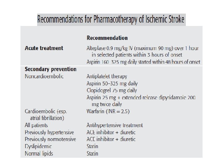
(1) IV tissue plasminogen activator (alteplase) within 3 hours of onset; and (2) aspirin within 48 hours of onset. Alteplase initiated within 3 hours of symptom onset has been shown to reduce the ultimate disability due to ischemic stroke. A head CT scan must be obtained to rule out hemorrhage before beginning therapy. The patient must also meet specific inclusion criteria and no exclusionary criteria. The dose is 0. 9 mg/kg (maximum 90 mg) infused IV over 1 hour after a bolus of 10% of the total dose given over 1 minute.

• Anticoagulant and antiplatelet therapy should be avoided for 24 hours, and the patient should be monitored closely for hemorrhage. • Aspirin 50 to 325 mg/day started between 24 and 48 hours after completion of alteplase has also been shown to reduce long-term death and disability. • The AHA/ASA guidelines recommend that antiplatelet therapy as the cornerstone of antithrombotic therapy for the secondary prevention of ischemic stroke and should be used in noncardioembolic strokes. • Aspirin, clopidogrel, and extended-release dipyridamole plus aspirin are all considered first-line antiplatelet agents

Warfarin: • is the antithrombotic agent of first choice for secondary prevention in patients with atrial fibrillation and a presumed cardiac source of embolism. • Elevated blood pressure is common after ischemic stroke, and its treatment is associated with a decreased risk of stroke recurrence. • The Joint National Committee and AHA/ASA guidelines recommend an angiotensin- converting enzyme inhibitor and a diuretic for reduction of blood pressure in patients with stroke or TIA after the acute period (first 7 days).

PHARMACOLOGIC THERAPY HEMORRHAGIC STROKE: OF • There are currently no standard pharmacologic strategies for treating intracerebral hemorrhage. Medical guidelines for managing blood pressure, increased intracranial pressure, and other medical complications in acutely ill patients in neurointensive care units should be followed. • Subarachnoid hemorrhage due to aneurysm rupture is associated with a high incidence of delayed cerebral ischemia in the 2 weeks after the bleeding episode. • Vasospasm of the cerebral vasculature is thought to be responsible for the delayed ischemia and occurs between 4 and 21 days after the bleed. • The calcium channel blocker nimodipine is recommended to reduce the incidence and severity of neurologic deficits resulting from delayed ischemia.

• Nimodipine 60 mg every 4 hours should be initiated on diagnosis and continued for 21 days in all subarachnoid hemorrhage patients. • If hypotension occurs, it can be managed by reducing the dosing interval to 30 mg every 2 hours (same daily dose), reducing the total daily dose (30 mg every 4 hours), and maintaining intravascular volume and pressor therapy.

ERDP/ASA, extended-release dipyridamole plus aspirin SAH, subarachnoid hemorrhage.
- Slides: 19