Stretch Reflex Golgi Tendon Reflex By Dr Salah

Stretch Reflex& Golgi Tendon Reflex By : Dr. Salah Elmalik Dept. of Physiology Email: saelmalik@Ksu, Edu. sa Ext 71608

Neuropsychiatry Block Chapter 55 Motor Functions of the Spinal Cord, The cord Reflexes (Guyton & Hall) Reference book/Ganong review of medical physiology

Objectives By the end of this lecture students are expected to : Describe the components of stretch reflex and its function Describe the structure , innervations and function of the muscle spindle. Explain the roles of alpha and gamma motor neurons in the stretch reflex Describe and explain muscle tone Discuss the spinal and supraspinal regulation of stretch reflex Describe the inverse stretch reflex and its function

What is a Stretch Reflex? It is a reflex contraction of a muscle when it is moderately stretched It is a monosynaptic reflex (also known as myotatic reflex) It has two components: dynamic stretch reflex (patellar-tendon or knee jerk reflex) static stretch ( muscle tone)
 Arc 1. Sensory receptor (muscle spindles) 2. Afferent")
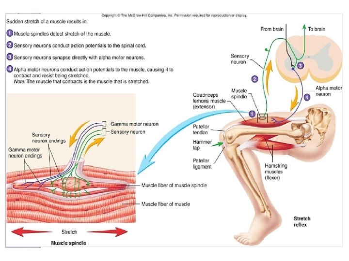
Pathway of Stretch Reflex (Reflex Arc) Arc 1. Sensory receptor (muscle spindles) 2. Afferent neuron (fast-conducting Ia and (II)nerve fibers ) 3. Integrating center (spinal cord) AHC ü alpha motor neurons synapse with the afferent sensory neurons in the spinal cord ( secrete glutamate) 4. Efferent (motor) neurons • alpha motor efferent arise from alpha motor neurons to supply extrafusal muscle fibers 5. Effector : extrafusal muscle fibers 6. Effect: Muscle contraction -Aim: To maintain muscle length

Pathway of Stretch Reflex

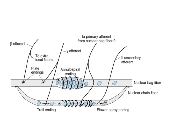
Muscle Spindles-1 Is located in the fleshy part of the muscle Consists of 3 -12 small intrafusal fibers within a capsule Each intrafusal fiber has a central (non-contractile) area (receptor), and a contractile area on each side. Intrafusal (spindle) Capsule muscle fibers Extrafusal (“ordinary”) muscle fibers Alpha motor neuron axon Contractile end of intrafusal fiber Gamma motor neuron axon Secondary (flowerspray) endings of afferent fibers (type II) Non-contractile central portion of intrafusal fiber Amani El Amin Primary (annulospiral) endings of afferent fibers (type Ia)
Nuclear bag fibres; Have")
Muscle Spindle-2 There are 2 types of intrafusal muscle fibers: 1)Nuclear bag fibres; Have a dilated central area filled with nuclei. Are 1 -3 of these fibres per spindle. 2)Nuclear chain fibres; Have nuclei which are arranged as a chain in the receptor area. Are 4 -9 of these fibres per spindle.

Muscle Spindle-3
 Innervation of the Muscle Spindle A/The primary ( annulospiral )")
Sensory ( Afferent ) Innervation of the Muscle Spindle A/The primary ( annulospiral ) ending; Group Ia endings(17 micrometers in diameter) encircle receptor areas of nuclear bag fibers mainly, but also nuclear chain fibres Send sensory signals to the CNS at the highest conduction velocity of 70 to 120 m/sec Discharge most rapidly if the muscle is suddenly stretched (dynamic response) and less rapidly (or not) during sustained stretch (static response) Measures the rate & or velocity of change in muscle length.
 Innervation of the Muscle Spindle-2 B/ Secondary ( Flower-spray )")
Sensory ( Afferent ) Innervation of the Muscle Spindle-2 B/ Secondary ( Flower-spray ) Afferents Group II fibers ( 8 micrometers in diameter), innervate ONLY the nuclear chain receptor Discharge at an increased rate throughout the period during which the muscle is being stretched , directly proportion to the degree of stretch (measure only muscle length , Static Response).
 efferent endings terminate on the peripheral contractile")
Efferent Innervation of Muscle Spindle Gamma (γ) efferent endings terminate on the peripheral contractile parts of the intrafusal muscle fibres as: • Plate endings: end mainly on the nuclear bag fibres (called dynamic gamma efferent) • Trail endings: end mainly on nuclear chain fibres ( called static gamma efferent) The function of γ- motor neurons is to regulate the sensitivity of the intrafusal muscle fibers, but HOW?

Efferent Innervation of Muscle Spindle-2 • They adjust ms spindle sensitivity • ↑ γ-MNs cause contraction of the peripheral parts of intrafusal fibers → stretch of central parts of ms spindle → ↑es the sensitivity of the ms spindle to stretch i. e. ms spindle needs a small amount of passive stretch to be stimulated

How Are Muscle Spindles Stimulated? -1 1. Passive stretch of the whole muscle: It causes stretch of the muscle spindle which lies parallel to muscle fibers.
Activation of the γ-MNs: By supraspinal centers It")
How Are Muscle Spindles Stimulated? -2 2)Activation of the γ-MNs: By supraspinal centers It causes contraction of the peripheral part the intrafusal fibres→ stretch of receptor area
. Co-activation of α- and γ- Motor Neurons:")
How Are Muscle Spindles Stimulated? -3 3). Co-activation of α- and γ- Motor Neurons: Signals from the motor cortex to the alpha motor neurons, mostly transmitted to the gamma motor neurons simultaneously, an effect called coactivation What is the significance of this coactivation? Is to keep the length of the central of reception portion of the muscle constant Oppose sudden changes in muscle length

Extrafusal skeletal muscle fiber Spinal cord Intrafusal Muscle spindle fiber Contractile end Afferent input from sensory endings of muscle spindle fiber Alpha motor neuron output to regular skeletal-muscle fiber Stretch reflex pathway (Arc) γ-motorneuron output to the contractile end of spindle fiber Am an. E Descending pathways co -a ci. Atlmvii nate α- and γ- motor neurons ? ? ?
 rapid stretch of a muscle causes synchronous strong")
Dynamic stretch reflex Sudden (phasic ) rapid stretch of a muscle causes synchronous strong burst of excitatory discharges in annulospiral afferents to the alpha motor neuron This causes the latter to send strong motor excitatory impulses to extrafusal fibers Causing sudden , jerky ( brief) muscle contraction ( jerky movement) As the muscle shortens the spindle becomes lax and ceases to discharge no more stimulation of alpha motor neuron no more excitatory impulses from alpha motor neuron to the extrafusal fibers muscle relaxes This is the basis of Tendon Jerks ( dynamic stretch reflexes ).
 Dynamic response")
Types of Responses of Ms spindle to Stretch (Types of Stretch Reflex) Dynamic response Nuclear bag (1 ry) Basal discharge Nuclear chain (1 ry & 2 ry endings Static response
 stretch of muscle Impulses from muscle spindle")
Static stretch reflex Maintained ( tonic ) stretch of muscle Impulses from muscle spindle travel through spindle afferents (mainly along secondary ending) to alpha motor neuron , stimulating it to produce muscle contraction Causing sustained ( continuous ) contraction of the muscle as long as it is stretched The Static Stretch Reflex is the basis of muscle tone which is defined clinically as resistance to muscle stretch
 Dynamic Response")
Types of Responses of Ms spindle to Stretch (Types of Stretch Reflex) Dynamic Response Stimulus Sudden stretch Receptors nuclear bag Afferents 1 ry endings Spinal cord Center Response Rapid contraction followed by rapid relaxation Examples e. g. tendon jerk Static Response Maintained (steady) stretch nuclear chain primary and secondary endings Spinal cord Maintained subtetanic contraction e. g. muscle tone

Muscle Tone Is defined as a state of continuous partial contraction of skeletal ms during rest. It is present in all skeletal ms but specially in the antigravity msmuscle (extensors of LL, back, neck, flexor of UL, muscle of abdominal wall and elevator of mandible ) Functions of Muscle Tone A)Postural control b) Help in heat production and maintain of body temperature c) It helps both the venous return & lymph flow d) Keeps viscera in position

Functions of stretch Reflex They function to oppose sudden changes in muscle length They help maintain a normal posture Damping or smoothing of muscle contraction Generation of muscle tone

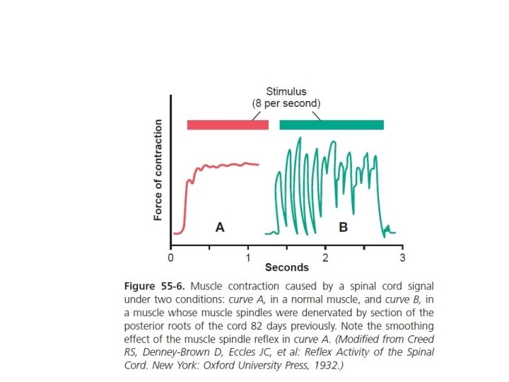
Damping or Smoothing Function of Stretch Reflex • Stretch reflex prevents oscillations or jerkiness of body movements • Motor signals from the motor areas are transmitted to the ms in an unsmooth form (↑ for few Sec and ↓ for another Sec) • This causes irregularities or oscillations of movements • The signals discharged from the ms spindles cause partial activity of αMNs of the ms • So, the motor signals find αMNs in state of partial activity, so they cause continuous activation of them → cause smooth ms contraction

Static stretch reflex AHCs 1 ry and 2 ry endings Alpha MNs Nuclear chain maintained stretch e. g. gravity

Clinical application of stretch reflex: Knee Jerk Reflex Contraction of the muscle being stretched (quadriceps) Reciprocal inhibition of the antagonistic muscle (hamstring) through reciprocal innervation inhibitory

Knee Jerk Reflex & Reciprocal Inhibition Antagonistic muscle is inhibited Significance of reciprocal Inhibition: El Amin Amani Vital in coordinating body movements

What is the Clinical Significance of Tendon Reflexes ? They are carried out clinically to test the integrity of reflex arc. A-reflexia or hypo-reflexia (hypo-tonia) indicates that the reflex arc is interrupted at one of its components by: Lesions of lower motor neuron e. g. poliomyelitis Peripheral nerve lesions e. g. peripheral neuropathy Neuromuscular junction disorder e. g. myasthenia gravis Primary muscle disorder e. g. myopathy Hyper-reflexia (hyper-tonia): exaggerated deep reflexes. Upper motor neuron lesion. Anxiety

Supraspinal control of Stretch Reflex-2

Factors that Influence Stretch Reflex Facilitation Inhibition 1. Supraspinal factors: I. II. III. IV. V. motor area 4 Vestibular nucleus Pontine Reticular Formation Neocerebellum 2. Noxious painful stimulus 3. Anxiety 4. Jendrassik-manuver suppressor area 4 and area 6 Basal ganglia , Red Nucleus. Medullary Reticular formation. Paleocerebellum ) 2. Excessive muscle stretch ( stimulation of Golgi tendon organ ). 3. Muscle contraction
 It is a reflex in which there is")
Golgi Tendon Reflex (Inverse Stretch Reflex) It is a reflex in which there is a reflex relaxation (or lengthening) of a muscles in response to excessive stretch or contraction of that muscles.

Inverse Stretch Reflex

Inverse Stretch Reflex-2 Neural pathway: Stimulus: ↑ed muscle tension by; Overstretch or Severe contraction Receptors: Golgi tendon organs 1) Site: tendons of skeletal ms in series with ms fibers 2) Structure: Are encapsulated sensory receptor 6 -20 elastic fibers 3) Innervations: Type Ib afferent fibres
 Receptors: GTOs • Stimulated by ↑ed muscle tension caused by")
Golgi Tendon Organs (GTOs) Receptors: GTOs • Stimulated by ↑ed muscle tension caused by passive overstretch or active contraction of the muscle • Afferents: • Ib fibers Center (spinal cord) : a)inhibitory interneurons→ inhibit the α-MNs supplying the same muscle b)excitatory interneurons→ excite the α-MNs supplying the antagonistic muscle Response: • Relaxation of the same muscle • Contraction of antagonistic group of muscles.

Significance This reflex protects muscle from rupture & tendon from avulsion and tear.

Comparison Between Stretch & Inverse Reflexes-1 STIMULUS Increased muscle length Inverse stretch reflex Increased muscle tension RESPONSE Muscle contraction Muscle relaxation RECEPTORS Muscle spindles Golgi tendon organs AFFERENTS Type Ia & II fibers Type Ib fibers Stretch reflex

Comparison Between Stretch & Inverse Reflexes-2 STRETCH REFLEX INVERSE STRETCH REFLEX SYNAPSES Mono-synaptic polysynaptic RECEPROCAL INNERVATION Inhibit antagonists through inhibitory interneurons Excites antagonistic muscles through excitatory interneurons PHYSIOLOGICAL SIGNIFICANCE Regulation of muscle length Genesis of muscle tone CLINICAL ASSESSMENT Sudden tap of muscle causes brisk contraction muscle jerk Regulation of muscle tension Prevent excessive increase in muscle tension & tendon avulsion (protective role) Overstretch of musclesudden muscle relaxation (lengthening reaction)

- Slides: 41