
STREPTOC OCCUS Dr R S Gopika M D
- Slides: 69
STREPTOC OCCUS Dr. R. S. Gopika M. D. (Hom. ) Professor & Ho. D Dept. of Pathology SKHMC
STREPTOCOCCUS ■ Gram-positive, ■ nonmotile, ■ catalase-negative, ■ facultatively ■ anaerobic cocci that occur in chains or pairs.
CLASSIFICATION ■ They are classified based - on their hemolytic capacity (α, ß, γ-hemolysis)
groups ■ Alpha -Colonies on blood agar are surrounded by a green zone- caused by H 2 O 2, which converts hemoglobin into methemoglobin. ■ Beta- Colonies on blood agar are surrounded by a large, yellowish hemolytic zone in which no more intact erythrocytes are present and the hemoglobin is decomposed ■ Gamma means there is no lysis.
LANCEFIELD ■ Based on the antigenicity of a C-carbohydrate occurring in their CLASSIFICATION cell walls (Lancefield antigen). ■ 20 serological types ■ Group A-V
GROUP A ■STREPTOCOCCUS S. pyogenes ■ Gram-positive cocci with a diameter of 1µm ■ chains ■ Colonies on blood agar show ß hemolysis caused by streptolysins
CULTURE ■ 37ºC ■ Grow in media containing carbohydrate or enriched with blood or serum. ■ NB – granular turbidity.
Blood agar ■ Small, . 5 - Imm , ■ circular , ■ semi transparent, ■ low convex, ■ clear haemolysis.
Selective media ■ Mannitol salt agar or MSA ■ Crystal voilet blood agar
Resistance ■ Heat – 54ºC- 30 sec
Virulence factors ■ Capsule ■ Cell wall Ag ■ Carbohydrate ■ Cytoplasmic mem Ag ■ Peptidoglycans
Capsule - hyaluronic acid – antiphagocytic Cellwall Ag – M proteins –inhibit phagocytosis, attachment T, R protein – react with myocardium Carbohydrate - react with myocardium , cardiac valves, toxic to connective tissue. Cytoplasmic mem Ag – Agenic relation with vascular intima. Peptidoglycans – exotoxins
Extracellular toxins and ■ Haemolysin- enzymes Streptolysin O, streptolysin S. Destroy the membranes of erythrocytes and other cells. Streptolysin O acts as an antigen. Past infections can be detected by measuring the antibodies to this toxin (ASO). ■ Pyrogenic streptococcal exotoxins (PSE) A, B, C. Responsible for fever, scarlet fever exanthem and enanthem, sepsis, and septic shock. The pyrogenic exotoxins are superantigens and induce production of large amounts of cytokines
Cont… ■ Streptokinase. Dissolves fibrin; facilitates spread of streptococci in tissues. ■ Hyaluronidase. Breaks down a substance that cements tissues together. ■ DNases. Breakdown of DNA, producing runny pus
Pathogenesis and clinical pictures ■ Streptococcal diseases can be classified as either acute non invasive infections, invasive infections or sequelae to them. opportunistic pathogen. It is part of the normal flora of the respiratory tract in many people.
Pathogenesis ■ diseases range from mild - like strep throat and impetigo, to severe, - like necrotizing fasciitis, and streptococcal toxic shock syndrome.
Most Common: ■ strep throat -strep pharyngitis – pharynx ■ impetigo, cellulitis, and erysipelas – infections of the skin which can be complicated by necrotizing fasciitis – skin, fascia and muscle
Less Common: Bacteremia can be associated with these infections ■ septic arthritis – joints ■ osteomyelitis – bones ■ vaginitis – in young girls ■ Meningitis – meninges ■ Sinusitis – sinuses ■ Pneumonia – pulmonary alveolus
Erysipelas Acute infection and inflammation of the dermal layer of skin. Painful red patches which enlarge and thicken
Diseases--The non-invasive infections ■ most common bacterial cause of sore throat. ■ A painful, red throat with white patches on tonsils is characteristic of pharyngitis/ strep throat. ■ It is usually accompanied by swollen lymph nodes, fever, and headache. ■ Rarely accompanied by nausea, vomiting, and abdominal pain.
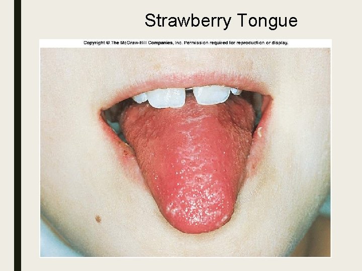
Strep Throat ■ Most common of all Strep diseases ■ Spread by saliva or nasal secretions ■ Incubation period 2 -4 days ■ Sore throat, slight fever (101ºF) ■ Rash appears and characteristic is the strawberry colored tongue
Strawberry Tongue
Scarlet fever If left untreated strep throat can lead to further complications, including scarlet fever. Scarlet fever is characterized by a red rash on the chest that may spread to the rest of the body. ■ The rash has a sandpaper-like texture and appears as tiny red pinpoints. ■ It is caused by the exotoxins ■ seen in children under ten.
Impetigo ■ Impetigo starts as a red sore and form crusty sores after a few days. ■ The sores most commonly occur on the face, but can also be found on the extremities. ■ They usually itch, but are extremely contagious though direct contact.
Cellulitus ■ begins when Streptococcus pyogenes infects the site of a minor injury, such as a bruise, burn, or wound. ■ It causes the skin to turn red, widespread pain, swelling, and tenderness of the infected area. ■ Other symptoms - fever and chills, swollen glands, blistering of the skin, and malaise
The invasive infections ■ more severe and less common. ■ This occurs when the bacteria is able to infect areas where bacteria are not usually found, such as blood and organs. ■ necrotizing fasciitis (NF), ■ streptococcal toxic shock syndrome (STSS), ■ pneumonia, ■ bacteremia.
Necrotizing fasciitis ■ is a rare, very serious infection -"flesh eating bacteria“/ Haemolytic Sterp gangrene ■ The bacteria enters the body through a minor trauma or surgical wound in immuno compromised persons ■ Causes infection just below the skin that spreads to deeper tissues. ■ acute onset, rapid ■ Early - Severe pain , minimal erythema, fever , chill, toxic apperance ■ Late – skin dusky , mottled erythema, anesthetized, extensive necrosis of subcutaneous tissue, fascia & muscles
Streptococcal Bacteremia ■ Occurs secondary to NF , rarely with pharygitis, cellulitis or pneumonia ■ it leads to endocarditis , septic arthritis, OM, viseral abcesses, TSS.
Streptococcal toxic shock syndrome ■ Due to pyrogenic exotoxins ■ Symptoms – pain, swelling, and redness of infected area, fever, dizziness, difficulty breathing, dangerously low blood pressure, and a weak, rapid pulse. ■ Shock & multi system failure
Sequelae Streprtococcal Ag show molecular mimicry with human Ag So Ab produced against previous Strep infection cross react with human tissues to produce lesions
Sequelae ■ Rheumatic fever ■ Post-streptococcal glomerulonephritis ■ PANDAS ■ Guttate psoriasis ■ Reactive arthritis
PANDAS ■ Pediatric Autoimmune Neuropsychiatric Disorders Associated With Streptococcal Infections. ■ Children ■ rapid onset of obsessive-compulsive disorder (OCD) and/or tic disorders
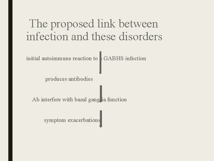
The proposed link between infection and these disorders initial autoimmune reaction to a GABHS infection produces antibodies Ab interfere with basal ganglia function symptom exacerbations.
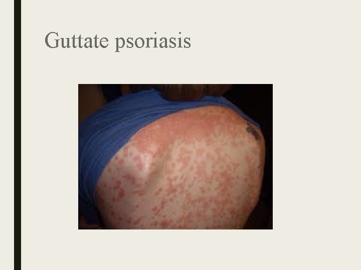
Guttate psoriasis ■ Guttate psoriasis is characterized by the acute onset of small, 110 mm diameter, erythematous, scaly, teardrop-shaped spots with a silvery scale , appear on the arms, legs, and middle of the body. ■ appears after an infection- strep throat
Guttate psoriasis
POST STREPTOCOCCAL REACTIVE ARTHRITIS ■ is a reactive arthritis characterized by a pharyngeal streptococcal infection ■ Relatively shorter latent period ( 7 to 10) days ■ May be persistent or relapsing ■ Slower response to aspirin ■ Not associated with other major manifestations ■ Symmetric invlnt. of large , small joints & axial skeleton
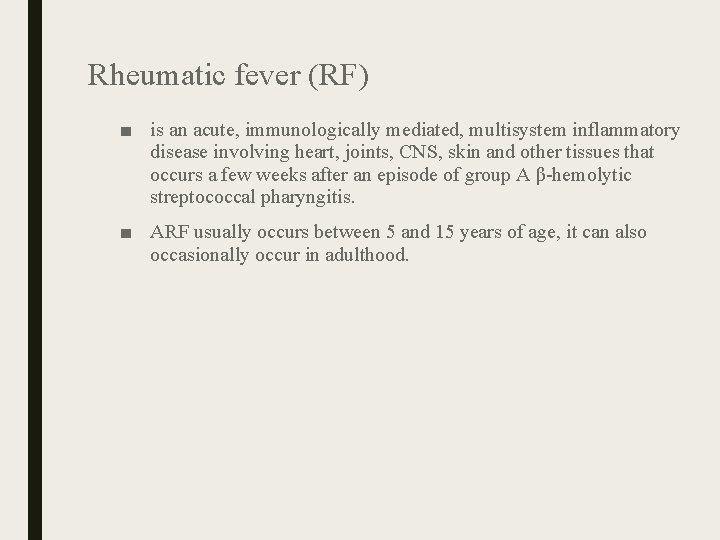
Rheumatic fever (RF) ■ is an acute, immunologically mediated, multisystem inflammatory disease involving heart, joints, CNS, skin and other tissues that occurs a few weeks after an episode of group A β-hemolytic streptococcal pharyngitis. ■ ARF usually occurs between 5 and 15 years of age, it can also occasionally occur in adulthood.
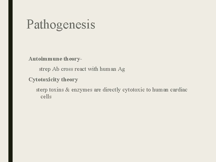
Pathogenesis Autoimmune theory strep Ab cross react with human Ag Cytotoxicity theory sterp toxins & enzymes are directly cytotoxic to human cardiac cells
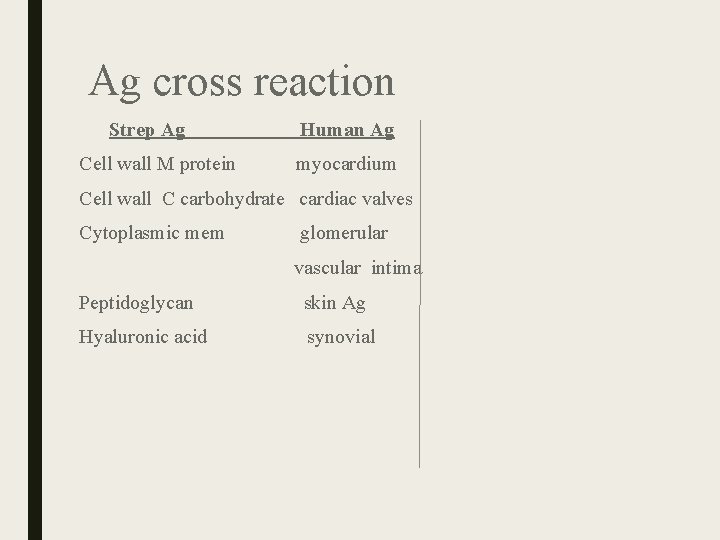
Ag cross reaction Strep Ag Human Ag Cell wall M protein myocardium Cell wall C carbohydrate cardiac valves Cytoplasmic mem glomerular vascular intima Peptidoglycan skin Ag Hyaluronic acid synovial
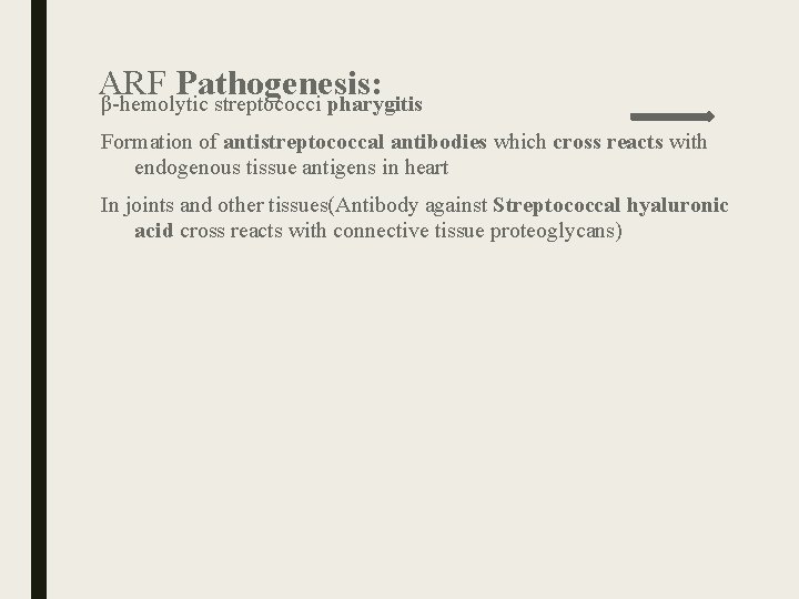
ARF Pathogenesis: β-hemolytic streptococci pharygitis Formation of antistreptococcal antibodies which cross reacts with endogenous tissue antigens in heart In joints and other tissues(Antibody against Streptococcal hyaluronic acid cross reacts with connective tissue proteoglycans)
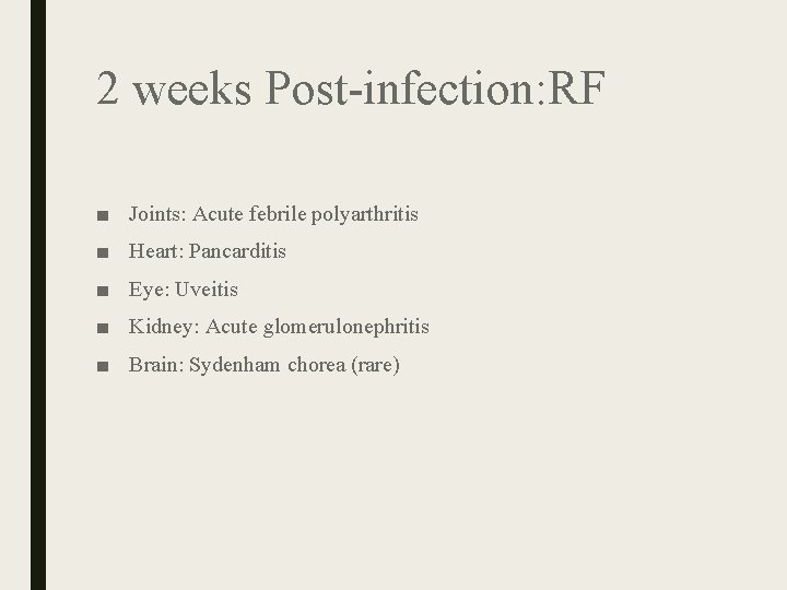
2 weeks Post-infection: RF ■ Joints: Acute febrile polyarthritis ■ Heart: Pancarditis ■ Eye: Uveitis ■ Kidney: Acute glomerulonephritis ■ Brain: Sydenham chorea (rare)
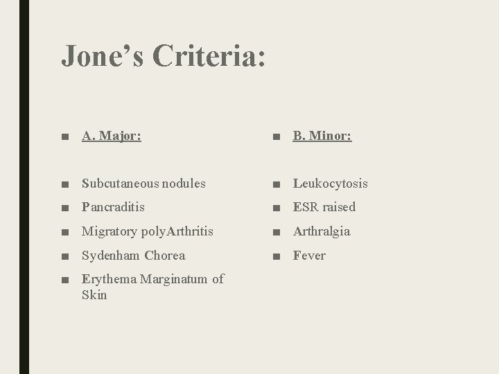
Jone’s Criteria: ■ A. Major: ■ B. Minor: ■ Subcutaneous nodules ■ Leukocytosis ■ Pancraditis ■ ESR raised ■ Migratory poly. Arthritis ■ Arthralgia ■ Sydenham Chorea ■ Fever ■ Erythema Marginatum of Skin
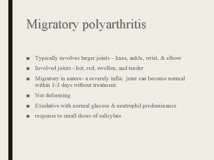
Migratory polyarthritis ■ Typically involves larger joints – knee, ankle, wrist, & elbow ■ Involved joints - hot, red, swollen, and tender ■ Migratory in nature- a severely infla: joint can become normal within 1 -3 days without treatment. ■ Not deforming ■ Exudative with normal glucose & neutrophil predominance ■ response to small doses of salicylate
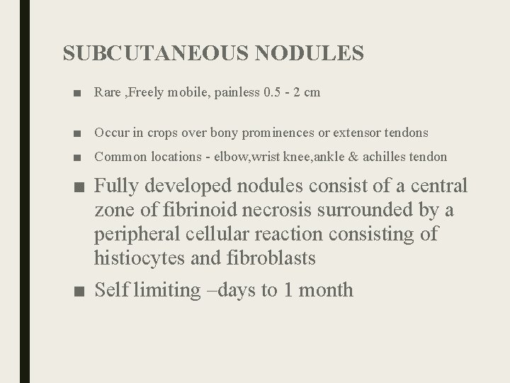
SUBCUTANEOUS NODULES ■ Rare , Freely mobile, painless 0. 5 - 2 cm ■ Occur in crops over bony prominences or extensor tendons ■ Common locations - elbow, wrist knee, ankle & achilles tendon ■ Fully developed nodules consist of a central zone of fibrinoid necrosis surrounded by a peripheral cellular reaction consisting of histiocytes and fibroblasts ■ Self limiting –days to 1 month
ERYTHEMA MARGINATUM ■ Erythematous, macular lesions with pale centers that are not pruritic ■ Multiple lesions primarily on the trunk or proximal extremities, rarely on distal extremities that recurs over weeks to months & never on face ■ It occurs early in course of RF
Uveitis ■ Uveitis is swelling of the middle layer of the eye, the uvea. ■ severe redness in the eye ■ pain ■ dark floating spots in your vision, called floaters ■ light sensitivity ■ blurred vision
Sydenham chorea/ St. Vitus dance ■ Mc in females. ■ rare > 20 yrs ■ A neurological disorder-can occur months after an initial attack, causing jerky involuntary movements, muscle weakness, slurred speech, and personality changes ■ Exacerbated by stress and disappears with sleep.
Diagnosis: Either of the following ■ Essential criteria (serologic evidence of a previous streptococcal infection) + 2 or more Major Criteria ■ 1 Major Criteria + 2 Minor Criteria
Difference between PSRA & ARF ■ PSRA begins sooner (approximately 10 days) after streptococcal infection . ■ ARF(approximately 21 days).
Rheumatic heart disease ■ is a consequence of acute rheumatic fever (RHD) ■ The risk of RHD is greater with repeated episodes of ARF. ■ RHD is particularly associated with damage to the valves of the heart. ■ Rheumatic fever is said to “lick the knee but bite the heart”.
Morphology: RHD is a sequelae of ARF A. Acute Rheumaic Heart Disease: 1. Aschoff bodies or Rheumatic granuloma 2. Pancarditis B. Chronic Rheumaic Heart Disease:
ARHD- Aschoff bodies or Rheumatic granuloma: Inflammatory myocardial lesions. Fibrinoid necrosis demarcated by: Antischkow cells (Specialized histiocytes resembling Epithelioid cells which appears catterpillar like in cross section and owl’s eye in longitudinal section) Lymphoplasmacytic infiltrate Rarely Aschoff cells (Inflammatory Giant cells)
Pancarditis: Diffuse inflammation and Aschoff Bodies in any of the 3 layers of heart ■ Pericardium: “Bread and Butter” Pericarditis (Fibrinous or Serofibrinous) ■ Myocardium: Myocarditis (Scattered Aschoff bodies within interstitial connective tissue) ■ Endocardium: Fibrinoid necrosis along the lines of closure of valves forming 1 to 2 mm vegetations (verrucae); Macculum plaques usually in left atrium
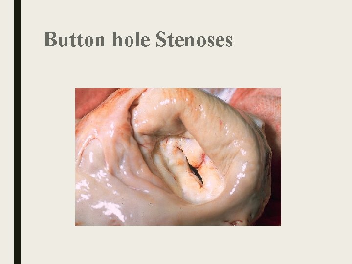
Chronic Rheumatic Heart Disease: Organization of Acute Inflammation and Subsequent Fibrosis: Valve leaflet thickening Commisural fusion and shortening Thickening and fusion of chordae tendinae “Fish mouth” or “Button hole” Stenoses: Fibrous bridging across the valvular commisures and calcification Microscopical Examination: Diffuse fibrosis and neovascularization Aschoff bodies replaced by fibrous scar
Button hole Stenoses
Functional Consequences: ■ Valvular stenosis and regurgitation ■ Stenosis > Regurgitation ■ Mitral valve alone: 70% ■ Long-standing congestive pulmonary changes: Leads to Right ventricular hypertrophy
Symptoms of RHD ■ Heart -palpitations ■ Chest pain ■ Breathlessness on exertion ■ Orthopnoea ■ paroxysmal nocturnal dyspnoea ■ oedema ■ Fainting ■ Stroke ■ Fever associated with infection of damaged heart valves.
HEART VALVES: vegetaions in ARHD Fibrosis in CRHD damaged heart valve cannot open or shut properly interferes with flow of blood through the heart failure
HEART VALVES: Complications ■ heart failure ■ The strain causes the heart to enlarge. ■ infection of damaged heart valves (infective endocarditis) ■ Clots formation in the enlarged heart & damaged valves. ■ Stroke. -clots then break off (embolise) and cause blockage in blood vessels in the brain.
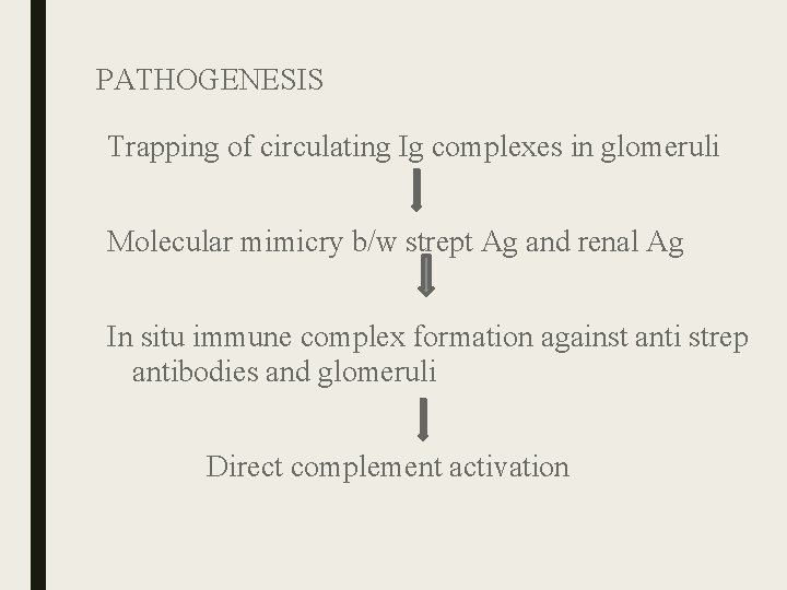
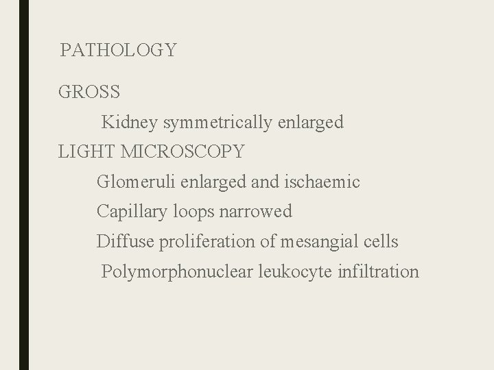
Post Streptococcal Glomerulo nephritis ■ Acute inflammation of renal glomerular parenchyma due to deposition of immune complexes characterized by sudden onset of Oliguria, Hematuria , Hypertension Edema ■ occurs 7 – 14 days after pharyngitis 2 wks – 6 wks after skin infection with group A β‐hemolytic streptococci ■ type III hypersensitivity reaction
PATHOGENESIS Trapping of circulating Ig complexes in glomeruli Molecular mimicry b/w strept Ag and renal Ag In situ immune complex formation against anti strep antibodies and glomeruli Direct complement activation
PATHOLOGY GROSS Kidney symmetrically enlarged LIGHT MICROSCOPY Glomeruli enlarged and ischaemic Capillary loops narrowed Diffuse proliferation of mesangial cells Polymorphonuclear leukocyte infiltration
Laboratory investigation ?
Group B b-Hemolytic Streptococcus ■ Staphylococcus agalactiae ■ Colonize the urogenital tract of pregnant women
invasive diseases ■ Cause invasive diseases in newborns Early-onset infection Late-onset disease Early-onset infection – – Occurs in neonates who are less than 7 days old Vertical transmission of the organism from the mother Manifests in the form of pneumonia or meningitis with bacteremia Associated with a high mortality rate
■ Late-onset infection – Occurs between 1 week and 3 months after birth – Usually occurs in the meningitis form – Mortality rate is not as high as early-onset In adults – Occurs in immunosuppressed patients or those with underlying diseases – Often found in a previously healthy adult who just experienced childbirth
Laboratory Diagnosis: Group B b-Hemolytic Streptococcus ■ Colony morphology – Grayish-white, mucoid, creamy, narrow zone of b-hemolysis ■ Presumptive Identification tests – Catalase-negative
Enterococcus ■ Members of the gut flora ■ Associated infections – – Bacteremia Urinary tract infections Wound infections Endocarditis
Peptostreptococci ■ Normal flora of skin, mouth, intestine & vagina ■ Puerperal sepsis, skin & soft tissue infections & brain abcesses ■ Necrotizing lesions- necrotizing fascitis, necrotizing pneumonitis. ■ Meleneys gangrene- rare infection of superfical fascia.
