Strabismus Amblyopia Leukocoria Saeed Alwadani MD Assistant Professor

Strabismus, Amblyopia & Leukocoria Saeed Alwadani, MD Assistant Professor Consultant Ophthalmologist Ophthalmology department King Saud University 28/9/17

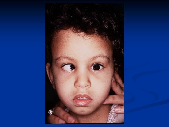
What is Strabismus ? Ocular misalignment due to abnormality in binocular vision or anomalies in neuromuscular control of ocular motility Strabismus has an inherited pattern, i. e. , it is much more likely to occur if one or both parents are affected. However, many cases occur without any family history of the disorder

Classification of Strabismus: According to fusion status 1. Phoria Latent tendency of the eye to deviate and controlled by fusional mechanism 2. Intermittent Phoria Fusion control is present part of the time 3. Tropia Manifest misalignment of the eye all the time

Classification of Strabismus: According to fixation 1. Alternating Spontaneous alternation of fixation from one eye to the other 2. Monocular Preference of fixation with one eye

Classification of Strabismus: According to type of deviation 1. Horizontal Esodeviation Exodeviation 2. Vertical Hyperdeviation Hypodeviation 3. Torsional Incyclodeviation Excyclodeviation 4. Combined

Classification of Strabismus: According to age of onset 1. Congenital 2. Acquired

Classification of Strabismus: According to variation of the deviation with gaze position or fixing eye 1. Comitant Same deviation in different direction of gaze 2. Incomitant Variable deviation in different direction of gaze usually in paralytic or restrictive type of strabismus

Examination 1. History 2. Inspection 3. Assessment of monocular eye function Visual acuity Preverbal children CSM OKN Preferential looking Visual evoked potential

Examination Assessment of monocular eye function Visual acuity Verbal children Symbol tests Single illiterate E Allen pictures H O T V letters
 3. Cover")
Examination Assessment of binocular eye function 1. Hirschberg test 2. Krimski’s test(measuring) 3. Cover test 4. Alternate cover test 5. Prism cover test

Examination Fundoscopy Cycloplegic refraction • Tropicamide • Cyclopentolate • Atropin

Type of Strabismus Esotropia • • Pseudoesotopia Infantile esotropia Accommodative esotropia Partially accommodative esotropia

Pseudoesotropia • Occur in patients with flat broad nasal bridge and prominent epicanthal fold • Gradually disappear with age • Hirschberg test differentiate it from true esotropia

Infantile Esotropia • Common comitant esotropia occur before six month of age • Deviation is often large more than 40 prism diopter • Frequently associated with nystagmus and inferior oblique over action • Treatment Correction of refractive error Treat amblyopia Surgical correction of strabismus

Accommodative Esotropia • Occur around 2 ½ years of age • Start as intermittent then become constant • High hypermetropia • Treatment Full cycloplegic correction Treat amblyopia

Partially Accommodative Esotropia • Improve partially with glasses • Treatment Full cycloplegic correction Treat amblyopia Surgical correction of strabismus

Type of Strabismus Exotropia • Intermittent exotropia • Constant exotropia • Sensory exotropia

Intermittent exotropia • • Onset of deviation within the first year of age Closing one eye in bright light Usually not associated with any refractive error Usually not associated with amblyopia • Treatment Correction of any refractive error Surgical correction of strabismus

Constant exotropia • Maybe present at birth or maybe progress from intermittent exotropia • Treatment Correction of any refractive error Correction of amblyopia Surgical correction of strabismus

Sensory exotropia • Constant exotropia that occur following loss of vision in one eye e. g trauma • Treatment Correction of any organic lesion of the eye Correction of amblyopia Surgical correction of strabismus

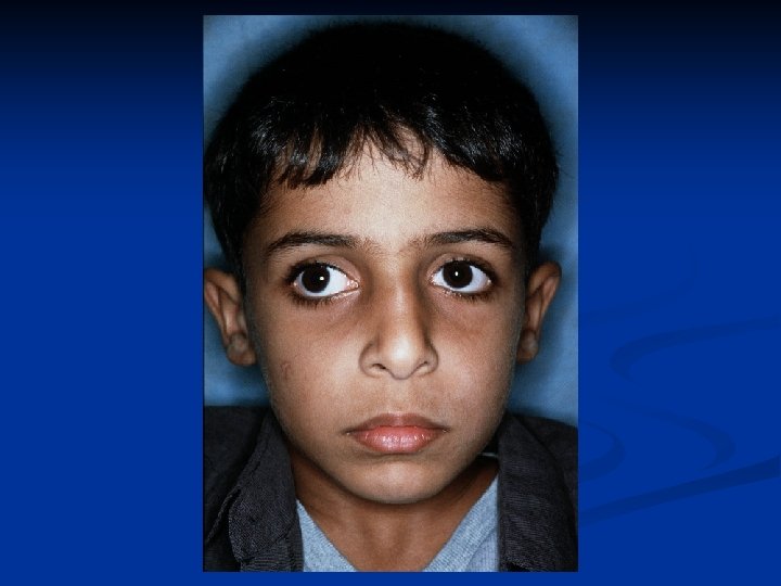
Types of Strabismus Paralytic strabismus • 6 th nerve palsy • 4 th nerve palsy • 3 rd nerve palsy

6 th Nerve Palsy • Incomitant esotropia • Limitation of abduction • Abnormal head position

4 th Nerve Palsy • Congenital or acquired • Hypertropia of the affected eye with excyclotropia • Abnormal head position

3 rd Nerve Palsy • Congenital or acquired • Exotropia with Hypotropia of the affected eye • In children caused by: trauma, inflammation, post viral and tumor • In adult caused by: aneurysm, diabetes, neuritis, trauma, infection and tumor

Special Types of Strabismus • Duane strabismus • Brown syndrome • Thyroid opthalmopathy

Duane Syndrome • Limitation of abduction • Mild limitation of adduction • Retraction of the globe and narrowing of the palpebral fissure on adduction • Upshoot or downshoot on adduction • Pathology faulty innervation of the lateral rectus muscle by fibers from medial rectus leading to co-contraction of the medial rectus and lateral rectus muscles

Brown Syndrome • Limitation of elevation on adduction • Restriction of the sheath of the superior oblique tendon • Treatment needed in abnormal head position or vertical deviation in primary position

Thyroid Ophthalmopathy • Restrictive myopathy commonly involving inferior rectus, medial rectus and superior rectus • Patients presents with hypotropia, esotropia or both

Surgery of Extraocular Muscle • Recession : weakening procedure where the muscle disinserted and sutured posterior to its normal insertion

Surgery of Extraocular Muscle • Resection : strengthening procedure where part of the muscle resected and sutured to its normal insertion

Complication of Extraocular Muscle Surgery • Perforation of sclera • Lost or slipped muscle • Infection • Anterior segment anesthesia • Post operative diplopia • Congectival granuloma and cyst

Amblyopia
 Amblyopia refers to a decrease of vision, either")
What is Amblyopia ? (Lazy Eye) Amblyopia refers to a decrease of vision, either unilaterally or bilaterally, for which no cause can be found by physical examination of the eye 2%– 4% of U. S. population affected

Amblyopia Three critical periods of human visual acuity development have been determined. During these time periods, vision can be affected by the various mechanisms to cause or reverse amblyopia. These periods are as follows: • The development of visual acuity from the 20/200 range to 20/20, which occurs from birth to age 3 -5 years. • The period of the highest risk of deprivation amblyopia, from a few months to 7 or 8 years. • The period during which recovery from amblyopia can be obtained, from the time of deprivation up to the teenage years or even sometimes the adult years

Amblyopia Diagnosis of amblyopia usually requires a 2 -line difference of visual acuity between the eyes

Causes of Amblyopia Many causes of amblyopia exist; the most important causes are as follows: Anisometropia • Inhibition of the fovea occurs to eliminate the abnormal binocular interaction caused by one defocused image and one focused image. • This type of amblyopia is more common in patients with anisohypermetropia than anisomyopia. Small amounts of hyperopic anisometropia, such as 1 -2 diopters, can induce amblyopia. In myopia, mild myopic anisometropia up to -3. 00 diopters usually does not cause amblyopia.

Causes of Amblyopia Strabismus The patient favors fixation strongly with one eye and does not alternate fixation. This leads to inhibition of visual input to the retinocortical pathways. Incidence of amblyopia is greater in esotropic patients than in exotropic patients Alternation with alternate suppression avoids amblyopia

Causes of Amblyopia Visual deprivation Amblyopia results from disuse or understimulation of the retina. This condition may be unilateral or bilateral. Examples include cataract, corneal opacities, ptosis, and surgical lid closure Deprivation Amblyopia Bilateral Deprivation Amblyopia

Causes of Amblyopia Organic Structural abnormalities of the retina or the optic nerve may be present. Functional amblyopia may be superimposed on the organic visual loss

Causes of Amblyopia Ametropic Amblyopia Uncorrected high hyperopia is an example of this bilateral amblyopia.

SCREENING: IMPORTANCE Amblyopia is usually preventable or treatable n Early detection is key to effective treatment n Life-threatening disorders may present as amblyopia n Screening responsibility rests with primary care physician n

AMBLYOPIA: EARLY DETECTION Assess red reflex n Determine visual acuity n Evaluate ocular alignment n

Direct ophthalmoscope

Direct ophthalmoscope: assessing red reflex

Direct ophthalmoscope: examining retina

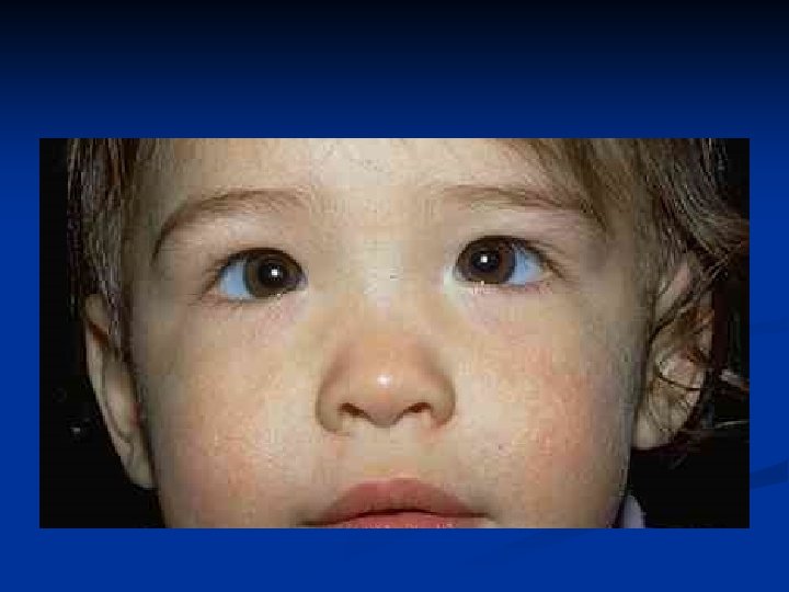
Normal red reflex

Asymetric red reflex

Treatment The clinician must first rule out an organic cause and treat any obstacle to vision (eg, cataract, occlusion of the eye from other etiologies). Remove cataracts in the first 2 months of life, and aphakic correction must occur quickly Treatment of anisometropia and refractive errors must occur next The next step is forcing the use of the amblyopic eye by occlusion therapy


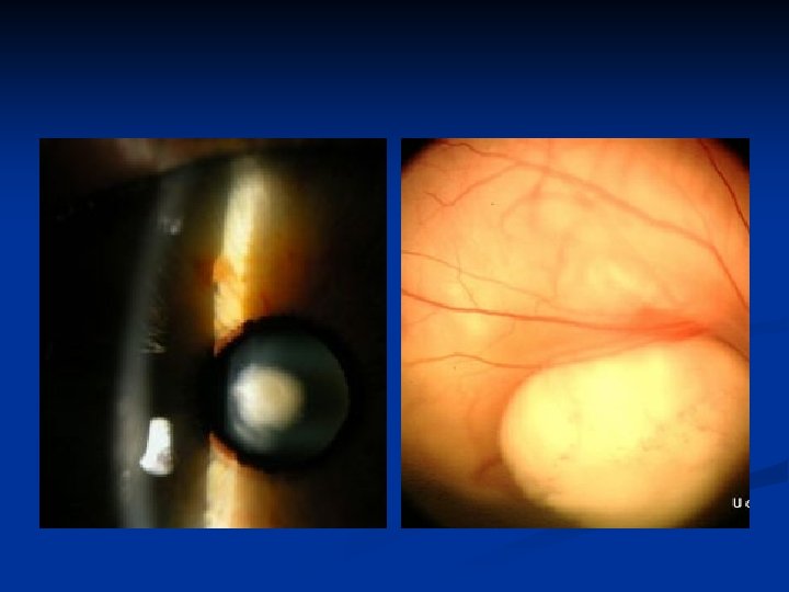
Leukocoria A white pupillary reflex

n Leukocoria in a child requires urgent attention , primarily because in most patients with retinoblastoma it is the first sign noticed.

n Secondarily , a white pupil indicates a severely amblyopiogenic condition , which may be treatable. n Anatomic location is important in the diffential diagnosis of Leukocoria

Causes of Leukocoria n n n n n Cataract Retinoblastoma Toxocariasis Coat´s disease ROP PHPV Retinal detachment Coloboma Retinal dysplasia Norrie´s disease

Work –up 1 - History 2 - Complete ocular examination 3 - B Scan ultrasonoghraphy 4 - Intravenous fluorescein angiogram (coats disease , ROP, retinoblastoma ) 5 - CT or MRI 6 - Serum ELISA

cataract • opacification of the lens. • Congenital cataracts usually are diagnosed at birth.

n Unilateral cataracts are usually isolated sporadic incidents

• Bilateral cataracts are often inherited and associated with other diseases. • They require a full metabolic, infectious, systemic, and genetic workup. • The common causes are hypoglycemia, trisomy (eg, Down, Edward, and Patau syndromes), myotonic dystrophy, infectious diseases (eg, toxoplasmosis, rubella, cytomegalovirus, and herpes simplex [TORCH]), and prematurity

RETINOBLASTOMA n Retinoblastoma is the most common intraocular tumor of childhood. CLINICAL MANIFESTATIONS n Leukocoria (60%) n Strabismus (20%) n OTHER- Uveitis, Orbital cellulitis, Hyphaema, Heterochromia, Glaucoma, Bupthalmos

RETINOBLASTOMA

Retinoblastoma Calcification is another feature of retinoblastomas, usually occurring in necrotic areas. Calcium stains with H&E. It is worth identifying calcium in suspect eyes by ultrasound, or CT scan to differentiate retinoblastomas from other tumours.

MANAGEMENT EMPIRICAL GENETIC COUNSELLING n ENUCLEATION n unilateral, poor visual prognosis n PLAQUE n 4 -12 mm +/- vitreous seeding n EXTERNAL BEAM n >12 mm, multiple foci, only eye n LASER n consider- indirect, xenon arc n cryotherapy if <2 dd in size n CHEMOTHERAPY, if intracranial extension
 n A gray-yellow retrolental membrane may produce leukocoria, with")
Persistent hyperplastic primary vitreous (PHPV) n A gray-yellow retrolental membrane may produce leukocoria, with the subsequent suspicion of retinoblastoma. n In PHPV, the globe is white and slightly microphthalmic. Patients have no history of prematurity or oxygen administration.
 n Vasoproliferative retinopathy affecting premature infants exposed to high oxygen")
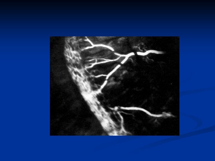
RETINOPATHY OF PREMATURITY (ROP) n Vasoproliferative retinopathy affecting premature infants exposed to high oxygen INCIDENCE n Prematurity (<32/40) n Birth weight (30% < 1000 gm affected) n Oxygen duration n 90% ROP regresses spontaneously, 5% blindness
 Signs include: • neovascularization, • fibrous bands • retinal detachments")
RETINOPATHY OF PREMATURITY (ROP) Signs include: • neovascularization, • fibrous bands • retinal detachments • vitreous hemorrhage • leukocoria

COLOBOMA n Optic Disc Coloboma n Due to failure of closure of foetal fissure inferiorly n May be isolated disc or associated chorioretinal coloboma n Usually sporadic, some AD n Can be bilateral n Visual acuity varies from normal to NPL.

- Slides: 73